Evaluating the Toxicological Complexity of New Psychoactive Substances through Metabolomics: A Review of the Advances, Challenges, and Perspectives
Abstract
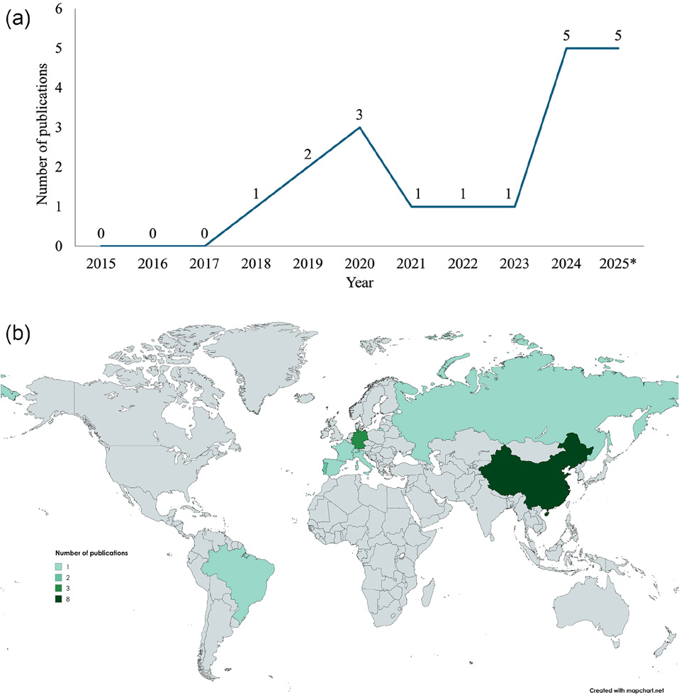
New psychoactive substances (NPS) have become a global challenge due to their rapid emergence, structural variability, and capacity to evade conventional drug regulations. Developed to mimic the effects of controlled substances, these compounds often exhibit unpredictable pharmacological properties and complex metabolic pathways, complicating forensic detection and toxicological assessment. In this context, metabolomics has emerged as a powerful analytical strategy to investigate the biochemical consequences of NPS exposure, allowing the identification of endogenous metabolic disturbances and affected pathways. When integrated with advanced chemometric methods, metabolomics offers valuable insights into mechanisms of toxicity, supports the discovery of exposure and effect biomarkers, and improves the understanding of interspecies variability in toxic responses. This review summarizes original research articles (n = 19) published between 2015 and 2025, evaluating several NPS classes such as synthetic cathinones, synthetic cannabinoids, tryptamines, synthetic opioids, and other emerging psychoactive compounds, demonstrating the current applications of metabolomics in NPS research. Therefore, this work emphasizes the contribution of metabolomics to toxicological, pharmacological, and analytical investigations of NPS, outlining key findings related to their mechanisms of action and toxicity, as well as perspectives for future research.
Keywords:
toxicometabolomics; liquid chromatography-mass spectrometry; drugs of abuse; in vivo; in vitro
1. Introduction
New psychoactive substances (NPS) have emerged as a global concern due to their rapid spread, structural diversity, and ability to evade traditional drug control laws.1 Designed to mimic the effects of conventional drugs while circumventing legal restrictions, these compounds encompass a wide range of chemical classes, including synthetic cannabinoids (SCRAs), synthetic cathinones (SCs), synthetic opioids, tryptamines, and phenethylamines.1-3 Their continuous synthesis and distribution pose major challenges for forensic and clinical toxicology, regulatory frameworks, and public health systems. According to the European Union Drugs Agency (EUDA), more than 930 NPS have been detected in Europe, while the United Nations Office on Drugs and Crime (UNODC) highlights the global nature of this phenomenon, reporting 50-100 newly identified NPS each year.1-3
The consumption of NPS is frequently associated with severe health risks, ranging from acute intoxications to unknown long-term effects. Synthetic opioids, for example, have been linked to numerous fatal overdoses due to their markedly higher potency compared with traditional opioids such as morphine and heroin.1,2 Synthetic cannabinoids and cathinones have been implicated in multiple intoxication events and serious clinical manifestations, including psychosis, seizures, and cardiovascular collapse.1,4,5 Even minor structural modifications can significantly alter toxicokinetics and toxicodynamics, producing metabolites with equal or greater toxicity than the parent compound and complicating comparisons with traditional drugs of the same class.1,3,4 Studies6-8 have also revealed unexpected metabolic pathways for NPS, such as the formation of amino acid adducts in cathinones, underscoring the need for comprehensive metabolic investigations.
Although marketed as “legal highs” or “research chemicals,” NPS are used across diverse populations, with particularly high prevalence among adolescents and young adults.1-3 Additionally, the coronavirus (COVID-19) pandemic reshaped drug markets and further reinforced the circulation of NPS as substitutes for traditional illicit drugs.2-4 Identifying NPS in routine clinical and forensic analyses (e.g., immunoassays and liquid chromatography-tandem mass spectrometry, LC-MS/MS) is challenging due to the limited availability of reference standards and validated analytical methods, their typically low concentrations in biological matrices such as blood, oral fluid, or urine, and the frequent absence of established metabolic information regarding phase I and phase II pathways.2-4 Together, these factors hinder risk assessment and complicate the development of effective diagnostic and therapeutic strategies.5,6,8,9
Metabolomics is the systematic study of metabolites in biological systems and of changes in metabolite concentrations or biochemical pathways resulting from genetic or environmental perturbations.10 This approach is particularly valuable in toxicological studies (toxicometabolomics), as it enables the characterization of biochemical alterations following different exposure scenarios, the detection of subtle physiological changes, and the elucidation of systemic responses.10 Consequently, metabolomics has become a powerful tool for investigating NPS toxicity, allowing the detection of both endogenous metabolic disruptions and xenobiotic metabolites.7,8,11 Metabolomics are commonly performed using liquid chromatography coupled to high-resolution mass spectrometry (LC-HRMS) or nuclear magnetic resonance (NMR) spectroscopy, in combination with advanced statistical and chemometric tools - such as principal component analysis (PCA) and partial least squares discriminant analysis (PLS-DA) - to support biomarker discovery and studies of toxicodynamics, toxicokinetics, and interspecies variability.8,9,12
Metabolomics can be broadly divided into targeted analysis, which quantifies predefined metabolites, and untargeted analysis, which identifies a wide range of metabolites without prior assumptions.8-10 Untargeted approaches are particularly valuable for NPS because they can reveal unexpected xenobiotic metabolites and key biomarkers of toxicity.8-10 Recent advances - including computational metabolomics and in silico modeling - have further expanded analytical capabilities through multiscale data integration.7,9,12 However, the structural elucidation of unknown metabolites remains a major challenge, often requiring the combined use of spectral libraries and in silico fragmentation prediction tools.10-12 These analytical workflows, although powerful, underscore the need for greater standardization to support their implementation in forensic, clinical, and regulatory contexts.9,11,12
Despite advances in the field, metabolomics applications in NPS toxicology remain fragmented. There is a clear need to consolidate current evidence, identify knowledge gaps, and outline the key challenges and future perspectives in this rapidly evolving area.1,6,12 In this sense, this review aimed to critically synthesize metabolomics-based investigations of the toxicology from different NPS classes.
2. Review Methodology
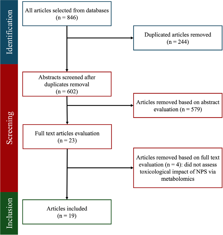
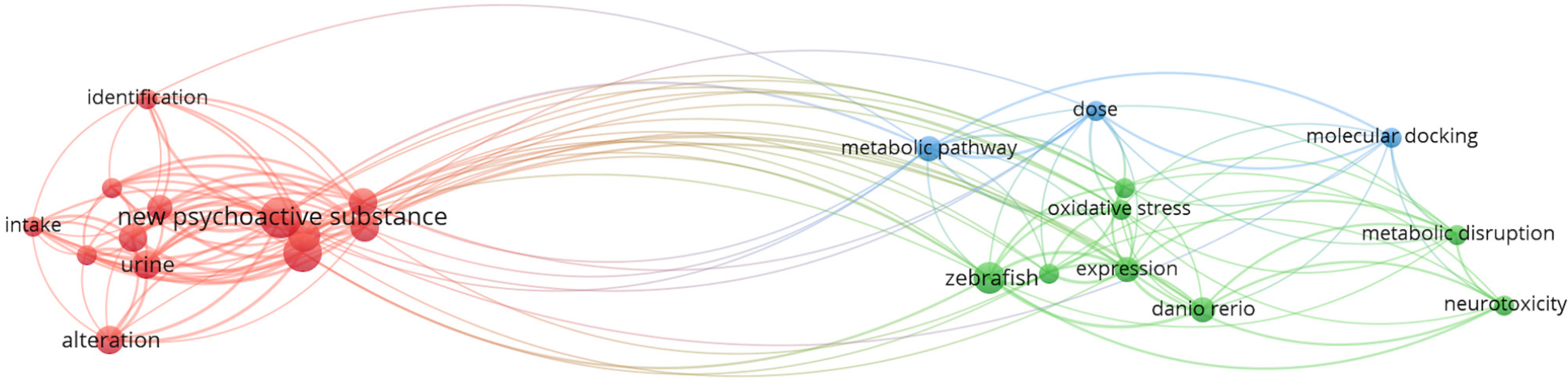
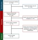
To compose this review, a total of 846 articles were retrieved through searches conducted in the Embase, Web of Science, ScienceDirect, Scopus, and PubMed databases, selected to ensure broad coverage of biomedical, toxicological, and analytical chemistry literature and to minimize database-specific bias. The search strategy employed the keywords “metabolomics”, “new psychoactive substances”, “in vivo”, “in vitro” and “human,” using the Boolean operators ‘AND’ and ‘OR’. These terms were chosen to capture studies investigating the biological and toxicological effects of NPS through metabolomics-based approaches across experimental and clinical contexts. The search period ranged from 2015 to November 2025, reflecting the period of significant expansion in both NPS emergence and high-resolution metabolomics applications. Only peer-reviewed articles published in English were included. Studies were considered eligible if they (i) investigated one or more NPS, (ii) applied metabolomics or untargeted metabolic profiling methodologies, and (iii) evaluated toxicological, pharmacological, or biological effects in in vitro, in vivo, or human models. Articles focusing exclusively on analytical method development for drug detection without interpretation of metabolic alterations or toxicological impact were excluded. After the removal of duplicates (n = 244), 602 records remained for title and abstract screening, from which 23 articles were selected for full-text evaluation. Four articles were subsequently excluded because they did not assess the toxicological impact of NPS within a metabolomics context. Consequently, 19 articles were included in this review. Figure 1 presents the flow diagram of the article selection process, while Figure 2 illustrates the keyword co-occurrence map, highlighting the predominant topics identified in the titles and abstracts. The most relevant terms cluster into distinct groups, representing different subfields within the topic. It is important to acknowledge inherent methodological limitations. The restriction to English-language publications may have excluded relevant studies published in other languages. Furthermore, the reliance on predefined keywords may have led to the omission of studies applying metabolomics approaches. Finally, the heterogeneity of experimental models, analytical platforms, and reporting standards across studies may limit direct comparability of findings.

Keyword co-occurrence map generated using VOSviewer (version 1.6.20; Centre for Science and Technology Studies, Leiden University, Netherlands, 2023) considering the titles and abstracts from the selected articles. The strength of the co-occurrence relationships between keywords is illustrated by the thickness of the connecting lines, highlighting conceptual associations. The frequency of each term is represented by the size of the corresponding node, reflecting its relative relevance within the analyzed literature. Related terms are grouped into thematic clusters based on their co occurrence patterns, with each cluster visually distinguished by a different color.
3. Toxicometabolomics of New Psychoactive Substances
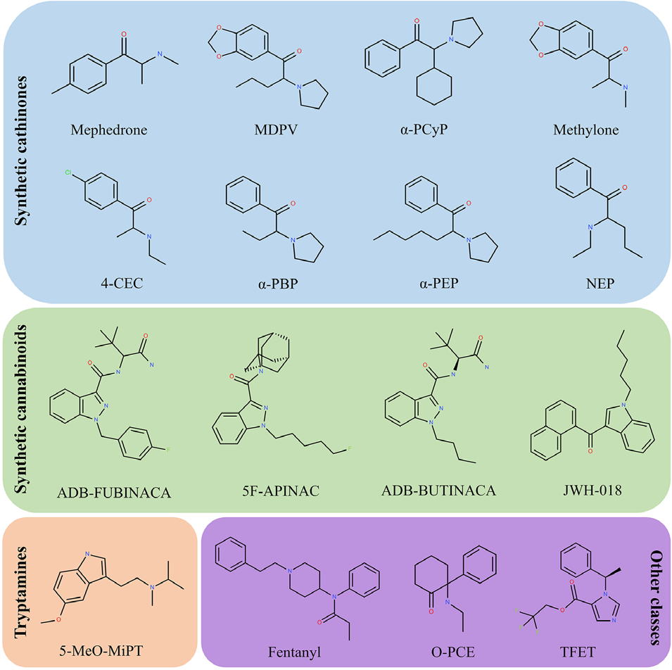
As described by Araújo et al.,13 toxicometabolomics is a subfield of metabolomics used to investigate toxic agents by characterizing their mechanisms of toxicity and identifying biomarkers, target organs, metabolic responses, and altered biological pathways.13 Among the 19 articles selected for this review, only three explicitly use the term “toxicometabolomics” to describe studies involving NPS; however, the remaining 16 studies also employ metabolomic techniques to investigate these substances and can therefore be considered toxicometabolomic evaluations.6,14,15Table 1 provides an overview of all 19 included studies, detailing the NPS investigated, experimental models and matrices used, study designs, analytical platforms, altered metabolites or metabolic pathways, and corresponding biological interpretations. Additionally, a comprehensive overview of the molecular structures of all substances evaluated in this review was illustrated in Figure 3.
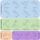
Molecular structures of the new psychoactive substances (NPS) evaluated in this study, organized according to their chemical class.
Synthetic cathinones constitute the most extensively evaluated class of NPS, appearing in nine of the 19 studies. Synthetic cannabinoid receptor agonists (SCRAs) follow, with six studies. Notably, one article examined both classes within the same investigation. Tryptamines were assessed in two studies, whereas synthetic opioids, ketamine derivatives, and sedatives/hypnotics were each represented by a single study. The most used analytical platforms were LC-HRMS, LC-MS/MS, and gas chromatography-mass spectrometry (GC-MS). In vitro studies employed primary mouse hepatocytes (PMH), pooled human liver microsomes (pHLM), and hepatic cell lines such as HepaRG, HepG2, and Huh7. In vivo studies included human, rat, and mouse samples, as well as zebrafish embryos, larvae, and adults.
3.1. Synthetic cathinones
Synthetic cathinones (SCs) are a growing public health concern due to their widespread popularity and the associated risks of morbidity and mortality.17,18,31-33 These compounds are chemically derived from cathinone, a naturally occurring alkaloid found in the Catha edulis plant, whose leaves have been used for millennia in African and Arabic regions for their psychostimulant effects.33 Cathinone’s structural core resembles that of a β-ketone phenethylamine, and synthetic derivatives typically involve modifications to both the phenyl ring and the alkylamine side chain.33,34 The first two synthetic derivatives-methcathinone (ephedrone) and its 4-methyl analogue, mephedrone-were synthesized in the late 1920s.33,34 Although discovered in the mid-20th century, these compounds only gained widespread attention in the recreational drug market decades later, emerging as “legal highs” because most were not yet regulated.33,34 This “cathinone wave,” particularly across the Eastern Hemisphere, led to significant public health concerns due to increasing cases of intoxication and death.33,34
SCs represent a major class of NPS and have been widely detected over the past decades across Europe, Asia, the United States, Canada, South America, Australia, and Africa.35 Notably, they have become established as relatively affordable alternatives within the illicit drug market.32 In Europe, for instance, 178 SCs were identified in 2023, and the volume of imported and seized SCs in that same year was three times higher than the combined quantity of amphetamine and methamphetamine seizures.32 In Brazil, following a decline in SC detections-from more than 200 cases in 2018 to fewer than 10 in 2021-a resurgence was observed in 2022 (37 detections) and 2023 (53 detections), accounting for 15% of all NPS identified in the most recent report from the Ministry of Justice and Public Security.31
Synthetic cathinones are known for their potent psychostimulant and entactogenic effects, which resemble those of classical recreational drugs such as cocaine and 3,4-methylenedioxymethamphetamine (MDMA).17,18 They are commonly encountered as crystal-like powders and are typically consumed orally, nasally, by smoking, or via intravenous administration.36,37 Their primary mechanism of action involves increasing extracellular concentrations of monoamines - such as serotonin, dopamine, and norepinephrine - by disrupting the normal function of their membrane reuptake transporters.38 The toxic effects of SCs are diverse and can be severe, including cardiovascular manifestations such as tachycardia and hypertension, as well as psychiatric and neurological symptoms such as psychosis and seizures.36,37
Given the rapid proliferation and structural diversity of SCs, understanding their toxicological profiles and mechanisms of action is essential for risk assessment and for the development of effective detection and treatment strategies.16 In this context, non-targeted analytical approaches such as metabolomics have proven highly valuable for detecting, characterizing, and elucidating the metabolic and toxicological effects of these substances.39-41
The application of metabolomics is a crucial strategy for navigating the complex and rapidly evolving landscape of SCs. Araújo et al.17 investigated the metabolic effects of 3,4-methylenedioxypyrovalerone (MDPV) in primary mouse hepatocytes (PMH) under normothermic (37 °C) and hyperthermic (40.5 °C) conditions using a sensitive untargeted GC-MS metabolomics approach.17 The metabolomic workflow applied in this study did not include the use of internal standards (IS) during sample preparation, nor did it report system suitability testing (SST) or injection randomization, potentially limiting the assessment of analytical robustness and increasing susceptibility to bias.17 However, pooled QC samples were incorporated into the analytical sequence, and features exhibiting a coefficient of variation (CV) greater than 30% were excluded.17 Metabolite annotation was supported by reference materials and the NIST 2014 library, with confidence levels assigned in accordance with the Metabolomics Standards Initiative (MSI) criteria.42
The results showed that under normothermic conditions - and even at subtoxic concentrations - MDPV exposure markedly disrupted ascorbate and pyruvate metabolism, as well as the tricarboxylic acid (TCA) cycle.17 Reductions in key metabolites, including fumarate, malate, and aspartate, were accompanied by increases in glucuronic acid and arabitol.17 These alterations suggest adenosine triphosphate (ATP) depletion, impaired antioxidant defenses, and β-oxidation favoring as a source of fatty acid production to obtain energy.17 Furthermore, dysfunction in the pentose phosphate pathway was indicated, while elevated levels of glycating agents in the extracellular volatilome suggested potential deoxyribonucleic acid (DNA) damage, cell death, and increased oxidative stress.17
Interestingly, several of these metabolic disturbances mirrored those seen in MDMA-induced hepatotoxicity, indicating possible shared mechanisms of toxicity.17 Importantly, co-exposure to hyperthermia exacerbated hepatic metabolic dysfunctions independently of concentration.17 Additional disruptions were observed in amino acid metabolism - including phenylalanine, alanine, glutamate, glutamine, arginine, and proline - as well as alterations in aminoacyl-transfer RNA (-tRNA) biosynthesis and butanoate metabolism.17 A particularly relevant finding was the significant decrease in intracellular glutamate, which suggests impaired glutathione (GSH) synthesis, leading to reduced antioxidant capacity and heightened oxidative stress.17 Collectively, these results highlight the sensitivity of the liver to MDPV-induced hepatotoxicity, especially under heat stress, and underscore the value of untargeted metabolomics for elucidating mechanisms of NPS toxicity.17
Steuer et al.16 applied an untargeted LC-HRMS metabolomics approach to plasma samples from controlled human administration studies to investigate the metabolic effects of mephedrone.16 Regarding the analytical pipeline, the authors used SST throughout the analytical sequence, incorporated IS during sample preparation, and used randomized injection sequences, with QC samples injected every five runs and data filtering based on a maximum 30% CV in QC samples.16 However, extraction blanks and conditioning QCs were not described.16
Both the parent compound and its metabolite, 4-carboxymephedrone, were detected, with peak concentrations observed three hours after administration and lower levels still measurable after six hours.16 Mephedrone intake significantly altered endogenous metabolites, particularly long-chain fatty acids and steroids, most of which showed increased levels.16 When compared with MDMA and amphetamine, the metabolic profiles revealed overlapping effects - especially regarding linoleic acid and pregnenolone sulfate - although mephedrone’s pattern more closely resembled that of amphetamine.16 Glycoursodeoxycholic acid levels decreased in a time-dependent manner following mephedrone administration, a trend similarly observed with amphetamine.16 In contrast, cortisol levels remained unchanged in mephedrone-treated individuals, differing from the increases typically seen after MDMA use.16 Metabolite annotation was supported by databases including METLIN, HMDB, NIST 2014, and LipidBlast, and the levels of confidence were reported following MSI recommendations.16
Pathway analysis indicated alterations in aminoacyl-tRNA biosynthesis, linoleic acid metabolism, unsaturated fatty acid biosynthesis, bile acid biosynthesis, steroid hormone biosynthesis, and tyrosine metabolism - collectively suggesting increased energy demand.16 These findings are valuable for improving toxicological screening procedures, particularly in matrices such as urine, where the parent compound may already be absent.16 Nevertheless, the authors emphasize that although their dataset is comprehensive, in vivo studies are still necessary to fully elucidate the physiological effects of these substances in living organisms.16
In a combined in vitro and in vivo toxicometabolomics study, Hemmer et al.14 investigated the metabolism of 2-cyclohexyl-1-phenyl-2-(1-pyrrolidinyl)-ethanone (PCYP), a synthetic cathinone with potent dopaminergic activity.14 Using pooled human liver microsomes (pHLM) and male Wistar rats, the authors conducted an untargeted LC HRMS analysis and successfully identified sixteen phase I metabolites and one phase II metabolite as exogenous biomarkers.14 Notably, PCYP itself was detected only in vitro, consistent with its rapid clearance in vivo.14 Major metabolic pathways included N,N bis dealkylation, pyrrolidine ring hydroxylation and oxidation, ring opening, and hexyl-ring hydroxylation.14 Several rat-specific biotransformations were also identified, including benzyl hydroxylation, multiple hydroxylations of the hexyl ring combined with pyrrolidine oxidation, and pyrrolidine cleavage to yield a carboxylic acid.14 The only phase II metabolite identified - a glucuronide conjugate - was substantially less abundant in rats than in humans, underscoring interspecies differences in drug-metabolizing enzymes.14
Through untargeted metabolomics, Hemmer et al.14 also reported significant alterations in five endogenous metabolites.14 From a methodological perspective, the workflow included IS and randomized injections with QC samples analyzed every five injections, assuring reproducibility along the injections.14 However, SST and extraction blanks were not reported, as well as explicit data filtering strategies.14 Metabolite annotation relied primarily on the NIST MS Search library, relating to the use of MSI-based levels of confidence.14 Plasma samples showed increased levels of adenosine and 3-methyladipic acid, suggesting disruptions in energetic and lipid metabolism.14 Quinoline-2-ol concentrations were elevated in both plasma and urine, whereas kynurenic acid decreased and dihydroxyquinoline increased in urine, indicating perturbations in tryptophan metabolism with potential consequences for neurotransmission.14 Collectively, these findings reveal the complex metabolic fate of PCYP, its impact on endogenous pathways associated with oxidative stress and neurotransmitter balance, and the value of toxicometabolomics for identifying biomarkers relevant to toxicological screening and the mechanistic understanding of NPS.14
Araújo et al.18 employed untargeted GC-MS metabolomics combined with chemometric analyses in primary mouse hepatocytes (PMH) to characterize early metabolic disruptions induced by subtoxic concentrations of 3,4-methylenedioxymethcathinone (methylone).18 Although IS were not employed, the workflow incorporated injection randomization, extraction blanks to account for background signals, and pooled QC samples analyzed every nine injections, with features filtered using a 30% CV cutoff.18 Metabolite annotation used MSI-based identification levels using NIST14 and SWGDRUG databases, as well as reference materials for the highest confidence level.18
The study evaluated both intracellular and extracellular metabolomic profiles and was particularly informative because, despite the absence of significant effects on cell viability or membrane integrity, it revealed distinct metabolic alterations - underscoring the sensitivity of metabolomics for detecting pre-cytotoxic changes.18 Intracellularly, methylone increased intermediates of the TCA cycle (malate, fumarate) and elevated levels of glucogenic amino acids (aspartate, cysteine, glutamate, phenylalanine, threonine, and tyrosine), suggesting compensatory replenishment of TCA cycle pools and enhanced ATP production.18 An increase in lactate further indicated a shift toward anaerobic glycolysis, consistent with the metabolic acidosis reported in fatal intoxications.18 Elevated cysteine and glutamate suggested activation of glutathione (GSH) synthesis as an adaptive antioxidant response, while alterations in the urea cycle indicated ammonia detoxification resulting from increased amino acid catabolism.18 In the extracellular volatilome, the researchers observed elevated levels of formaldehyde and glyoxal - potent glycating agents associated with oxidative stress.18 Additionally, they reported a general decrease in alcohols, alkanes, alkenes, and benzenoids, alongside an increase in acetophenone.18 The release of both formaldehyde and glyoxal, mirroring findings reported for MDMA, suggests shared hepatotoxic mechanisms among amphetamine-like stimulants.18 Overall, these results highlight methylone’s capacity to disrupt energy and redox homeostasis even at subtoxic levels, identify promising candidate biomarkers for early hepatotoxicity, and provide valuable mechanistic insight into the toxicity of synthetic cathinones.18
Wang et al.19 provided important insights into the pharmacokinetics and toxicological aspects of 4-chloroethcathinone (4-CEC) using a metabolomics-based approach.19 The study employed non-targeted LC-HRMS to identify altered serum metabolites and metabolic pathways following intraperitoneal administration in mice.19 Internal standards were included during sample preparation, and QC samples were generated to monitor batch performance - however, the frequency of QC injections was not reported.19 Three groups were compared: a 7-day administration group, a 2 h post-administration group, and a control group.19 Through univariate and multivariate analyses, as well as metabolic pathway mapping, the authors observed significant alterations in the metabolism of various amino acids, including tryptophan, arginine, proline, alanine, aspartate, glutamate, glycine, serine, and threonine, in addition to branched-chain amino acids (BCAA) such as valine, leucine, and isoleucine.19 Metabolite annotation relied primarily on HMDB and no confidence level criteria were reported in the study, limiting the transparency of metabolite identification reliability.19 Pathways involving glutathione and vitamins - including niacin, nicotinamide, and retinol - as well as sphingolipids and glycerophospholipids, were also perturbed.19 Furthermore, several energy-related metabolic pathways were disrupted following 4-CEC exposure.19 The pharmacokinetic evaluation revealed that 4-CEC is extensively distributed, with an apparent volume of distribution of 52.2 L kg-1.19 The highest concentrations were detected in the brain, lung, kidney, and liver, identifying these organs as potential toxicity targets - a finding supported by biochemical assays and histopathological analysis.19 Additionally, 4-CEC was rapidly absorbed and eliminated: after intraperitoneal injection, peak plasma concentration was reached within 10 min, the clearance rate was 0.36 L min-1 kg-1, and the elimination half-life was approximately 100 min.19 These pharmacokinetic and toxicometabolomic findings are crucial for risk assessment, as well as for the monitoring and management of intoxications involving 4-CEC.19
Manier et al.6,20 applied LC-HRMS-based untargeted metabolomics to investigate two synthetic cathinones, α-pyrrolidinobutiophenone (α-PBP) and α-pyrrolidinoheptaphenone (α-PEP), using different in vitro models.6,20 In the first approach, pooled human liver microsomes (pHLM) were used to study and predict the biotransformation of these NPS.20 Employing both reverseand normal-phase chromatographic separations and operating in positive and negative MS ionization modes, the authors compared highand low-concentration incubations with negative controls.20 Through multivariate analysis, they identified the main metabolites previously reported for these compounds in other in vitro systems, as well as three potentially novel metabolites for α-PBP: a dihydroxy metabolite, a ring-opened dihydro-hydroxy metabolite, and a dihydro-oxo metabolite.20 In contrast, four α-PEP metabolites previously detected in primary human hepatocytes (PHH) and human urine were not observed in pHLM incubations, likely reflecting enzymatic differences between these metabolic models.20
Beyond insights into in vitro metabolism, an untargeted approach was also performed, incorporating IS and randomized injection order, along with QC samples injected at the beginning and periodically at each five runs.20 However, extraction blanks and SST were not reported.20 QC-based filtering was performed by only maintaining features detected in every QC sample.20 This analysis revealed an unexpected enrichment of docosahexaenoic acid (DHA) - a crucial structural component of the human brain - following incubation with α-PEP, accompanied by a significant decrease in two non-annotated features.20 This metabolite annotation relied on NIST14 and METLIN databases, using MSI confidence levels.20 Despite this annotation, the mechanism underlying DHA enrichment in α-PEP incubations was not interpreted by the authors.20
Metabolomic alterations produced by the same compounds were further investigated by the authors in a subsequent study using the hepatic HepaRG cell line.6 In this study, the authors reported the use of QC samples at the beginning of the analytical sequence and after every five sample injections, as well as feature filtering based on their presence in all QC samples.6 However, IS were added during sample preparation, and extraction blanks were not included for background removal.6 Analyses of intracellular extracts and cell culture media were performed using both reverseand normal-phase chromatography and in positive and negative MS ionization modes.6 As before, lowand high-concentration exposure groups, as well as negative controls, were compared.6 Statistical analyses revealed several significantly altered metabolites among the group.6 However, most detected features could not be annotated, even though metabolite annotation was achieved using spectral comparison to METLIN and HMDB databases.6 Authors also reported using metabolites confidence levels of annotation following MSI parameters.6 Among the features with significant changes in the cell media following α-PEP exposure, only cholesterol sulfate and 25-hydroxycholesterol were annotated, suggesting an upregulation of cholesterol metabolism.6 In contrast, N-methylnicotinamide appeared elevated in cells exposed to α-PBP, potentially reflecting hepatotoxic effects associated with α-PBP.6
Beyond these metabolic insights, the authors described an intriguing and unexpected mechanism of adduct formation for the two investigated NPS.6 Specifically, they reported the formation of imines resulting from condensation reactions between the synthetic cathinones and the amino acids glycine and alanine through a pH dependent nucleophilic substitution occurring in the incubation medium, followed by possible cyclization within the electrospray ionization source.6 This finding is highly relevant for future studies involving these and other SCs, as it establishes a now-recognized phenomenon that may occur in both incubation media and the MS ion source-and potentially in vivo as well.6
Godoi et al.15 employed an in vivo zebrafish model to investigate both the metabolism of N-ethyl pentedrone (NEP) and its neurotoxicological mechanisms using an untargeted LC-HRMS-based toxicometabolomics approach.15 The experiment was conducted following the Zebrafish Water Tank protocol, in which adult zebrafish were exposed to NEP (0.5 µg mL-1) for 8 h.43 NEP metabolites were characterized in both the exposure water and zebrafish brain tissue, enabling the authors to monitor the time-dependent formation of metabolites and assess their accumulation in the central nervous system (CNS).15 Remarkably, this study was the first to report synthetic cathinone metabolites directly in brain tissue, corroborating previous findings that have demonstrated the presence of metabolic enzymes in the CNS capable of producing both phase I and phase II metabolites locally.44,45
Beyond metabolite identification, untargeted LC HRMS-based toxicometabolomics of zebrafish brains revealed six significantly altered endogenous metabolites following NEP exposure.15 The workflow employed by the authors incorporated IS, randomized injection order, extraction blanks, and structured QC procedures, including conditioning QCs and QC samples injected every four runs.15 Data filtering was performed using a maximum 30% CV threshold in QC samples combined with feature presence criteria across all groups, ensuring robust signal reliability and data robustness.15 Four metabolites were upregulated - propionylcarnitine, L-kynurenine, cytidine, and adenylyl(3’-5’)cytidine - while two were downregulated, including a putative phosphatidylinositol, PI(19:1(9Z)/0:0), and an unknown feature.15 Metabolites annotation was supported by several MS-FINDER databases, as well as HMDB and MassBank.15 Identification confidence followed MSI recommendations.15 These metabolic perturbations suggested that NEP induces mitochondrial dysfunction, oxidative stress, and disruptions in neurotransmitter biosynthesis and lipid homeostasis.15 Pathway enrichment analyses revealed alterations in NAD+ biosynthesis, tryptophan and pyrimidine metabolism, the TCA cycle, and phospholipid pathways-processes intimately linked to neuronal energy imbalance, excitotoxicity, and neuroinflammation.15 Collectively, these findings provide unprecedented insight into NEP-induced neurotoxicity and highlight zebrafish toxicometabolomics as a powerful tool for elucidating CNS effects of synthetic cathinones.15
An important point emerging from these studies is the complex variability that must be considered when selecting an experimental model for metabolomic investigations. While pHLM incubations provide broad coverage of metabolic pathways, and HepaRG cells allow a more physiologically relevant assessment of liver-related toxic responses, in vivo approaches such as the zebrafish model offer a more comprehensive perspective by enabling simultaneous evaluation of metabolic alterations and associated multi-organ toxic effects.
3.2. Synthetic cannabinoids
Synthetic cannabinoid receptor agonists (SCRAs) are psychoactive substances synthesized to functionally mimic the effects of ∆9-tetrahydrocannabinol (∆9-THC), the primary psychoactive compound in Cannabis.21,46 They constitute a heterogeneous and highly prevalent group among NPS.24,47
This class first emerged in the 1970s during research on the endocannabinoid system for potential therapeutic applications, including treatment of cancer-related pain.23,48 However, this scientific groundwork ultimately facilitated their entry into the recreational drug market in the early 2000s, when SCRAs were disseminated across Europe as “legal highs,” frequently sold as dried plant material sprayed with the active compounds.23,48
Epidemiological studies estimate that approximately 5.8% of U.S. students report lifetime use of SCRAs, and data collected by the São Paulo city government in Brazil indicate that SCRAs accounted for 14% of all suspected exogenous intoxication notifications in 2023.23,49 Their rapid structural diversification leads to largely unknown metabolic profiles, which complicates their detection in routine toxicological analyses and contributes to their continued widespread availability.46 Pharmacologically, SCRAs act as potent agonists of cannabinoid receptors, exhibiting higher affinity and potency than ∆9-THC and thereby posing unpredictable and potentially severe adverse effects.21,24
Wu et al.21 identified the presence of N-(1-amino-3,3-dimethyl-1-oxobutan-2-yl)-1-(4-fluorobenzyl)-1H-indazole-3-carboxamide (ADB-FUBINACA)-an indazole-based SCRA approximately 140 times more potent than ∆9-THC-in wastewater effluents from treatment plants in the United States, demonstrating the widespread use of this compound.21 Zebrafish embryos were exposed to sublethal concentrations of ADB-FUBINACA, and multiple morphological deformities were observed, including cardiac blockage, reduced body length, spinal curvature, and pericardial edema, along with evidence of cardiac apoptosis and impaired neuronal development.21 These alterations were associated with elevated oxidative stress markers, such as increased reactive oxygen species and malondialdehyde levels.21 Untargeted LC-HRMS analysis revealed disruptions in amino acid and nucleotide metabolic pathways, particularly those involving alanine metabolism, purine and pyrimidine metabolism, and arginine biosynthesis-all of which are closely linked to the observed developmental and physiological impairments.21 Despite the fact that untargeted metabolomics pipeline included random sample injection and structured QC procedures using maximum 30% CV threshold of QCs for features filtering, authors did not describe the use of IS or extraction blanks during sample preparation.21 Moreover, metabolites annotation did not follow any confidence level standardization guide, even though databases such as, HMDB, ChemSpider, and mzCloud were employed.21 Nevertheless, the study concluded that ADB-FUBINACA induced toxicity in zebrafish through mechanisms involving oxidative stress, disruption of energy and nucleotide metabolism, and altered neurotransmitter production.21
Luo et al.22 also investigated the effects of ADB-FUBINACA in zebrafish using an untargeted metabolomics pipeline including the use of IS in sample preparation and pooled QC samples injection every six samples throughout the sequence - demonstrating a structured approach for analytical robustness evaluation.22 The authors reported that exposure to this SCRA resulted in developmental toxicity, oxidative stress, lipid metabolism disorders, and locomotor impairments in zebrafish larvae.22 Additionally, LC-HRMS analysis identified 45 significantly altered metabolites, revealing five disrupted metabolic pathways: biosynthesis of unsaturated fatty acids; arachidonic acid metabolism; alanine, aspartate, and glutamate metabolism; glutathione metabolism; and pyrimidine metabolism.22 However, metabolite annotation confidence was not systematically classified according to MSI levels, illustrating an important limitation in this step.22 The authors concluded that ADB-FUBINACA toxicity is associated with activation of the arachidonic acid metabolic pathway, disturbances in lipid metabolism, and the induction of inflammatory responses and oxidative damage.22
Markin et al.23 evaluated both acute and chronic exposure to the cannabinoid 5F-APINAC (adamantan-1-yl 1-(5-fluoropentyl)-1H-indazole-3-carboxylate) in zebrafish larvae (0.001-10 μM).23 Targeted LC-MS/MS analysis of neurotransmitters revealed that the highest chronic concentration (10 μM) induced morphological alterations and embryotoxicity.23 The metabolomic pipeline included internal standards (IS) during sample preparation as the sole quality control measure, which may limit the comprehensive assessment of analytical robustness.23 Metabolically, gamma-aminobutyric acid (GABA) concentrations were markedly reduced at high doses under both acute and chronic exposure, while glutamine showed a decreasing trend in acute exposure and an increasing trend during chronic exposure.23 This modulation of GABA suggests that SCRAs can induce neuronal hyperexcitability, a phenomenon that may lead to severe complications.23 Tryptophan generally decreased across exposures, with a notable increase at 10 μM under chronic conditions, while tryptamine declined at high acute doses.23 Dopamine and acetylcholine increased at moderate concentrations but decreased at higher ones.23 Furthermore, xanthurenic acid levels decreased, whereas picolinic acid levels increased in the high-dose chronic groups.23 These findings indicate dysfunction within the GABAergic/glutamatergic, dopaminergic/adrenergic, and cholinergic systems, as well as perturbations of the kynurenine pathway.23 In particular, reductions in GABA and alterations in kynurenine metabolites suggest that 5F-APINAC can profoundly modulate central nervous system function.23
Beyond zebrafish-based investigations, rodent models - particularly rats - have also been widely employed to study the toxicological effects of SCRAs, especially regarding hepatotoxicity and the underlying metabolic mechanisms. Fan et al.24 used an untargeted metabolomic approach combined with molecular docking to evaluate the hepatotoxicity of ADB-BUTINACA (N-(1-amino-3,3-dimethyl-1-oxobutan-2-yl)-1-pentyl-1H-indazole-3 carboxamide) in Sprague Dawley (SD) rats.24 Animals were exposed to three doses (0.1, 1, and 5 mg kg-1), and livers collected after two days of treatment were analyzed by LC-HRMS.24 In the untargeted workflow, IS and pooled QC samples were included, with QC injections performed every six samples.24 Features exceeding a 30% CV in QC samples were excluded to improve analytical reproducibility and confidence.24 However, injection randomization, extraction blanks, and SST were not reported.24
Metabolomics revealed 42 significantly altered metabolites and 11 disrupted metabolic pathways, particularly those associated with taurine/hypotaurine metabolism, β-alanine metabolism, and arachidonic acid metabolism.24 Metabolites annotation was supported by database matching to HMDB, ChemSpider, and mzCloud, using confidence level guide proposed by Schymanski et al.50 High-dose exposure resulted in reduced hepatic concentrations of asparagine, aminobutyric acid, glucosamine-6-phosphate, valine, alanine, pantothenic acid, 2-oxobutyric acid, and arachidonic acid, while levels of 5-hydroxyindole-3-acetic acid, kynurenine, adenosine, uric acid, and ribose-1-phosphate increased.24 This metabolic signature indicates dysfunction in hepatic energy metabolism and lipid regulation.24 Molecular docking studies further demonstrated that ADB-BUTINACA exhibits competitive affinity for pantetheinase, thereby inhibiting coenzyme A biosynthesis and contributing to imbalances in energy and lipid metabolism.24 In vitro experiments in HepG2 and Huh7 cell lines showed decreased cell viability and increased reactive oxygen species, corroborating the toxic effects.24 Thus, Fan et al.24 concluded that ADB-BUTINACA induces hepatotoxicity via oxidative stress and disruption of energy and lipid metabolism.24 Notably, the untargeted LC-HRMS workflow enabled the detection of polar metabolites (e.g., amino acids and fatty acids) that might be overlooked by analytical methods focusing solely on lipophilic compounds or neurotransmitters.24
Using a mouse model, Zheng et al.25 investigated the long-term toxic effects of ADB-BUTINACA through an oral exposure experiment (0.1-10 mg kg-1) conducted over 30 days.25 A comprehensive non-targeted metabolomics, combined with reverse transcription quantitative polymerase chain reaction (RT-qPCR) and molecular docking, identified 60 significantly altered metabolites and 12 perturbed metabolic pathways.25 Metabolomic workflow included IS and structured QC procedures.25 Nevertheless, key Quality Assurance (QA) measures-such as injection randomization, extraction blanks, and comprehensive feature filtering criteria-were not reported.25 The absence of these elements may reduce confidence in the generated data, as the analytical strategy does not fully comply with widely accepted QA parameters required to ensure methodological robustness and reproducibility.25
These included the TCA cycle, taurine and hypotaurine metabolism, β-alanine metabolism, and the biosynthesis of unsaturated fatty acids.25 Among the most affected metabolites were eicosapentaenoic acid (EPA), DHA, taurine, and citric acid, all of which showed pronounced dose-dependent decreases.25 Histopathological examination revealed hepatocellular swelling, lipid droplet accumulation, and inflammatory infiltration, findings consistent with biochemical evidence of increased TNF-α and IL-6, as well as dysregulated lipid profiles (elevated total cholesterol, triglycerides, and LDL-C).25 At the molecular level, ADB-BUTINACA was shown to competitively bind to the enzymes fatty acid desaturase 2 (FADS2) and elongation of very long-chain fatty acids protein 2 (ELOVL2), key regulators of unsaturated fatty acid biosynthesis.25 This interaction likely inhibited enzymatic activity, resulting in disrupted lipid metabolism and oxidative imbalance.25 RT-qPCR analysis further confirmed downregulation of genes involved in energy metabolism (Cpt-α, Acox-1) and fatty acid desaturation (Fads1), corroborating the metabolomic findings.25 Although metabolite annotation was strengthened by complementary RT-qPCR validation and molecular docking analyses, standardized confidence levels of metabolites annotation were not systematically assigned, underscoring the need for greater transparency in reporting identification.25 Nevertheless, these results indicate that chronic ADB-BUTINACA exposure induces metabolic reprogramming characterized by impaired mitochondrial energy production, inhibition of unsaturated fatty acid biosynthesis, and persistent inflammation.25
The studies discussed herein provide complementary findings that highlight recurrent metabolic patterns. Wu et al.,21 Fan et al.,24 and Zheng et al.,25 identified perturbations in amino acid pathways linked to oxidative stress and energy metabolism.21,24,25 Specifically, Wu et al.21 reported disturbances in alanine, arginine, and nucleotide metabolism in zebrafish embryos, while Fan et al.24 and Zheng et al.25 emphasized dysregulation in taurine/hypotaurine and β-alanine metabolism in rodent liver, among others.21,24,25 In both cases, oxidative overload and increased cellular energy demand emerged as key features of SCRA-induced toxicity.21,24,25
The kynurenine pathway also appeared consistently affected in studies involving 5F-APINAC: Markin et al.23 identified alterations in kynurenine pathway metabolites (including xanthurenic and picolinic acids) in chronically exposed zebrafish larvae.23 Moreover, the authors uniquely quantified neurotransmitters, reporting marked reductions in GABA and increases in dopamine and acetylcholine at moderate exposure levels.23 These findings reinforce that SCRAs modulate the central nervous system through distinct biochemical alterations.23
In summary, no single metabolite has emerged as a universal biomarker across studies, reflecting differences in compounds, biological models, and analytical methodologies.21,23-25 However, convergent patterns - particularly oxidative stress and dysfunction in energy metabolism and neurotransmitter pathways - appear to characterize the toxicity of these NPS.21,23-25
A collective analysis of the studies reveals that SCRAs trigger extensive metabolic alterations across biological systems. Consistent evidence indicates the involvement of oxidative stress and disruptions in amino acid, energy, and lipid metabolism.21,24 For example, both ADB FUBINACA and ADB-BUTINACA affected amino acid and energy metabolic pathways, suggesting increased production of reactive species and depletion of key energy-cycle intermediates.21,24 In contrast, 5F-APINAC primarily disrupted neurological markers (such as GABA and dopamine) and profoundly altered the kynurenine pathway.23 Together, these findings converge to illustrate the toxic nature of SCRAs, with each study contributing unique insights into the broader metabolomic landscape.
3.3. Tryptamines
Tryptamines and their derivatives constitute a group of natural and synthetic compounds. While several endogenous tryptamine derivatives, such as serotonin and melatonin, play essential physiological roles, other naturally occurring and synthetic tryptamines are known for their psychedelic properties, primarily mediated through 5-HT2A receptor agonism.51 The use of natural tryptamines dates back millennia, particularly in spiritual and ritual contexts, such as the traditional consumption of Ayahuasca by several Indigenous groups of the Amazon.51 This brew is prepared from a decoction of the liana Banisteriopsis caapi, which is rich in β-carboline alkaloids that act as monoamine oxidase (MAO) inhibitors, and the leaves of Psychotria viridis, which contain N,N-dimethyltryptamine (DMT), a 5-HT2A serotonin receptor agonist.51 In addition to their natural occurrence in plants, other tryptamines are also found in certain fungal species, such as mushrooms of the genus Psilocybe, which produce the metabolites 4-phosphoryloxy-N,N-dimethyltryptamine (psilocybin) and 4-hydroxy-N,N-dimethyltryptamine (psilocin).51
Following the 1971 Convention on Psychotropic Substances, access to many of these compounds became restricted or prohibited, which contributed to a decline in clinical research for several decades and stimulated the development of synthetic analogues designed to mimic the effects of natural tryptamines.51,52 One of the main strategies used to circumvent these restrictions was the introduction of a methoxy group into the tryptamine structure, although it is important to note that 5-substituted tryptamines also occur naturally.51-54 Despite their increasing prevalence, limited toxicological data are available for synthetic tryptamine derivatives.53,54 However, isolated reports of rhabdomyolysis and fatal intoxications associated with 5-MeO-DiPT have been published.53,54 In this context, metabolomics plays an essential role in elucidating the effects that these substances exert on endogenous metabolic pathways, thereby improving the understanding and interpretation of intoxication cases, particularly for newly synthesized derivatives with poorly characterized toxicological profiles.
Zhao et al.26 conducted an untargeted metabolomics of the alterations induced by the tryptamine derivative 5 MeO MiPT (5-methoxy-N-methyl-N isopropyltryptamine), an analogue of 5-MeO-DiPT, using adult zebrafish as a model.26 The zebrafish were divided into three experimental groups that received intraperitoneal injections of 10 μL of different concentrations (1, 10, and 50 μg mL-1) of 5 MeO MiPT every two days for thirty days, and a control group that received injections of deionized water.26 Whole-body homogenates were extracted and subsequently analyzed by LC-HRMS.26 Metabolomic pipeline employed few QA procedures including pooled QC every six injections and features filtering using ± 2 standard deviation range.26 Other steps, such as injection randomization, extraction blanks, IS for normalization, or SST-were not comprehensively described.26
A total of 22 biomarkers were identified as significantly altered in the exposed groups, with six upregulated and sixteen downregulated.26 These biomarkers were categorized into five major groups: 5-oxoproline-related metabolites (5-oxoproline and glutathione); glycerophosphocholines (lysophosphatidylcholines: LPC 18:2, LPC 22:6, LPC 16:1, LPC 16:0, LPC 18:3, LPC 20:5, LPC 17:1, LPC 15:0; phytosphingosine; sphinganine; sphingosine); tauroursodeoxycholic acid-related metabolites (TUDCA and TX-taurine); BCAAs and amino acids (L-leucine, L-alanine, D-aspartate, L-aspartate); and other metabolites (pantothenic acid, 3-methyl-2-oxovaleric acid, methylsuccinic acid).26 Based on these changes, the metabolic pathways identified as significantly altered included sphingolipid metabolism; alanine, aspartate, and glutamate metabolism; aminoacyl-tRNA biosynthesis; valine, leucine, and isoleucine biosynthesis; and taurine and hypotaurine metabolism.26 Although significantly altered metabolites were identified and biologically interpreted, the article did not systematically classify metabolite annotations according to any confidence level guidance.26 As observed in other NPS-related untargeted studies, greater transparency regarding analytical validation and annotation confidence would further strengthen reproducibility and cross-study comparability.26
Furthermore, behavioral alterations were observed in zebrafish from the experimental groups, which were associated with the upregulation of 5-oxoproline.26 Increased 5-oxoproline can lead to glutamate depletion, thereby impacting the TCA cycle and disrupting normal GABA expression, potentially inducing symptoms such as lethargy, headaches, muscle tremors, and schizophrenia-like behavioral patterns.26 In addition, glutathione synthesis was compromised by the elevated levels of 5-oxoproline, reducing cellular antioxidant capacity and increasing susceptibility to cell death.26 A marked downregulation of lipid metabolites was also observed in zebrafish exposed to 5-MeO-MiPT.26 Specifically, eight lysophosphatidylcholines (LPCs) exhibited decreased expression, suggesting potential immune dysfunction and associations with neuropsychiatric conditions such as major depression and schizophrenia.26 Likewise, the sphingoid bases sphinganine, sphingosine, and phytosphingosine-crucial for neural signaling and inhibition of apoptosis-were significantly reduced in drug-treated groups, likely reflecting neuronal apoptosis.26 These alterations mirror lipid dysregulation reported in disorders such as schizophrenia and cirrhosis.26 Moreover, the concentration-dependent decline of lipid metabolites indicates an increased risk of nervous system and hepatic disturbances, consistent with the anxiety-like behavior and cognitive impairments observed in zebrafish.26
Branched-chain amino acids (BCAAs) play a crucial role in energy generation through the mitochondrial branched-chain aminotransferase (BCATm) pathway.26 A significant increase in BCAA levels was observed in zebrafish exposed to the drug, suggesting an inhibition of their normal energy metabolism and consequent accumulation.26 Given that leucine contributes up to 50% of cerebral glutamate synthesis, disruptions in BCAA metabolism likely impaired neurotransmission, ultimately resulting in reduced locomotor activity.26 Collectively, the identified metabolic disturbances help explain the symptoms observed in drug-treated zebrafish, including anxiety-like behavior and cognitive impairment.26 Moreover, these findings reinforce the potential risks of both neurological and hepatic dysfunction associated with 5-MeO-MiPT exposure.26
In a subsequent study, Zhao et al.27 validated the oxidative stress previously reported by quantifying oxidative stress-related enzymes, including superoxide dismutase (SOD), catalase (CAT), glutathione peroxidase (GSH-Px), and monoamine oxidase (MAO) in zebrafish liver and brain.27 Enzyme levels were measured using specific assay kits, and zebrafish were divided into four groups following the same experimental design as the prior study. 27 Compared with the control group, all four enzymes exhibited significant dose-dependent increases, further supporting the hypothesis that 5-MeO-MiPT exposure enhances oxidative stress in both hepatic and neural tissues.27 Histopathological analysis of the liver revealed that zebrafish exposed to 5-MeO-MiPT developed cirrhosis and fatty liver, conditions typically associated with excess reactive oxygen species (ROS) and oxidative stress.27 Extensive neuronal damage was also observed in brain tissue, likewise attributed to ROS accumulation and oxidative injury.27 Overall, the inhibition of antioxidant defenses caused by 5-MeO-MiPT exposure led to marked ROS accumulation, resulting in severe hepatic and neural injury.27 As a targeted enzymatic assessment was employed rather than a global metabolomic profiling strategy, typical untargeted workflow elements-such as pooled QC samples, feature filtering strategies, or annotation confidence classification-were not applicable.27 The analytical design was hypothesis-driven and centered on predefined biomarkers of oxidative stress, aligning with targeted validation principles. Nevertheless, detailed reporting of analytical performance parameters (e.g., intraand inter-assay variability) would further enhance methodological transparency.27
3.4. Multiple NPS
Olesti et al.11 performed a targeted metabolomics using Wistar rats exposed to various traditional drugs and two NPS, aiming to develop an in silico model for metabolomic interpretation.11 The NPS investigated were JWH-018, a SCRA, and mephedrone, a synthetic cathinone.11 Short-term (samples collected 1 h after intraperitoneal injection) and long-term (samples collected 4 days post-administration) experiments were conducted.11 After these exposure periods, the animals were euthanized, and brain tissue and blood samples were collected.11 From each brain, the prefrontal cortex, cerebellum, hippocampus, and striatum were dissected.11 All samples were analyzed by LC-MS/MS, focused on predefined neurochemical and hormonal biomarkers.11 The targeted nature of the method implies the use of analytical standards, providing high confidence in metabolite identification consistent with MSI level 1 criteria.11 Despite the fact that the authors did not mention sample randomization and the use of SST, other steps generally evaluated in untargeted approaches, such as pooled QC, extraction blanks, and QC-based feature filtering were not performed in this study.11
Nevertheless, a total of 168 biomarkers were evaluated, encompassing metabolites from serotonergic, dopaminergic, and noradrenergic pathways, as well as sex hormones and corticosteroids.11 Mephedrone produced a metabolomic alteration profile similar to MDMA in rat brains, with both substances causing serotonin depletion in the short-term experiments and increasing progesterone and corticosteroid release during the same period.11 In contrast, JWH-018 exhibited a metabolomic signature resembling ∆9-THC, characterized by reduced levels of 3-methoxytyramine, normetanephrine, and metanephrine - metabolites generated by catechol-O-methyltransferase (COMT).11 Regarding hormonal effects, JWH-018 induced only a slight reduction in testosterone.11 Overall, the patterns of hormonal alterations detected in plasma and brain samples were consistent across matrices.11
3.5. Other NPS
It is common for medications initially developed for clinical use to later emerge as recreational substances due to their abuse potential. Ketamine, for example, was originally synthesized as a general anesthetic, but its use in human medicine has declined because of its dissociative and cardiovascular side effects and the availability of newer, more effective anesthetics.55,56 Today, ketamine remains employed in specific emergency situations and in veterinary practice, and has also been approved for the treatment of drug-resistant depression by regulatory agencies such as the U.S. Food and Drug Administration (FDA), the European Medicines Agency (EMA), and Brazil’s Agência Nacional de Vigilância Sanitária (ANVISA).57-59
In the 1990s, ketamine was classified as a Schedule III controlled substance in the United States and similarly regulated in the European Union.56 These restrictions prompted the illicit market to seek alternatives that could circumvent existing legislation, leading to the introduction of ketamine-derived compounds into the illegal drug market.60 Although these derivatives reportedly produce effects similar to those of ketamine, scientific studies describing their pharmacodynamic potency and pharmacokinetic properties remain scarce.60
Opioids also exemplify a class of substances originally intended for therapeutic use that, due to their high abuse potential, have increasingly been adopted as recreational drugs.61 Although the use of opium dates back to over 3000 B.C., the primary alkaloids present in its sap morphine, codeine, and thebaine-were not isolated until the 19th century.61 This milestone initiated efforts to synthesize opioid derivatives with reduced dependence liability, ultimately leading to the creation of diamorphine (heroin).61 Contrary to early expectations, however, heroin exhibited not only greater potency but also a markedly higher capacity to induce dependence.62 Despite its removal from clinical practice in the early 20th century, heroin uses as a recreational drug continued to expand, becoming a major public health concern.62
The search for new opioid derivatives did not cease with heroin. In 1959, fentanyl was synthesized with the clinical objectives of achieving greater potency, a faster onset of action, and a shorter half-life compared to morphine.61,63 These pharmacological attributes also made fentanyl highly attractive to the illicit drug market, as its extreme potency allows trafficking of smaller quantities while simultaneously heightening the risk of intoxication and fatal overdose.63 Over subsequent decades, additional fentanyl analogues-such as alfentanil, remifentanil, and carfentanil - were developed, many of which are substantially more potent than fentanyl itself.63 Carfentanil, for example, is estimated to be approximately 100 times more potent than fentanyl and is primarily used in veterinary settings to immobilize large animals, including elephants and rhinoceroses.64 A striking indicator of its lethality occurred in Argentina in 2022, when 24 individuals died from severe intoxication after consuming cocaine adulterated with carfentanil.64
Additionally, sedatives and hypnotics represent another category of substances originally developed for therapeutic purposes that have demonstrated a high potential for abuse.30 One notable example is etomidate, a non-barbiturate sedative widely used for hypnotic induction and clinical anesthesia.30 According to UNODC reports, misuse of etomidate has been documented in several countries, with detections in illicit drug samples and toxicology casework.65 Analogues of etomidate - including metomidate, isopropoxate, and propoxate-have also been identified in Asia.65 Moreover, detections of etomidate in regions such as North America, Europe, and Oceania further support its emergence as a substance of non-medical use. The frequent co-detection of etomidate with other psychoactive substances - such as fentanyl, medetomidine, protonitazene, protonitazepyne, and diazepam - underscores the heightened risks associated with its consumption, particularly in the context of polydrug exposure.66-68
Francesco et al.28 employed untargeted metabolomics to analyze the toxicity of morphine, a natural opioid, and fentanyl, a synthetic opioid, using male and female CD-1 mice as experimental models.28 The study followed important QA steps such as IS during sample preparation, injection randomization, extraction blanks, as well as QC injections at the beginning and throughout the batch every six samples.28 Moreover, data filtering criteria followed a maximum threshold of 30% CV of QC injections.28
Study design included four groups-two male and two female.28 All animals initially received an intraperitoneal injection of saline as a control, and urine samples were collected at 0-12 h and 12-24 h post-administration.28 Following these baseline collections, one male group and one female group were administered 30 mg kg-1 morphine, while the remaining two groups received 6 mg kg-1 fentanyl.28 A previous study28 demonstrated that these doses are pharmacologically equivalent in CD-1 mice. After statistical analysis and data normalization, ten metabolites were identified as significantly altered, primarily related to the TCA cycle, and tyrosine, phenylalanine, and lipid metabolism.28 Metabolite annotation relied on mzCloud, ChemSpider, and HMDB database matching, yet any confidence level guidance was not systematically followed or described.28 Aconitic acid, 5-aminopentanoic acid, 5-hydroxyhexanoic acid, and 3-phenylacetic acid were downregulated relative to controls, whereas creatine and alanine were upregulated.28 Notably, fentanyl induced more pronounced metabolomic perturbations than morphine, despite equivalent pharmacological dosing.28 Although metabolomic studies on opioids in peripheral biofluids are limited, the findings align with previous evidence showing that opioids accelerate energy metabolism by stimulating the TCA cycle and fatty acid oxidation.28 The observed increase in creatine may reflect an adaptive mechanism to support heightened energy production and mitigate oxidative stress generated during cellular respiration.28 Alanine, in turn, participates in the glucose-alanine cycle, essential for glycolysis and gluconeogenesis.28
Sex-specific differences were also observed.28 Shortand medium-chain acylcarnitines were significantly increased in male mice, particularly in the fentanyl-treated group.28 Acylcarnitines are critical for maintaining intracellular glucose-lipid balance and regulating fatty acid oxidation; thus, their upregulation in males suggests an elevated energy demand and enhanced fatty acid metabolism in response to opioid exposure.28 Additionally, males exhibited elevated levels of 7,8-dihydro-8-oxoguanine (8-oxoG), a biomarker of oxidative stress, likely reflecting intensified fatty acid β-oxidation and associated reactive oxygen species formation.28 These observations are consistent with reports of heightened oxidative damage in males, potentially linked to decreased estradiol levels observed in opioid-treated male mice compared with controls.28 In summary, this study reinforces the substantial impact of opioids on systemic energy metabolism, which becomes markedly dysregulated under drug exposure.28 Fentanyl exhibited a greater capacity to alter the metabolomic profile compared to morphine, even at pharmacologically equivalent doses.28 Furthermore, male mice displayed higher levels of shortand medium-chain acylcarnitines and 8-oxoG than females, indicating a greater energy demand and oxidative stress burden in males under opioid exposure.28
Magny et al.29 reported a case involving a multidrug user, including the ketamine derivative 2-deschloro-N-ethylketamine (O-PCE).29 The patient was found comatose at home and transported to the emergency department.29 Toxicological screening revealed the presence of multiple licit drugs-such as antidepressants, antipsychotics, and benzodiazepines, as well as several NPS, including synthetic cathinones (methylmethcathinone (MMC) and chloromethcathinone (CMC)), phenethylamines (methyl-(2-aminopropyl)-benzofuran (MAPB) and (2-aminopropyl)benzofuran (APB)), ketamine, and O-PCE.29 Blood samples collected at different time points were subjected to LC-HRMS analysis, focusing on acylcarnitines.29 Blood was selected as the matrix because previous studies have reported pronounced changes in plasma carnitine (Car) metabolism induced by ketamine use.69 Structured metabolomics pipeline elements, such as pooled QC samples, injection randomization, extraction blanks, or systematic QC-based filtering, were not comprehensively detailed.29 Metabolite annotation was supported by library matching to GNPS and in house databases, but confidence level guidance was not explicitly mentioned, limiting the transparency of this step.29
Given the presence of multiple psychoactive substances, Pearson correlation analyses were performed to evaluate whether fluctuations in O-PCE concentrations across time points were associated with changes in endogenous metabolites.29 Among the 15 lysophospholipids detected, positive correlations were observed between O-PCE concentrations and Car(14:0), Car(16:0), and Car(18:1).29 To reduce bias arising from polydrug consumption, additional correlation analyses were conducted for ketamine, MAPB, and APB.29 Positive correlations with the same lipid metabolites were found for these substances as well.29 As a result, it was not possible to delineate metabolomic alterations specifically attributable to O-PCE.29 Nonetheless, the combined exposure to these drugs clearly induced changes in carnitine levels, suggesting a direct impact on the patient’s energy metabolism and/or mitochondrial function.29
In recent years, a class of highly potent benzimidazole-derived opioids, colloquially known as “nitazenes,” has emerged on the illicit market.70 Substances such as isotonitazene and etonitazene are structurally distinct from fentanyl but act as powerful µ-opioid receptor agonists, with some exhibiting potency comparable to, or even exceeding, that of fentanyl.70,71 These compounds were first synthesized in the 1950s for research purposes but were never approved for medical use.70,71 Their resurgence exemplifies the exploitation of historical pharmaceutical literature by clandestine chemists to generate substances capable of evading current drug legislation and routine toxicological screening.70,71 To date, no studies have described metabolomic alterations associated with nitazene exposure.
Xu et al.30 investigated the effects of 2,2,2-trifluoroethyl 1-(1-phenylethyl)-1H-imidazole-5-carboxylate (TFET), an etomidate analogue, using zebrafish larvae.30 TFET exposure reduced larval activity and induced abnormal behavioral patterns in this in vivo model.30 Moreover, LC-HRMS-based metabolomics were also performed to describe mechanisms of toxicity of TFET.30 Study workflow incorporated IS and QC measures every five samples to support data reliability.30 However, the study did not describe the use of injection randomization, extraction blanks, SST and any features filtering.30 Metabolite annotation relied on database matching using KEGG, NIKKAJI, ChEBI, PubChem, and HMDB, and confidence levels were assigned in accordance with the MSI guidelines.30 The study described 30 significantly altered metabolites associated with TFET treatment, primarily linked to disruptions in arginine and proline metabolism, glycine, serine, and threonine metabolism, and pyrimidine metabolism.30 Targeted metabolomics further revealed a reduction in GABA levels.30
Overall, by integrating behavioral assays, metabolomic profiling, and molecular docking analyses, the study demonstrated that TFET disrupts neurotransmitter homeostasis and amino acid metabolism, ultimately leading to inhibition of GABA receptor signaling and impairment of neurodevelopmental processes.30
4. Challenges
The application of metabolomics to NPS research faces a series of scientific, technical, and practical challenges that limit its implementation across laboratories and institutions. One major limitation concerns the high cost of analysis. Chemical reference standards for NPS are often unavailable for purchase due to drug scheduling restrictions or are produced only in limited quantities. Moreover, the use of specialized biological models, such as HepaRG cells, hepatocytes derived from induced pluripotent stem cells (iPSC)-derived hepatocytes, or organoids, further increases operational costs and limits accessibility, particularly in laboratories located in emerging countries. Additionally, the analytical instrumentation required for LC-MS, GC-MS, and NMR-based studies-including mobile phases, chromatographic columns, spectrometer probes, and routine maintenance-remains expensive to acquire and sustain.2,17,18,31
Another major challenge is the continuous emergence of new NPS. Novel analogues are synthesized every year, often intentionally designed to evade existing legislation, which means that analytical and metabolomics workflows must constantly adapt.72 This structural dynamism, driven by systematic molecular modifications, results in compounds with entirely new pharmacological and toxicological properties.73 Conducting metabolomics studies on these emerging substances is particularly demanding: the workflows are not only costly but also time-consuming, often requiring months to generate comprehensive datasets. Meanwhile, new compounds may appear on the illicit market within weeks and rapidly gain popularity among users.74 As a result, toxicological research progresses far more slowly than the pace of change in the illicit drug supply, leaving critical toxicological questions unanswered at the very moment when these substances are already being consumed on a large scale. This delay hampers the identification of biomarkers, mechanisms of toxicity, and dose-response relationships, ultimately limiting the capacity of forensic, clinical, and regulatory systems to respond effectively and in a timely manner.
Animal models have been widely recognized as valuable tools for toxicometabolomic assessments of NPS, although their use inevitably increases both methodological complexity and overall costs. Rodent models, while the most employed, frequently exhibit interspecies differences in the metabolome, resulting in discrepancies in the concentrations of several metabolites when compared with humans.6,20,75 These divergences complicate the extrapolation of metabolomic findings to human physiology and hinder the identification of translational biomarkers. Zebrafish models, in contrast, have emerged as an attractive alternative due to their lower cost, high-throughput capability, and the ability to simultaneously evaluate xenobiotic metabolism and systemic biochemical alterations in vivo. However, they also present limitations, including the small size of the organisms-which restricts sample volumes-and potential divergences in metabolic pathways relative to mammals.76,77 Together, these constraints highlight the difficulty of selecting an animal model that is simultaneously cost-effective, experimentally feasible, and physiologically relevant for metabolomic studies of NPS.
Another important barrier is the highly specialized knowledge required to perform metabolomics studies. Beyond expertise in pharmacology and toxicology, these workflows demand advanced skills in analytical chemistry, bioinformatics, and statistics. From a pharmacological and toxicological standpoint, metabolomics of NPS requires not only familiarity with general toxicology but also a detailed understanding of the structural classes most prevalent on the illicit market, such as SCRAs, SCs, synthetic opioids, tryptamines, and benzodiazepine analogues. Each class exhibits unique pharmacokinetic and pharmacodynamic properties that must be considered when designing experiments, as even subtle modifications in functional groups can dramatically alter physicochemical characteristics that directly affect absorption, potency, receptor selectivity, and ultimately toxicity.73
Beyond structural knowledge, it is also essential to contextualize patterns of use-including routes of administration (oral, inhaled, intranasal, or intravenous), frequency of intake, and co-consumption with other substances. These factors strongly influence the distribution, metabolism, and toxic manifestations of NPS in real-world scenarios. Dose ranges and toxic concentrations, which are often poorly characterized for newly emerging analogues, add further complexity to study design, since extrapolations from structurally related compounds may not accurately reflect actual exposure levels. The selection of the biological matrix is another critical consideration. Depending on the substance and the toxicological endpoint under investigation, the optimal sample may vary: brain tissue is indispensable for probing neurotoxicity; liver and kidney tissues provide insight into biotransformation and elimination; whereas plasma, serum, and urine are more suitable for systemic biomarker discovery and translational applications in clinical toxicology.78,79 Thus, even before analytical measurement begins, researchers must navigate a highly complex landscape of pharmacological variables.
Equally challenging are the analytical chemistry requirements necessary for robust metabolomic workflows. Sample preparation represents a decisive step, as the choice of extraction method directly affects recovery rates and metabolite stability. Solid-phase extraction, liquid-liquid extraction, and protein precipitation are among the most widely used approaches; however, their efficiency depends heavily on the physicochemical properties of both endogenous metabolites and the xenobiotic compounds under investigation. The choice of solvent systems and clean-up strategies must be carefully optimized to minimize matrix effects and ion suppression while preserving metabolite diversity.78
Chromatographic conditions, such as the selection of stationary phase (reversedor normal-phase), gradient composition, and run time, also determine the ability to separate structurally similar compounds or isomers, a recurrent challenge in metabolomics.80 Similarly, the choice of experimental design (targeted vs. untargeted approaches), analytical platform (e.g., MS vs. NMR), and data acquisition strategy (e.g., data-dependent acquisition, DDA, vs. data-independent acquisition, DIA) defines the balance between sensitivity, dynamic range, and metabolite coverage.81 For NMR-based metabolomics, optimizing pulse sequences for specific matrices is critical to enhance resolution and suppress background signals, ensuring that low-abundance metabolites are not overlooked.82
An additional and often underappreciated challenge concerns the heterogeneity in QA and QC practices across some published metabolomics studies involving NPS. As observed throughout this review, the implementation of essential QA parameters is inconsistent. While some studies incorporated IS and structured QC strategies - including pooled QC injections at the beginning and throughout the analytical sequence and CV-based feature filtering - others did not report the use of injection randomization, extraction blanks, SST, or background correction procedures. In several cases, metabolomic pipelines relied on limited quality control measures, potentially restricting the evaluation of analytical robustness and increasing susceptibility to batch effects or instrumental drift. Each of these methodological decisions requires substantial expertise, as even minor variations can result in significant differences in metabolomic profiles, making cross-laboratory comparability particularly challenging.42,83
Once the main pharmacological, toxicological, and analytical challenges are addressed, attention must then be directed to the complex tasks of data preprocessing and bioinformatics. At this stage, the wide range of available open-source preprocessing platforms (e.g., XCMS,84 MZmine,85 MS-DIAL)86 and the absence of standardized pipelines often result in divergent outputs, thereby limiting reproducibility.42 Additionally, advanced chemometric analyses and multi-omics integration increasingly rely on programming skills in R or Python, as well as on machine learning and artificial intelligence methodologies. Although user-friendly platforms such as MetaboAnalyst help mitigate part of this complexity, they remain insufficient for more advanced analytical needs, further reinforcing disparities between well-established laboratories and less experienced research groups.16,33,34,87
Metabolite annotation and subsequent biomarker discovery also add substantial complexity to metabolomics studies involving NPS. Many xenobiotic derivatives appear at very low abundance, often below the detection thresholds of automated identification tools, making it difficult to distinguish true metabolites from background noise or analytical artifacts.8-10 Publicly available databases such as HMDB, mzCloud, METLIN, and GNPS provide valuable reference spectra, but coverage for emerging synthetic compounds remains limited. As a result, researchers must frequently rely on in silico fragmentation algorithms, which themselves are constrained by the quality and diversity of the training datasets.88-90 Consequently, manual curation of metabolite annotations remains indispensable in many cases, requiring expert interpretation of mass spectral patterns and fragmentation behavior.
Another bottleneck regarding metabolomics studies and metabolites annotation is related to the reporting of identification confidence levels. Although several studies followed the MSI or Schymanski et al.50 guidelines for classifying metabolite identification, few of them relied primarily on database matching without clearly specifying confidence criteria, limiting their annotation confidence. Therefore, in these cases, failure to rigorously define identification levels (e.g., MSI levels 1-4 or analogous confidence frameworks) may increase the risk of misannotation. Without transparent reporting of identification confidence and validation procedures, the translation of metabolomic findings into forensic and clinical applications remains limited. Even after confident annotation, biological interpretation of metabolomic signatures presents an additional challenge, particularly when xenobiotic metabolism intersects with complex endogenous pathways.91
To address these limitations, advanced computational tools have increasingly been applied. MS/MS-based molecular networking and both supervised and unsupervised machine learning (ML) models have proven valuable for annotating unknown compounds and their analogues and hold significant promise for NPS characterization. Examples include: MS2LDA for unsupervised substructure discovery;92 MS2Query for analogue searching;93 DreaMS for pattern recognition in MS/MS data;94 in silico molecular-network annotation propagation (NAP);95 and the Mass Spectrometry Query Language (MassQL)96 for flexible querying of MS datasets. Together, these computational innovations expand analytical capabilities but also highlight the high degree of specialization required to interpret the metabolomics of emerging psychoactive substances.
Toxicometabolomics frequently relies on pathway enrichment analyses (e.g., KEGG, Reactome, MetaboAnalyst) to contextualize metabolite perturbations resulting from NPS exposure. However, pathway-based interpretation of metabolomics data requires careful consideration, as metabolite levels are influenced by dynamic fluxes, transport processes, tissue-specific distribution, and multiple biochemical origins. Unlike transcriptomics - where co-regulation often enhances pathway coherence - metabolomics lacks such consistency, and most enrichment tools were originally developed for gene expression data, relying on assumptions that may not fully apply to metabolites. Consequently, linking altered pathways to mechanistic toxicology often requires integration with complementary OMICs approaches.12,97 Taken together, these complex analytical steps, combined with the lack of harmonization across studies, reduce reproducibility and hinder the integration of findings into forensic, clinical, and regulatory contexts.14,16
5. Future Perspectives
This review encompasses 19 studies published between 2019 and November 2025 involving metabolomics and NPS. A notable increase in publications occurred in 2024 and 2025, with five articles published in each year, as illustrated in Figure 4. Despite the challenges discussed and the relatively limited number of studies, several opportunities could significantly expand and strengthen the application of metabolomics in NPS research.
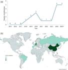
Number of publications involving toxicometabolomics studies on New Psychoactive Substances (NPS) regarding their (a) temporal distribution from 2015 to 2025, and (b) geographic distribution according to the corresponding authors’ country.98 Data for 2025 include studies published between January and November.
An emerging priority is the development of collaborative and integrative platforms supported by public health and public safety institutions. Shared repositories containing both endogenous and xenobiotic metabolites - ideally linked to standardized metadata on sample preparation, analytical acquisition methods, and biological context - would facilitate interlaboratory validation, support biomarker discovery, and reduce redundant efforts.16,39 Expanding curated spectral libraries to incorporate NPS-related metabolites, along with promoting community-driven annotation and curation practices, will further accelerate metabolite identification and reduce the likelihood of false positives.91
Ongoing advances in analytical techniques promise to reduce costs and improve throughput in the future. Miniaturized systems, automation, and high-throughput screening workflows may render metabolomics more accessible, allowing toxicological information on emerging substances to be generated more rapidly and increasing the synchrony between the identification of new NPS and the assessment of their impacts on human health.41,75 In addition to harmonized standard operating procedures, broader adoption of rigorous QA/QC parameters will be fundamental for consolidating toxicometabolomics as a reliable tool in NPS research. Future studies should consistently incorporate necessary steps such as sample preparation standardization using IS, pooled QC samples and SST analyzed throughout the batch, injection randomization, extraction blanks for background correction, and transparent feature-filtering criteria based on QC performance. Equally important is the systematic reporting of metabolite identification confidence levels, following internationally recognized frameworks. The implementation of these parameters across laboratories would promote methodological homogenization, strengthen inter-study comparability, and enhance the analytical robustness and reproducibility of findings. Ultimately, greater adherence to standardized QA/QC practices will increase confidence in metabolomic signatures associated with NPS exposure, facilitating biomarker validation, mechanistic elucidation of toxicity, and translation into forensic, clinical, and regulatory applications.99
Integrating computational and in silico tools with experimental metabolomics can further accelerate the annotation of unknown metabolites. New approaches, particularly machine learning tools for fragmentation prediction and metabolic pathway inference, are being developed and updated frequently, complementing experimental validation and reducing the need for exhaustive manual curation.17,21 Additionally, the development of containerized and user-friendly pipelines has the potential to democratize access to robust bioinformatics workflows, benefiting laboratories with limited computational expertise or infrastructure.
Advancing the biological interpretation of metabolomic signatures associated with NPS mechanisms of action is also essential. Incorporating xenobiotic reactions into pathway databases, combined with multi-omics integration, will provide greater mechanistic depth and enable metabolic alterations to be contextualized within toxicologically relevant pathways. Such approaches may help bridge the current gap between descriptive metabolite profiles and actionable insights into mechanisms of toxicity.
Finally, regulatory engagement and standardization efforts are crucial. Harmonizing sample preparation protocols and data-analysis pipelines would consolidate the field and facilitate the integration of NPS metabolomics into forensic, clinical, and regulatory toxicology.9,14 Closer alignment with early-warning systems and toxicovigilance networks will ensure that metabolomics contributes to the timely identification of high-risk compounds, while interdisciplinary training programs will prepare new researchers to navigate the complex intersection of pharmacology, toxicology, analytical chemistry, and bioinformatics.
6. Conclusion
The emergence of NPS represents a persistent and global public health threat, driven by their structural diversity, rapid turnover in illicit markets, and unpredictable toxicological profiles. Metabolomics studies demonstrate that, despite substantial heterogeneity among these compounds, including SCs, SCRAs, opioids, tryptamines, and other emerging analogues, common toxicological patterns can be discerned. Disruptions in oxidative balance, energy metabolism, neurotransmitter systems, and lipid metabolism consistently emerge as hallmarks of NPS toxicity, underscoring the profound biochemical impact of these substances and reinforcing the urgency of expanding mechanistic insight.
Metabolomics has proven particularly valuable in this context. By simultaneously capturing xenobiotic metabolites and endogenous biochemical perturbations, it enables the identification of biomarkers of exposure and effect, while also elucidating toxicokinetic and toxicodynamic processes that remain inaccessible through conventional toxicology. This ability to link biochemical signatures to mechanisms of toxicity positions metabolomics as a powerful complement to forensic, clinical, and regulatory frameworks. Importantly, the approach also offers translational potential, providing candidate biomarkers that can inform both early-warning systems and the clinical management of intoxications.
However, the remarkable pace at which new NPS emerge far outstrips the capacity of laboratories to generate reference data, while interspecies variability complicates the extrapolation of experimental findings to human physiology. High analytical costs, limited availability of reference standards, and the absence of standardized workflows further constrain reproducibility and slow the translation of results into applied contexts. These limitations underscore the need for collaborative infrastructures, standardized protocols, and curated repositories of endogenous and xenobiotic metabolites.
Looking forward, progress will depend on integrative strategies that combine metabolomics with other omics platforms, computational prediction tools, and high-throughput screening technologies. Such advances will accelerate the annotation of unknown metabolites, refine mechanistic understanding, and strengthen the identification of clinically relevant biomarkers. Integrating metabolomics into toxicovigilance networks and regulatory strategies will help bridge the gap between descriptive biochemical profiles and actionable toxicological knowledge. Ultimately, addressing these challenges will consolidate metabolomics as a central tool for unraveling the toxicological complexity of NPS and mitigating their impact on public health and society worldwide.
Acknowledgments
The authors gratefully acknowledge the financial support provided by (FAPESP, grants: 2024/10492-2, 2022/00037-0, 2021/04768-7, 2024/08366-9, and 2024/14936-2), (CNPq, grants: 403396/2024-7; 309124/2025-5; 140266/2022-4 and 406958/2022-0 - INCT-SP) and (CAPES/PROCAD-DROGAS; INSPEQT 2.0 Project, grant 2/2024).
Data Availability Statement
The authors declare that all data presented herein are available in this article.
References
-
1 Shafi, A.; Berry, A. J.; Sumnall, H.; Wood, D. M.; Tracy, D. K.; Ther. Adv. Psychopharmacol. 2020, 10 [Crossref]
» Crossref -
2 European Union Drugs Agency (EUDA); New psychoactive substances-the current situation in Europe (European Drug Report 2024); 2024. [Link] accessed in March 2026
» Link -
3 United Nations Office on Drugs and Crime (UNODC); The Challenge of New Psychoactive Substances - A technical update 2024 [Link] accessed in March 2026
» Link -
4 Lo Faro, A. F.; Berardinelli, D.; Cassano, T.; Dendramis, G.; Montanari, E.; Montana, A.; Berretta, P.; Zaami, S.; Busardò, F. P.; Huestis, M. A.; Biology 2023, 12, 273. [Crossref]
» Crossref -
5 da Cunha, K. F.; Oliveira, K. D.; Huestis, M. A.; Costa, J. L.; J. Anal. Toxicol. 2020, 44, 697. [Crossref]
» Crossref -
6 Manier, S. K.; Wagmann, L.; Flockerzi, V.; Meyer, M. R.; Arch. Toxicol. 2020, 94, 2047. [Crossref]
» Crossref -
7 Li, X.; Ren, J.; Li, T.; Han, Y.; Dong, H.; Sun, H.; Yan, G.; Wang, X.; Nat. Prod. Commun. 2024, 19 [Crossref]
» Crossref -
8 Sillé, F.; Hartung, T.; Metabolites 2024, 14, 98. [Crossref]
» Crossref -
9 Olesti, E.; González-Ruiz, V.; Wilks, M. F.; Boccard, J.; Rudaz, S.; Analyst 2021, 146, 1820. [Crossref]
» Crossref -
10 Dunn, W. B.; Broadhurst, D. I.; Atherton, H. J.; Goodacre, R.; Griffin, J. L.; Chem. Soc. Rev. 2011, 40, 387. [Crossref]
» Crossref -
11 Olesti, E.; de Toma, I.; Ramaekers, J. G.; Brunt, T. M.; Carbó, M.; Fernández-Avilés, C.; Robledo, P.; Farré, M.; Dierssen, M.; Pozo, Ó. J.; de la Torre, R.; J. Psychopharmacology 2018, 33, 347. [Crossref]
» Crossref -
12 Nafie, M. S.; Abu-Elsaoud, A. M.; Diab, M. K.; Comput. Struct. Biotechnol. J. 2025, 27, 3191. [Crossref]
» Crossref -
13 Araújo, A. M.; Carvalho, F.; de Pinho, P. G.; Carvalho, M.; Metabolites 2021, 11, 692. [Crossref]
» Crossref -
14 Hemmer, S.; Wagmann, L.; Pulver, B.; Westphal, F.; Meyer, M. R.; Metabolites 2022, 12, 1209. [Crossref]
» Crossref -
15 Godoi, A. B.; Rodrigues, L. C.; Alves, M. F.; Fais, V. C.; Maurer-Morelli, C. V.; Costa, J. L.; ACS Omega 2025, 10, 50626. [Crossref]
» Crossref -
16 Steuer, A. E.; Kaelin, D.; Boxler, M. I.; Eisenbeiss, L.; Holze, F.; Vizeli, P.; Czerwinska, J.; Dargan, P. I.; Abbate, V.; Liechti, M. E.; Kraemer, T.; Metabolites 2020, 10, 306. [Crossref]
» Crossref -
17 Araújo, A. M.; Bastos, M. L.; Carvalho, F.; Guedes de Pinho, P.; Carvalho, M.; Toxicology 2020, 441, 152503. [Crossref]
» Crossref -
18 Araújo, A. M.; Carvalho, M.; Bastos, M. L.; Carvalho, F.; de Pinho, P. G.; Arch. Toxicol. 2019, 93, 3277. [Crossref]
» Crossref -
19 Wang, Y.; Yang, Y.; Zhan, Y.; Yin, J.; Zhou, X.; Xu, C.; Gao, F.; Liu, J.; Wu, C.; Liu, S.; Zhang, J.; Shu, C.; Arabian J. Chemistry 2023, 16, 105039. [Crossref]
» Crossref -
20 Manier, S. K.; Keller, A.; Schäper, J.; Meyer, M. R.; Sci. Rep. 2019, 9, 2741. [Crossref]
» Crossref -
21 Wu, Y.; Fan, F.; Zhou, L.; Shen, Y.; Wang, A.; Qin, Y.; Wang, J.; Yao, W.; Environ. Res. 2025, 276, 121517. [Crossref]
» Crossref -
22 Luo, Y.; Ying, L. R.; Liu, Y. Z.; Fan, Y. L.; Wang, K.; Lan, Y. F.; Chian, S.; Ecotoxicol. Environ. Saf. 2025, 303, 118782. [Crossref]
» Crossref -
23 Markin, P. A.; Brito, A.; Moskaleva, N. E.; Tagliaro, F.; La Frano, M. R.; Savitskii, M. V.; Appolonova, S. A.; Comp. Biochem. Physiol., Part C: Toxicol. Pharmacol. 2021, 243, 109000. [Crossref]
» Crossref -
24 Fan, Y.; Huang, Y.; Zhou, Y.; Ke, X.; Tian, Y.; Zheng, S.; Sun, Y.; Huang, Z.; Zhou, J.; Wu, L.; Ecotoxicol. Environ. Saf. 2024, 288, 117375. [Crossref]
» Crossref -
25 Zheng, S.; Zhou, Y.; Song, X.; Ke, X.; Wu, H.; Huang, Z.; Fan, Y.; Chem. Biol. Interact. 2025, 421, 111754. [Crossref]
» Crossref -
26 Zhao, S.; Chen, J.; Zhong, C.; Meng, L.; Wang, Y.; Ecotoxicol. Environ. Saf. 2024, 272, 116044. [Crossref]
» Crossref -
27 Zhao, S.; Liu, M.; Chen, J.; Meng, L.; Wang, Y.; Ecotoxicol. Environ. Saf. 2024, 283, 116969. [Crossref]
» Crossref -
28 Francesco, G.; Montesano, C.; Vincenti, F.; Bilel, S.; Corli, G.; Petrella, G.; Cicero, D. O.; Gregori, A.; Marti, M.; Sergi, M.; Sci. Rep. 2024, 14, 9432. [Crossref]
» Crossref -
29 Magny, R.; Mégarbane, B.; Chevillard, L.; Roulland, E.; Bardèche-Trystram, B.; Dumestre-Toulet, V.; Labat, L.; Houzé, P.; J. Pharm. Biomed. Anal. 2024, 243, 116086. [Crossref]
» Crossref -
30 Xu, R.; Huang, C.; Li, G.; Liu, B.; Lai, H.; Jin, T.; Toxicol. Appl. Pharmacol. 2025, 506, 117621. [Crossref]
» Crossref -
31 Ministério da Justiça e Segurança Pública (MJSP); Relatório 2023 - Drogas Sintéticas [Link] accessed in March 2026
» Link -
32 European Union Drugs Agency (EUDA); European Drug Report 2025: Trends and Developments [Link] accessed in March 2026
» Link -
33 Soares, J.; Costa, V. M.; Bastos, M. L.; Carvalho, F.; Capela, J. P.; Arch. Toxicol. 2021, 95, 2895. [Crossref]
» Crossref -
34 Kelly, J. P.; Drug Test. Anal. 2011, 3, 439. [Crossref]
» Crossref -
35 United Nations Office on Drugs and Crime (UNODC); World Drug Report 2013; 2013. [Link] accessed in March 2026
» Link -
36 Drug Enforcement Administration (DEA); Drugs of Abuse: A DEA Resource Guide; 2024. [Link] accessed in March 2026
» Link -
37 United Nations Office on Drugs and Crime (UNODC); Synthetic Cathinones [Link] accessed in March 2026
» Link -
38 Nadal-Gratacos, N.; Pazos, M. D.; Pubill, D.; Camarasa, J.; Escubedo, E.; Berzosa, X.; Lopez-Arnau, R.; ACS Pharmacol. Transl. Sci. 2024, 7, 2588. [Crossref]
» Crossref -
39 Goncalves, R.; Pelletier, R.; Couette, A.; Gicquel, T.; Le Daré, B.; Toxicologie Analytique et Clinique 2022, 34, 29. [Crossref]
» Crossref -
40 Matey, J. M.; Zapata, F.; Menéndez-Quintanal, L. M.; Montalvo, G.; García-Ruiz, C.; Talanta 2023, 265, 124816. [Crossref]
» Crossref -
41 Dixit, A.; Soni, A.; Pandey, A.; Rapid Commun. Mass Spectrom. 2025, 39, e10116. [Crossref]
» Crossref -
42 Sumner, L. W.; Amberg, A.; Barrett, D.; Beale, M. H.; Beger, R.; Daykin, C. A.; Fan, T. W. M.; Fiehn, O.; Goodacre, R.; Griffin, J. L.; Hankemeier, T.; Hardy, N.; Harnly, J.; Higashi, R.; Kopka, J.; Lane, A. N.; Lindon, J. C.; Marriott, P.; Nicholls, A. W.; Reily, M. D.; Thaden, J. J.; Viant, M. R.; Metabolomics 2007, 3, 211. [Crossref]
» Crossref -
43 de Souza Anselmo, C.; Sardela, V. F.; Matias, B. F.; de Carvalho, A. R.; de Sousa, V. P.; Pereira, H. M. G.; de Aquino Neto, F. R.; Drug Test. Anal. 2017, 9, 1685. [Crossref]
» Crossref -
44 Christen, V.; Fent, K.; Aquat. Toxicol. 2014, 150, 93. [Crossref]
» Crossref -
45 Goldstone, J. V.; Jönsson, M. E.; Behrendt, L.; Woodin, B. R.; Jenny, M. J.; Nelson, D. R.; Stegeman, J. J.; Arch. Biochem. Biophys. 2009, 482, 7. [Crossref]
» Crossref -
46 Bijlsma, L.; Gil-Solsona, R.; Hernández, F.; Sancho, J. V.; Anal. Bioanal. Chem. 2018, 410, 5107. [Crossref]
» Crossref -
47 Castaneto, M. S.; Gorelick, D. A.; Desrosiers, N. A.; Hartman, R. L.; Pirard, S.; Huestis, M. A.; Drug Alcohol Depend. 2014, 144, 12. [Crossref]
» Crossref -
48 Alves, V. L.; Gonçalves, J. L.; Aguiar, J.; Teixeira, H. M.; Câmara, J. S.; Crit. Rev. Toxicol. 2020, 50, 359. [Crossref]
» Crossref -
49 Centro de Estudos sobre Drogas e Desenvolvimento Social Comunitário (Cdesc); Canabinoides Sintéticos: Estratégias Globais de Prevenção e Ação [Link] accessed in March 2026
» Link -
50 Schymanski, E. L.; Jeon, J.; Gulde, R.; Fenner, K.; Ruff, M.; Singer, H. P.; Hollender, J.; Environ. Sci. Technol. 2014, 48, 2097. [Crossref]
» Crossref -
51 Araújo, A. M.; Carvalho, F.; Bastos, M. L.; Guedes de Pinho, P.; Carvalho, M.; Arch. Toxicol. 2015, 89, 1151. [Crossref]
» Crossref -
52 Tittarelli, R.; Mannocchi, G.; Pantano, F.; Romolo, F. S.; Curr. Neuropharm. 2015, 13, 1875. [Crossref]
» Crossref -
53 Tanaka, E.; Kamata, T.; Katagi, M.; Tsuchihashi, H.; Honda, K.; Forensic Sci. Int. 2006, 163, 152. [Crossref]
» Crossref -
54 Alatrash, G.; Majhail, N. S.; Pile, J. C.; Mayo Clinic Proceedings. 2006, 81, 550. [Crossref]
» Crossref -
55 Varì, M. R.; Ricci, G.; Cavallo, M.; Pichini, S.; Sirignano, A.; Graziano, S.; Curr. Pharm. Des. 2022, 28, 1213. [Crossref]
» Crossref -
56 Pelletier, R.; Le Daré, B.; Le Bouëdec, D.; Kernalléguen, A.; Ferron, P. J.; Morel, I.; Gicquel, T.; Int. J. Mol. Sci. 2022, 23, 15574. [Crossref]
» Crossref -
57 Serviços e Informações do Brasil; Anvisa autoriza primeiro medicamento inalável para tratar depressão grave [Link] accessed in March 2026
» Link -
58 U.S. Food and Drug Administration; SPRAVATO® (esketamine) nasal spray [Link] accessed in March 2026
» Link -
59 European Medicines Agency. Spravato [Link] accessed in March 2026
» Link -
60 Hofer, K. E.; Grager, B.; Müller, D. M.; Rauber-Lüthy, C.; Kupferschmidt, H.; Rentsch, K. M.; Ceschi, A.; Ann. Emerg. Med. 2012, 60, 97. [Crossref]
» Crossref -
61 Shafi, A.; Berry, A. J.; Sumnall, H.; Wood, D. M.; Tracy, D. K.; Ther. Adv. Psychopharmacol. 2022, 12 [Crossref]
» Crossref -
62 Cicero, T. J.; Ellis, M. S.; Surratt, H. L.; Kurtz, S. P.; JAMA Psychiatry 2014, 71, 821. [Crossref]
» Crossref -
63 Stanley, T. H.; J. Pain 2014, 15, 1215. [Crossref]
» Crossref -
64 Ferrari, L.; Gerardo, B.; Rodriguez, C. R.; Cases, G. G.; Escudero, P.; Zar, G.; Pomies, D.; Giannuzzi, L.; Toxicologie Analytique et Clinique 2022, 34, S39. [Crossref]
» Crossref -
65 United Nations Office on Drugs and Crime (UNODC); March 2025 - Increasing Detections of Etomidate and Analogues on Illicit Drug Markets is Becoming a Global Concern [Link] accessed in March 2026
» Link -
66 Canadian Drug and Substance Watch New Emerging Substances: Canadian Drug and Substance Watch. [Link] accessed in March 2026
» Link -
67 Philadelphia Department of Public Health Etomidate detected in Philadelphia’s drug supply and involved in overdose deaths. [Link] accessed in March 2026
» Link -
68 WEDINOS Sample results. [Link] accessed in March 2026
» Link -
69 Dinis-Oliveira, R. J.; Forensic Sci. Res. 2017, 2, 2. [Crossref]
» Crossref -
70 Pergolizzi Jr., J.; Raffa, R.; LeQuang, J. A. K.; Breve, F.; Varrassi, G.; Cureus 2023 [Crossref]
» Crossref -
71 Stangeland, M.; Dale, O.; Skulberg, A. K.; Clin. Toxicol. 2025, 63, 393. [Crossref]
» Crossref -
72 Zawilska, J. B.; Andrzejczak, D.; Drug Alcohol Depend. 2015, 157, 1. [Crossref]
» Crossref -
73 Weinstein, A. M.; Rosca, P.; Fattore, L.; London, E. D.; Front. Psychiatry 2017, 8, 156. [Crossref]
» Crossref -
74 Wishart, D. S.; Nat. Rev. Drug Discovery 2016, 15, 473. [Crossref]
» Crossref -
75 Wu, H. Y.; Chen, Y. C.; Hsu, J. F.; Lu, H. T.; Pan, Y. Y.; Ma, M. C.; Liao, P. C.; J. Food Drug Anal. 2023, 31, 137. [Crossref]
» Crossref -
76 Rodrigues, L. C.; de Godoi, A. B.; Fais, V. C.; Peterson, R. T.; Maurer-Morelli, C. V.; Costa, J. L.; Toxicol. Rep. 2025, 14, 102018. [Crossref]
» Crossref -
77 de Souza Anselmo, C.; Sardela, V. F.; de Sousa, V. P.; Pereira, H. M. G.; Comp. Biochem. Physiol., Part C: Toxicol. Pharmacol. 2018, 212, 34. [Crossref]
» Crossref -
78 Li, S.; Looby, N.; Chandran, V.; Kulasingam, V.; Metabolites 2024, 14, 200. [Crossref]
» Crossref -
79 Godoi, A. B.; Antunes, N. J.; Cunha, K. F.; Martins, A. F.; Huestis, M. A.; Costa, J. L.; Pharmaceutics 2024, 16, 257. [Crossref]
» Crossref -
80 Johnson, C. H.; Ivanisevic, J.; Siuzdak, G.; Nat. Rev. Mol. Cell Biol. 2016, 17, 451. [Crossref]
» Crossref -
81 Trivedi, D. K.; Hollywood, K. A.; Goodacre, R.; New Horiz. Transl. Med. 2017, 3, 294. [Crossref]
» Crossref -
82 Johnson, C. H.; Gonzalez, F. J.; J. Cell. Physiol. 2012, 227, 2975. [Crossref]
» Crossref -
83 Fernie, A. R.; Aharoni, A.; Willmitzer, L.; Stitt, M.; Tohge, T.; Kopka, J.; Carroll, A. J.; Saito, K.; Fraser, P. D.; de Luca, V.; Plant Cell 2011, 23, 2477. [Crossref]
» Crossref -
84 Smith, C. A.; Want, E. J.; O’Maille, G.; Abagyan, R.; Siuzdak, G.; Anal. Chem. 2006, 78, 779. [Crossref]
» Crossref -
85 Schmid, R.; Heuckeroth, S.; Korf, A.; Smirnov, A.; Myers, O.; Dyrlund, T. S.; Bushuiev, R.; Murray, K. J.; Hoffmann, N.; Lu, M.; Sarvepalli, A.; Zhang, Z.; Fleischauer, M.; Dührkop, K.; Wesner, M.; Hoogstra, S. J.; Rudt, E.; Mokshyna, O.; Brungs, C.; Ponomarov, K.; Mutabdžija, L.; Damiani, T.; Pudney, C. J.; Earll, M.; Helmer, P. O.; Fallon, T. R.; Schulze, T.; Rivas-Ubach, A.; Bilbao, A.; Richter, H.; Nothias, L.-F.; Wang, M.; Orešič, M.; Weng, J.-K.; Böcker, S.; Jeibmann, A.; Hayen, H.; Karst, U.; Dorrestein, P. C.; Petras, D.; Du, X.; Pluskal, T.; Nat. Biotechnol. 2023, 41, 447. [Crossref]
» Crossref -
86 Tsugawa, H.; Cajka, T.; Kind, T.; Ma, Y.; Higgins, B.; Ikeda, K.; Kanazawa, M.; VanderGheynst, J.; Fiehn, O.; Arita, M.; Nat. Methods 2015, 12, 523. [Crossref]
» Crossref -
87 Pang, Z.; Lu, Y.; Zhou, G.; Hui, F.; Xu, L.; Viau, C.; Spigelman, A. F.; Macdonald, P. E.; Wishart, D. S.; Li, S.; Xia, J.; Nucleic Acids Res. 2024, 52, W398. [Crossref]
» Crossref -
88 Wishart, D. S.; Guo, A. C.; Oler, E.; Wang, F.; Anjum, A.; Peters, H.; Dizon, R.; Sayeeda, Z.; Tian, S.; Lee, B. L.; Berjanskii, M.; Mah, R.; Yamamoto, M.; Jovel, J.; Torres-Calzada, C.; Hiebert Giesbrecht, M.; Lui, V. W.; Varshavi, D.; Varshavi, D.; Allen, D.; Arndt, D.; Khetarpal, N.; Sivakumaran, A.; Harford, K.; Sanford, S.; Yee, K.; Cao, X.; Budinski, Z.; Liigand, J.; Zhang, L.; Zheng, J.; Mandal, R.; Karu, N.; Dambrova, M.; Schiöth, H. B.; Greiner, R.; Gautam, V.; Nucleic Acids Res. 2022, 50, D622. [Crossref]
» Crossref -
89 Guijas, C.; Montenegro-Burke, J. R.; Domingo-Almenara, X.; Palermo, A.; Warth, B.; Hermann, G.; Koellensperger, G.; Huan, T.; Uritboonthai, W.; Aisporna, A. E.; Wolan, D. W.; Spilker, M. E.; Benton, H. P.; Siuzdak, G.; Anal. Chem. 2018, 90, 3156. [Crossref]
» Crossref -
90 Wang, M.; Carver, J. J.; Phelan, V. V.; Sanchez, L. M.; Garg, N.; Peng, Y.; Nguyen, D. D.; Watrous, J.; Kapono, C. A.; Luzzatto Knaan, T.; Porto, C.; Bouslimani, A.; Melnik, A. V.; Meehan, M. J.; Liu, W. T.; Crüsemann, M.; Boudreau, P. D.; Esquenazi, E.; Sandoval-Calderón, M.; Kersten, R. D.; Pace, L. A.; Quinn, R. A.; Duncan, K. R.; Hsu, C. C.; Floros, D. J.; Gavilan, R. G.; Kleigrewe, K.; Northen, T.; Dutton, R. J.; Parrot, D.; Carlson, E. E.; Aigle, B.; Michelsen, C. F.; Jelsbak, L.; Sohlenkamp, C.; Pevzner, P.; Edlund, A.; McLean, J.; Piel, J.; Murphy, B. T.; Gerwick, L.; Liaw, C. C.; Yang, Y. L.; Humpf, H. U.; Maansson, M.; Keyzers, R. A.; Sims, A. C.; Johnson, A. R.; Sidebottom, A. M.; Sedio, B. E.; Klitgaard, A.; Larson, C. B.; Boya, C. A. P.; Torres-Mendoza, D.; Gonzalez, D. J.; Silva, D. B.; Marques, L. M.; Demarque, D. P.; Pociute, E.; O’Neill, E. C.; Briand, E.; Helfrich, E. J. N.; Granatosky, E. A.; Glukhov, E.; Ryffel, F.; Houson, H.; Mohimani, H.; Kharbush, J. J.; Zeng, Y.; Vorholt, J. A.; Kurita, K. L.; Charusanti, P.; McPhail, K. L.; Nielsen, K. F.; Vuong, L.; Elfeki, M.; Traxler, M. F.; Engene, N.; Koyama, N.; Vining, O. B.; Baric, R.; Silva, R. R.; Mascuch, S. J.; Tomasi, S.; Jenkins, S.; Macherla, V.; Hoffman, T.; Agarwal, V.; Williams, P. G.; Dai, J.; Neupane, R.; Gurr, J.; Rodríguez, A. M. C.; Lamsa, A.; Zhang, C.; Dorrestein, K.; Duggan, B. M.; Almaliti, J.; Allard, P. M.; Phapale, P.; Nothias, L. F.; Alexandrov, T.; Litaudon, M.; Wolfender, J. L.; Kyle, J. E.; Metz, T. O.; Peryea, T.; Nguyen, D. T.; VanLeer, D.; Shinn, P.; Jadhav, A.; Müller, R.; Waters, K. M.; Shi, W.; Liu, X.; Zhang, L.; Knight, R.; Jensen, P. R.; Palsson, B.; Pogliano, K.; Linington, R. G.; Gutiérrez, M.; Lopes, N. P.; Gerwick, W. H.; Moore, B. S.; Dorrestein, P. C.; Bandeira, N.; Nat. Biotechnol. 2016, 34, 828. [Crossref]
» Crossref -
91 Chang, H. Y.; Colby, S. M.; Du, X.; Gomez, J. D.; Helf, M. J.; Kechris, K.; Kirkpatrick, C. R.; Li, S.; Patti, G. J.; Renslow, R. S.; Subramaniam, S.; Verma, M.; Xia, J.; Young, J. D.; Anal. Chem. 2021, 93, 1912. [Crossref]
» Crossref -
92 van der Hooft, J. J. J.; Wandy, J.; Barrett, M. P.; Burgess, K. E. V.; Rogers, S.; Proc. Natl. Acad. Sci. 2016, 113, 13738. [Crossref]
» Crossref -
93 de Jonge, N. F.; Louwen, J. J. R.; Chekmeneva, E.; Camuzeaux, S.; Vermeir, F. J.; Jansen, R. S.; Huber, F.; van der Hooft, J. J. J.; Nat. Commun. 2023, 14, 1752. [Crossref]
» Crossref -
94 Bushuiev, R.; Bushuiev, A.; Samusevich, R.; Brungs, C.; Sivic, J.; Pluskal, T.; Nat. Biotechnol. 2025 [Crossref]
» Crossref -
95 da Silva, R. R.; Wang, M.; Nothias, L.-F.; van der Hooft, J. J. J.; Caraballo-Rodríguez, A. M.; Fox, E.; Balunas, M. J.; Klassen, J. L.; Lopes, N. P.; Dorrestein, P. C.; PLoS Comput. Biol. 2018, 14, e1006089. [Crossref]
» Crossref -
96 Damiani, T.; Jarmusch, A. K.; Aron, A. T.; Petras, D.; Phelan, V. V.; Zhao, H. N.; Bittremieux, W.; Acharya, D. D.; Ahmed, M. M. A.; Bauermeister, A.; Bertin, M. J.; Boudreau, P. D.; Borges, R. M.; Bowen, B. P.; Brown, C. J.; Chagas, F. O.; Clevenger, K. D.; Correia, M. S. P.; Crandall, W. J.; Crüsemann, M.; Fahy, E.; Fiehn, O.; Garg, N.; Gerwick, W. H.; Gilbert, J. R.; Globisch, D.; Gomes, P. W. P.; Heuckeroth, S.; James, C. A.; Jarmusch, S. A.; Kakhkhorov, S. A.; Kang, K. B.; Kessler, N.; Kersten, R. D.; Kim, H.; Kirk, R. D.; Kohlbacher, O.; Kontou, E. E.; Liu, K.; Lizama-Chamu, I.; Luu, G. T.; Luzzatto Knaan, T.; Mannochio-Russo, H.; Marty, M. T.; Matsuzawa, Y.; McAvoy, A. C.; McCall, L.-I.; Mohamed, O. G.; Nahor, O.; Neuweger, H.; Niedermeyer, T. H. J.; Nishida, K.; Northen, T. R.; Overdahl, K. E.; Rainer, J.; Reher, R.; Rodriguez, E.; Sachsenberg, T. T.; Sanchez, L. M.; Schmid, R.; Stevens, C.; Subramaniam, S.; Tian, Z.; Tripathi, A.; Tsugawa, H.; van der Hooft, J. J. J.; Vicini, A.; Walter, A.; Weber, T.; Xiong, Q.; Xu, T.; Pluskal, T.; Dorrestein, P. C.; Wang, M.; Nat. Methods 2025, 22, 1247. [Crossref]
» Crossref -
97 Pan, Y. Y.; Chen, Y. C.; Chang, W. C. W.; Ma, M. C.; Liao, P. C.; Chemom. Intell. Lab. Syst. 2021, 210, 104271. [Crossref]
» Crossref -
98 MapChart. [Link] accessed in MArch 2026
» Link -
99 Di Minno, A.; Gelzo, M.; Caterino, M.; Costanzo, M.; Ruoppolo, M.; Castaldo, G.; Int. J. Mol. Sci. 2022, 23, 5213. [Crossref]
» Crossref
Edited by
-
Editor handled this article:
João Henrique Ghilardi Lago (Associate)