Synthesis, Photophysical Behavior, DFT Analysis, Electrochemical and Investigative Optical Sensor Properties of C3-Functionalized 4-Hydroxycoumarins
Abstract
Coumarins are versatile heterocyclic scaffolds widely explored for their photophysical properties and applications in fluorescence-based sensing. In this study, a series of C3-functionalized sulfur-substituted 4-hydroxycoumarins 2a-2k was investigated as potential fluorescent probes for metal ion detection. The compounds were comprehensively characterized using spectroscopic techniques, electrochemical analysis, and density functional theory (DFT) calculations. Photophysical studies revealed intense UV absorption and fluorescence emission in the violet region, with moderate Stokes shifts consistent with intramolecular charge transfer (ICT) transitions. Time-resolved measurements indicated nanosecond-scale excited-state lifetimes, while theoretical calculations supported the ICT nature of the electronic transitions. Preliminary aggregation studies demonstrated aggregation-induced emission (AIE) behavior for derivative 2a, suggesting potential applications in solid-state luminescent systems. Among the investigated compounds, derivative 2k exhibited the most promising sensing performance, showing selective fluorescence quenching toward Hg2+ ions through heavy atom effects and ligand-to-metal charge transfer interactions. Quantitative fluorescence titration revealed a Stern-Volmer constant on the order of 104 M-1, with limits of detection and quantification of 3.66 and 11.10 μM, respectively. Furthermore, the probe demonstrated rapid response, good anti-interference capability, and successful detection of Hg2+ in cosmetic samples. These results highlight sulfur-substituted 4-hydroxycoumarins as promising fluorescent platforms for the development of optical sensors for heavy metal monitoring in environmental and consumer products.
Keywords:
coumarin; sulfur derivatives; fluorescent probe; optical sensor; Hg2+ detection
Introduction
Coumarins have attracted significant interest in recent years due to their photophysical properties. The coumarin nucleus can be visualized as a fused system of the electronrich benzene ring and an electron-deficient pyrone ring. In an unsubstituted coumarin, the benzene ring acts as a donor, while the pyrone ring acts as an acceptor. Due to this weak charge transfer from benzene to the pyrone ring, unsubstituted coumarin absorbs and emits at 280 and 330 nm. However, several methods are adopted that involve substitution at the benzene and pyrone nucleus and the inclusion of groups at certain positions give the compound strong light absorption and luminescence.1
Over the years, researchers have delved into the synthesis, characterization and application of coumarin derivatives, discovering their potential as indispensable tools for fluorescence-based studies and applications.2-7 Endowed with a broad therapeutic profile, coumarins have challenged researchers for decades to improve this structure according to their needs. Thus, the coumarin skeleton is seen as an advantageous structure for the design and synthesis of molecules aimed at expanding their applications, improving their performance characteristics and addressing the challenges associated with specific applications in different fields.
They are considered good examples of privileged structures, usually rigid, heteroatomic polycyclic systems that are capable of binding to multiple pharmacological targets, thus providing a viable starting point in the search for new drugs. Substances containing the coumarin framework exhibit various interesting biological properties such as anticoagulant, anti-neurodegenerative, antioxidant, anticancer, antifungal, antispasmodic and antimicrobial.8 In addition, coumarins have demonstrated several applications in the development of fluorescent chemosensors for molecular, analytical, bioorganic and material chemistry imaging. These substances have also been used to produce low-cost fluorescent materials. Other applications include the use of coumarins in fluorescence labeling processes, metal ion detection, microenvironmental polarity detection and pH sensing.9 Therefore, this work focuses on the investigation of sulfur-substituted 4-hydroxycoumarins as potential optical sensors for cation detection. Their absorption and emission properties, theoretical calculations, and electrochemical behavior were also evaluated.
Experimental
General
Melting points were obtained on a Thomas Hoover apparatus (Philadelphia, USA) and are uncorrected. Analytical grade solvents were used. Column chromatography was performed using silica gel (Acros Organics 0.035-0.070 mm, pore diameter ca. 6.0 nm). Infrared spectra were recorded on a Shimadzu IR Prestige-21 FTIR spectrometer (Kyoto, Japan). 1H and 13C nuclear magnetic resonance (NMR) were recorded at room temperature using a VNMRSYS-500 or a Varian MR 300 instrument, using the solvents indicated, with tetramethylsilane (TMS) as internal standard. Chemical shifts (d) are given in ppm and coupling constants (J) in hertz (Hz). High-resolution mass spectra (HRMS) were recorded on a MICROMASS Q-TOF mass spectrometer (Waters). All structural characterization data were previously reported in our studies.10,11
General procedures for synthesis of coumarins 2a-2k
In a 125 mL round-bottom flask containing a solution of coumarins (1 g, 6.2 mmol), formalin (12.3 mmol) and 20 mL of ethanol, the appropriate thiophenol (12.3 mmol) was added. The mixture was then externally heated at 90 °C for 12 h. The obtained solid was filtered and washed with petroleum ether iced. Then, the product was purified by recrystallization in ethanol.12
4-Hydroxy-3-((phenylthio)methyl)-2H-chromen-2-one (2a)
White solid, 83% yield; mp 157-159 °C; IR (KBr) ν / cm-1 1649, 1626, 1562, 1391, 1252, 1170, 1143, 1107, 1072, 932, 762,735, 683, 642; 1H NMR (500.00 MHz, DMSO-d6) d 7.95 (1H, dd, J 8.2, 1.5 Hz), 7.62 (1H, tt, J 8.1, 1.7 Hz), 7.40 (2H, dd, J 8.2, 0.9 Hz), 7.38-7.34 (m, 2H), 7.30 (2H, t, J 7.7 Hz), 7.19 (1H, t, J 7.4 Hz), 4.18 (2H, s); 13C NMR/APT (125.00 MHz, DMSO-d6) d 161.8, 161.3, 151.9, 136.5, 132.2, 129.0, 128.7, 125.8, 123.9, 123.3, 116.1, 115.8, 101.0, 27.6; HRMS-ESI m/z, calcd. for C16H13O3S+ [M + H]+: 285.0580, found: 285.0588; D = 2.8 ppm.
4-Hydroxy-3-((p-tolylthio)methyl)-2H-chromen-2-one (2b)
Yellow solid, 78% yield; mp 230-232 °C; IR (KBr) ν / cm-1 1597, 1511, 1455, 1404, 1105, 1066, 801, 758, 680; 1H NMR (500.00 MHz, DMSO-d6) d 7.92 (1H, dd, J 8.1, 1.3 Hz), 7.47 (1H, tt, J 8.2, 2.3 Hz), 7.28 (2H, d, J 7.9 Hz), 7.23-7.18 (2H, m), 7.07 (2H, d, J 7.9 Hz), 4.09 (2H, s), 2.24 (3H, s); 13C NMR/APT (125.00 MHz, DMSO-d6) d 167.9, 163.5, 153.3, 135.4, 134.8, 131.2, 129.7, 129.1, 124.7, 123.0, 120.4, 116.2, 102.9, 97.1, 46.4, 29.6, 20.9; HRMS-ESI m/z, calcd. for C17H15O3S+ [M + H]+: 299.0736, found: 299.0737; D = 0.3 ppm.
4-Hydroxy-3-(((4-hydroxyphenyl)thio)methyl)-2H-chromen2-one (2c)
Pale yellow solid, 81% yield; mp 180-182 °C; IR (KBr) ν / cm -1 1669, 1620, 1571, 1495, 1432, 1403, 1270, 1223, 1197, 1155, 1105, 1064, 905, 835, 761, 675, 634; 1H NMR (500.00 MHz, DMSO-d6) d 9.44 (1H, s), 7.91 (1H, dd, J 8.2, 1.6 Hz), 7.60 (1H, tt, J 8.2, 2.0 Hz), 7.34 (2H, ddd, J 5.7, 3.6, 1.2 Hz), 7.24 (2H, dd, J 8.7, 7.9 Hz), 6.68 (2H, dd, J 8.7, 7.9 Hz), 4.01 (2H, s); 13C NMR/APT (125.00 MHz, DMSO-d6) d 161.8, 160.8, 156.9, 151.9, 133.9, 132.0, 123.8, 123.5, 123.3, 116.1, 115.9, 115.7, 101.8, 29.8; HRMS-ESI m/z, calcd. for C16H13O4S+ [M + H]+: 301.0529, found: 301.0531; D = 0.7 ppm.
4-Hydroxy-3-(((4-methoxyphenyl)thio)methyl)-2H-chromen2-one (2d)
Yellow solid, 84% yield; mp 168-170 °C; IR (KBr) ν / cm-1 1669, 1614, 1567, 1492, 1280, 1247, 1216, 1181, 1133, 1102, 1053, 1019, 829, 756, 640; 1H NMR (500.00 MHz, DMSO-d6) d 7.92 (1H, dd, J 8.1, 1.3 Hz), 7.60 (1H, t, J 7.8 Hz), 7.35-7.30 (4H, m), 6.85 (2H, d, J 8.7 Hz), 4.06 (2H, s), 3.72 (3H, s); 13C NMR/APT (125.00 MHz, DMSO-d6) d 161.8, 160.8, 158.6, 151.8, 133.2, 132.0, 125.9, 123.7, 123.2, 116.0, 115.8, 114.3, 101.7, 55.1, 29.4; HRMS-ESI m/z, calcd. for C17H15O5S+ [M + H]+: 315.0686, found: 315.0685; D = 0.3 ppm.
4-Hydroxy-3-(((4-(methylthio)phenyl)thio)methyl)-2Hchromen-2-one (2e)
White solid, 76% yield; mp 170-173 °C; IR (KBr) ν / cm-1 1669, 1605, 1563, 1499, 1310, 1276, 1234, 1183, 1103, 1050, 952, 910, 813, 758, 705, 655; 1H NMR (500.00 MHz, DMSO-d6) d 7.95 (1H, dd, J 8.2, 1.6 Hz), 7.62 (1H, tt, J 7.8, 2.0 Hz), 7.38-7.33 (4H, m), 7.19 (2H, dd, J 8.7, 1.5 Hz), 4.14 (2H, s), 2.45 (3H, s); 13C NMR/ APT (125.00 MHz, DMSO-d6) d 161.8, 161.2, 151.9, 136.3, 132.3, 132.2, 130.5, 126.4, 123.9, 123.3, 116.1, 115.8, 101.2, 28.3, 14.8; HRMS-ESI m/z, calcd. for C17H15O3S2+ [M + H]+: 331.0457, found: 331.0467; D = 3.0 ppm.
3-(((4-Fluorophenyl)thio)methyl)-4-hydroxy-2H-chromen2-one (2f)
White solid, 64% yield; mp 230-233 °C; IR (KBr) ν / cm-1 1667, 1632, 1488, 1452, 1390, 1276, 1218, 1173, 1148, 1107, 937, 903, 835, 812, 758, 681, 630; 1H NMR (500.00 MHz, DMSO-d6) d 7.93 (1H, dd, J 12.9, 4.7 Hz), 7.62 (1H, td, J 8.1, 1.6 Hz), 7.47-7.43 (2H, m), 7.38-7.30 (2H, m), 7.15-7.11 (2H, m), 4.12 (2H, s); 13C NMR/APT (125.00 MHz, DMSO-d6) d 207.1, 163.7, 162.1, 161.9, 161.2, 160.1, 151.9, 132.7, 132.6, 132.2, 131.4, 131.4, 123.9, 123.4, 116.2, 115.8, 115.8, 115.6, 101.2, 81.9, 28.8; HRMS-ESI m/z, calcd. for C16H12FO3S+ [M + H]+: 303.0486, found: 303.0491; D = 1.6 ppm.
3-(((4-Chlorophenyl)thio)methyl)-4-hydroxy-2H-chromen2-one (2g)
White solid, 66% yield; mp 170-172 °C; IR (KBr) ν / cm-1 1666, 1627, 1249, 1177, 1093, 1066, 935, 905, 817, 753, 715, 682, 637; 1H NMR (500.00 MHz, DMSO-d6) d 7.95 (1H, dd, J 8.3, 1.4 Hz), 7.62 (1H, td, J 8.1, 1.5 Hz), 7.41 (2H, d, J 8.7 Hz), 7.36 (2H, d, J 8.2 Hz), 7.34 (2H, d, J 8.7 Hz), 4.17 (2H, s); 13C NMR/APT (125.00 MHz, DMSO-d6) d 161.7, 161.3, 151.9, 135.4, 132.1, 130.9, 130.7, 128.5, 123.8, 123.3, 116.1, 115.7, 102.2, 100.9, 27.9; HRMS-ESI m/z, calcd. for C16H11ClO3S+ [M + H]+: 319.0190, found: 319.0194; D = 1.2 ppm.
4-Hydroxy-3-((2-naphthylthio)methyl)-2H-chromen-2-one (2h)
White solid, 89% yield; mp 140-142 °C; IR (KBr) ν / cm-1 1665, 1623, 1567, 1497, 1452, 1341, 1305, 1269, 1191, 1107, 1069, 937, 896, 863, 812, 736, 629; 1H NMR (500.00 MHz, DMSO-d6) d 8.11 (1H, d, J 1.4 Hz), 7.93 (2H, d, J 8.9 Hz), 7.91-7.85 (2H, m), 7.66 (1H, dd, J 8.7, 1.9 Hz), 7.61-7.56 (1H, m), 7.55-7.48 (2H, m), 7.37-7.31 (2H, m), 3.86 (2H, s); 13C NMR/APT (125.00 MHz, DMSO-d6) d 164.00, 162.51, 151.76, 133.06, 132.86, 131.97, 131.50, 129.00, 127.45, 127.13, 126.78, 126.34, 126.33, 125.21, 123.66, 123.18, 116.69, 115.81, 102.37, 19.23; HRMS-ESI m/z, calcd. for C20H15O3S+ [M + H]+: 335.0736, found: 335.0742; D = 1.8 ppm.
4-Hydroxy-3-(phenyl(phenylthio)methyl)-2H-chromen-2one (2i)
Yellow solid, 74% yield; mp 123-125 °C; IR (KBr) ν / cm-1 2957, 1648, 1596, 1535, 1492, 1422, 1345, 1203, 1169, 1085, 1027, 926, 849, 810, 740, 719, 690, 666; 1H NMR (500.00 MHz, DMSO-d6) d 8.03 (1H, dd, J 8.2, 1.5 Hz), 7.62 (3H, dd, J 11.0, 4.5 Hz), 7.39-7.34 (4H, m), 7.29 (4H, dt, J 11.8, 7.7 Hz), 7.20 (2H, dt, J 13.5, 7.3 Hz), 6.05 (1H, s); 13C NMR/APT (125.00 MHz, DMSO-d6) d 160.7, 160.0, 151.9, 140.0, 136.9, 132.0, 129.3, 128.7, 127.7, 127.4, 126.47, 126.1, 123.6, 123.4, 116.0, 115.6, 106.5, 47.2; HRMS-ESI m/z, calcd. for C22H17O3S+ [M + H]+: 361.0893, found: 361.0886; D = 1.9 ppm.
3-((Cyclohexylthio)methyl)-4-hydroxy-2H-chromen-2-one (2j)
Yellow solid, 56% yield; mp 130-132 °C; IR (KBr) ν / cm-1 1653, 1625, 1567, 1494, 1447, 1415, 1386, 1345, 1305, 1257, 1175, 1108, 1061, 934, 904, 749, 697, 631; 1H NMR (500.00 MHz, DMSO-d6) d 7.96 (1H, dd, J 8.3, 1.5 Hz), 7.61 (1H, td, J 8.2, 1.6 Hz), 7.38-7.31 (2H, m), 3.78 (1H, s), 3.71 (2H, s), 2.78-2.72 (1H, m), 1.99-1.92 (2H, m), 1.68 (2H, d, J 4.2 Hz), 1.57-1.52 (1H, m), 1.30-1.17 (4H, m); 13C NMR/APT (125.00 MHz, DMSO-d6) d 162.7, 160.7, 152.4, 152.3, 132.4, 131.9, 124.3, 124.1, 123.9, 123.7, 116.6, 116.5, 116.3, 104.3, 102.7, 43.7, 33.8, 25.9, 25.8, 23.7, 19.9; HRMS-ESI m/z, calcd. for C16H18O3S+ [M + H]+: 291.1049, found: 291.1047; D = 0.7 ppm.
3-(((Furan-2-ylmethyl)thio)methyl)-4-hydroxy-2H-chromen2-one (2k)
Yellow solid, 45% yield; mp 123-125 °C; IR (KBr) ν / cm-1 3261, 1663, 1618, 1569, 1496, 1414, 1384, 1315, 1254, 1228, 1160, 1109, 1076, 987, 950, 909, 855, 770, 724, 701; 1H NMR (500.00 MHz, DMSO-d6) d 7.96 (1H, dd, J 7.7, 0.8 Hz), 7.65-7.60 (1H, m), 7.37 (2H, d, J 8.1 Hz), 6.11 (1H, d, J 3.1 Hz), 5.89 (1H, d, J 3.0 Hz), 4.59 (2H, s), 3.79 (2H, s); 13C NMR/APT (125.00 MHz, DMSO-d6) d 162.4, 160.9, 152.0, 152.0, 150.0, 131.9, 123.8, 123.3, 116.1, 116.0, 108.0, 105.9, 100.8, 63.9, 25.2, 22.8; HRMS-ESI m/z, calcd. for C15H12NaO4S+ [M + Na]+: 311.0349, found: 311.0344; D = 1.6 ppm.
Luminescence
Absorption UV-Vis spectroscopy for studied derivatives 2a-2k was performed using a Shimadzu UV2600 spectrophotometer (data interval of 1.0 nm and slit 1.0 nm) across various solvents: dichloromethane (DCM), tetrahydrofuran (THF), acetonitrile (MeCN) and dimethyl sulfoxide (DMSO). All spectra were recorded in the 250-400 nm range at a fixed concentration of 20 μM. Steady-state fluorescence emission spectra of derivatives 2a-2k in the same solvents were measured using a Horiba Yvon-Jobin Fluoromax Plus spectrofluorometer (Em/Exc; slit 5.0 mm) in the 350-550 nm range at a fixed concentration of 20 μM. Fluorescence quantum yield values (QY) of the coumarin derivatives were determined by comparing their corrected fluorescence spectra with that of a 9,10-diphenylanthracene (DPA) standard molecule in chloroform solution (QY = 65%) using the equation described in the literature.13 Fluorescence lifetime decay (τf) profiles were recorded using the Time-Correlated Single Photon Counting (TCSPC) method with a DeltaHub controller and a Horiba spectrofluorometer. Data were processed with DAS6 and OriginPro 8.5 software (OriginLab, Northampton, USA, 2020) using exponential (mono-exponential) fitting of raw data. A NanoLED (Horiba) source (1.0 MHz; pulse width < 1.2 ns; 284 nm) served as the excitation source.
DFT calculations
The density functional theory (DFT) has been applied to study the geometric and spectroscopic properties of compounds 2a-2k. The hybrid long-range corrected wB97XD functional has been used to describe the exchange and correlation interactions.12 The molecular orbitals were represented by linear combinations of split-valence atomic basis set incremented with polarization functions 6-311(d,p). The influence of the solvent environment was considered implicitly using the Polarizable Continuum Model (PCM).14 This implicit model is computationally advantageous, and gives a good description of dielectric screening and overall solvent polarization effects on the solute, but it has limitations in describing situations involving explicit solvent-solute interactions, such as in hydrogen bonds and coordination to metal centers. All calculations were made using the Gaussian9 code (Gaussian, Inc., Wallingford CT, 2016).
Electrochemistry
Cyclic voltammetry analysis (CV) was conducted for derivatives 2a-2k using an AutoLab Eco Chemie PGSTAT 128 N potentiostat/galvanostat system at room temperature and under an argon atmosphere. Dry DCM was employed as the solvent, with electrochemical grade tetrabutylammonium hexafluorophosphate (0.1 M TBAPF6) as the supporting electrolyte. The setup included a glassy carbon electrode as the working electrode, a platinum wire as the auxiliary electrode, and another platinum wire as a pseudo-reference electrode. The reference electrode potential was calibrated using the ferrocene redox couple as an internal reference (E1/2 = +0.40 V).15
Optical sensor preparation
For all spectroscopic experiments, the solution of derivative 2k (1.0 equiv.) was prepared by dissolving in pure DMSO. The cation solutions (Na+, Mg2+, Zn2+, Cu2+, Ni2+, Co2+, Pb2+, Hg2+, Cd2+, Fe3+, Al3+ and Cr3+; 5.0 equiv.) were prepared by dissolving the corresponding nitrate salts using deionized water. The quantitative titration of compound 2k with Hg2+ ions were performed in a DMSO/H2O mixture (9:1, v/v) solution by absorption and emission techniques.
All experiments were performed at room temperature and carried out three times at least to ensure the experimental reproducibility. The absorption analysis was carried out between 250 and 500 nm range, in the absence and in the presence of each cation species. The steady-state fluorescence emission measurements were performed by scanning the spectra between 350 and 550 nm range with excitation wavelength at 310 nm, with slit (Em/Exc = 5.0 nm).
For interactive parameters by steady-state fluorescence emission analysis, Stern-Volmer (KSV) constant, association (Ka) constant, binding (Kb) constant and, Gibbs free-energy (DGº) values are determined according to equations 1-4:
where F0 and F are the initial and final emission intensities, [Q] is the concentration of Hg2+ ions, f is the fraction of the initial fluorescence intensity corresponding to the fluorophore that is accessible by the quencher (f ca. 1.0), R is the gas constant (1.9872 call-1 mol-1 K-1) and T is the temperature in Kelvin (298.15 K), respectively.
Limit of detection (LOD) and limit of quantification (LOQ) values are obtained by equations 5 and 6:
where σ is the standard deviation value of the intercept and S is the slope value of fitting curve.
Results and Discussion
Photophysical analysis
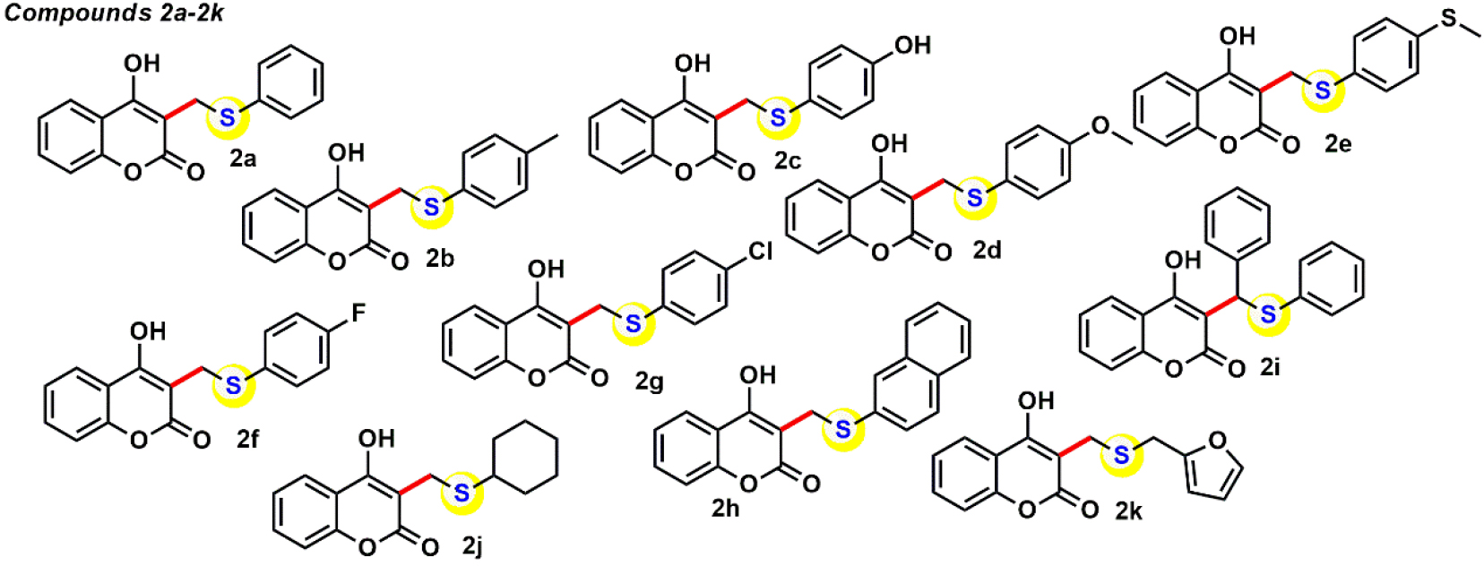
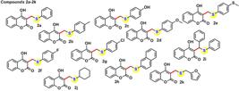
The coumarin derivatives 2a-2k (Figure 1), previously reported by our research group through a methodology that provides efficient access to a diverse set of sulfursubstituted 4-hydroxycoumarins under mild conditions, were prepared for the experiments described herein, and their photophysical properties were investigated in this study.10,11,16 All 1H and 13C NMR spectra analysis of compounds 2a-2k are listed in the Supplementary Information (SI) section (Figures S1-S11).
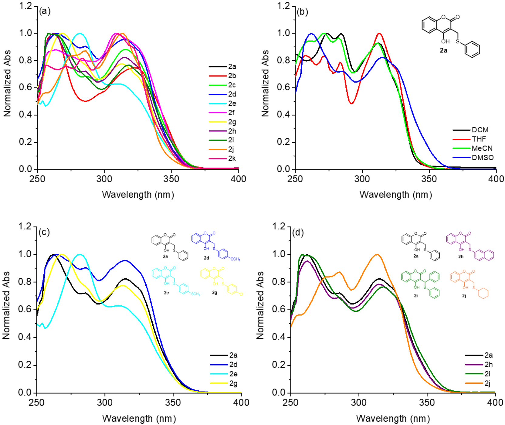
Compounds 2a-2k were analyzed for their absorption properties in DCM, THF, MeCN, and DMSO solutions, with a fixed concentration of 20 µM. The absorption peaks as well as the molar extinction coefficients (ɛ) values are listed in Table 1, and the absorption spectra in all solvents can be found in the SI section (Figures S12-S15). The derivatives studied were evaluated according to: (i) absorption peaks in the same solvent, (ii) differences in absorption peaks in different solvents, (iii) influence of donor-acceptor substituents in the same solvent, and (iv) influence of aliphatic or aromatic substituents in the same solvent, respectively. Firstly, the absorptive behavior of the derivatives 2a-2k in DMSO was analyzed, in the 250 to 400 nm range (Figure 2a). All derivatives showed absorption peaks in the UV range, between 260 and 325 nm, and these bands can be attributed to π→π* and intramolecular charge transfer (ICT) transitions, typical of coumarin derivatives.17 Furthermore, no significant change in the transitions bands was observed according to the electronic properties of the substituents attached to the peripheral sulfur atom of coumarin.
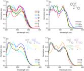
(a) Normalized absorption spectra of coumarin derivatives 2a-2k in DMSO. (b) Normalized absorption spectra of derivative 2a in all tested solvents. (c) Comparative normalized absorption spectra of coumarin derivatives 2a, 2d, 2e and 2g in DMSO according electronic properties of substituent and (d) comparative normalized absorption spectra of coumarin derivatives 2a, 2h, 2i and 2j in DMSO according aliphatic or aromatic peripheral rings attached in the sulfur atom. All spectra used fixed concentration at 20 µM.
Figure 2b shows the behavior of derivative 2a used as an example according to the change in solvent polarity. It was observed that in solutions of DCM, THF, and MeCN, the absorption bands maintained the same profile with slightly shifts. In DMSO solution, a broadening of the absorption peaks was observed, a fact that may be attributed to a greater interaction of the more polar solvent in the ground state due to secondary forces.18 The evaluation of the donor-acceptor influence of the groups inserted in the peripheral S-aromatic rings can be seen in Figure 2c. Thus, it was observed that only the derivative 2e (SCH3 unit) showed a change in the absorption spectrum profile, suggesting that heavier atoms may influence the absorptive properties of the derivatives,19 but without drastic changes in the absorption range. Finally, in Figure 2d, a comparison was made between the derivatives with aromatic or aliphatic groups inserted in the S-portion of the coumarin ring. It was observed that phenyl (2a), bis-phenyl (2i), and naphthyl (2h) units do not directly affect the absorption profile, with a difference only in 2j containing a 6-membered aliphatic ring. The absence of electronic conjugation in this unit results in a slight alteration in the spectral profile of this compound.
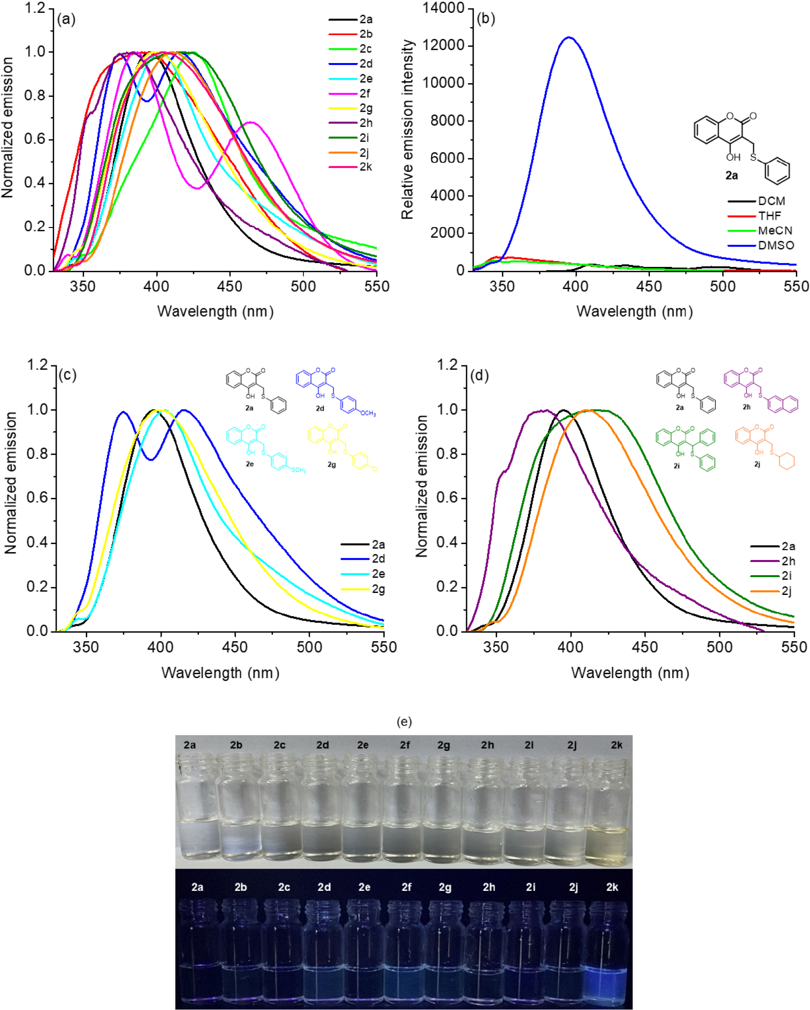
About emission properties, coumarin derivatives 2a-2k were studied in the same solvents, with a fixed concentration of 20 µM. The emission peaks as well as the steady-state emission parameters are listed in Table 1, and the emission spectra of the derivatives in all solvents are listed in the SI section (Figures S16-S18). As with absorption measurements, the derivatives were evaluated using the same factors regarding the influence of the solvent and its electronic properties present in the molecules. DMSO was chosen as the solvent because it was in this solution that the derivatives showed emission peaks.
According to Figure 3a, it was observed that the derivatives 2a-2k showed emission peaks in the range between 370 to 465 nm range (violet region), with some shifts depending on the substituent inserted in the coumarin skeleton. It was also noted that some derivatives showed more than one emission peak, and this fact may be related to the stabilization of different resonance structures in the excited state. Analyzing the influence of the solvent using derivative 2a as an example, it was observed that only in DMSO solution is an emission peak of the derivatives noted, and this property can be attributed to a better stabilization of the resonance structures in the excited state in a more polar medium (Figure 3b).20 Another finding observed in these derivatives is that there is no significant influence of the absence or presence of donor-acceptor groups on the emissive properties of the derivatives, with only a difference observed in the derivative 2d with OCH3 unit (Figure 3c). Finally, as exemplified in Figure 2d, we can observe some changes regarding the change of the aliphatic or aromatic moiety inserted in the peripheral S atom of the coumarin system. The presence of fused aromatic rings (naphthyl, 2h) in relation to two phenyl units (bis-phenyl, 2i) causes an emission peaks shift, with broadening of the emission bands, as well as altering the shift in the absence of aromaticity of the cyclohexyl unit in derivative 2j.
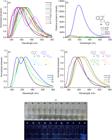
(a) Normalized steady-state fluorescence emission spectra of coumarin derivatives 2a-2k in DMSO. (b) Normalized steady-state fluorescence emission spectra of derivative 2a in all tested solvents. (c) Comparative normalized steady-state fluorescence emission spectra of coumarin derivatives 2a, 2d, 2e and 2g in DMSO according electronic properties of substituent. (d) Comparative steady-state fluorescence emission spectra of coumarin derivatives 2a, 2h, 2i and 2j in DMSO according aliphatic or aromatic peripheral rings attached in the sulfur atom and (e) comparative photography of compound 2a-2k DMSO solutions in daylight and UV365nm irradiation. All spectra used fixed concentration at 20 µM.
All compounds showed fluorescence emission with low quantum yields (QY between 2.0 to 18.0%) in DMSO solutions. The QY values observed in the coumarin derivatives are directly related to the coumarin ring present in all structures since variations in the observed QY values are in accordance with the electronic properties of the substituents inserted into the molecule.15 Also, moderate Stokes shifts (60 to 110 nm range) were observed in all derivatives in DMSO solution due to the intramolecular charge transfer (ICT) states in these compounds, which are dependent on the electronic effects of the substituent inserted in the coumarin ring (Table 1). As another relevant optical parameter, the brightness of compounds 2a-2k was determined by the product of the extinction coefficient and the fluorescence quantum yield (Table 1). In this way, derivatives showed weak brightness in DMSO solution when compared with other coumarin systems,21 being the weak intensity due to its worst charge separation in the excited state from the high directionality in its ICT transition.
Finally, time-resolved fluorescence analysis was recorded for coumarin derivatives 2a-2k in DMSO, with a fixed concentration of 20 µM. The time-resolved emission parameters are listed in Table 1, and the fluorescence decay plots of the derivatives in DMSO are listed in the SI section (Figure S30). In general, short lifetimes values were observed for all derivatives, ranging from 3.60 to 4.70 ns, with variations depending on the substituent inserted in the S-aryl portion of coumarin structure. This behavior is likely due to a reduction in radiative (kr) relaxations resulting from less restricted molecular motion, with losses due to internal conversion processes, thus favoring longer radiative lifetimes (τr) values.
DFT calculations by natural transition orbitals (NTOs) analysis
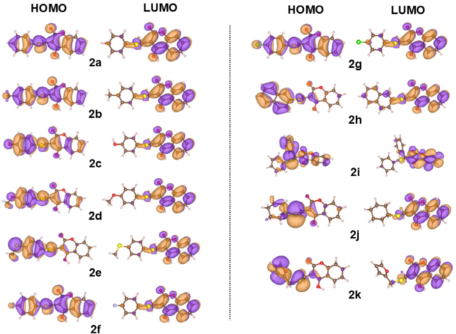
The electronic absorption spectra in DMSO of derivatives 2a-2k have been calculated, and the main theoretical results are shown in Table 2. The calculated absorption spectra of derivatives are shown in the SI section (Figure S31). The theoretical absorption spectra show a blue-shift with respect to the experimental ones, with absorption peaks in the 240-280 nm range. This can be attributed to the fact that the dipole moments of the ground states are greater than those of the excited states, leading to a greater stabilization of the ground states by the solvent, and increasing the excitation energies,23 or to the absence of the description of explicit solute-solvent interactions by implicit solvation models as the polarizable continuum model. The dipole moments of the ground and the first excited states, which are the states involved in the lowest energy electronic transitions, are reported on Table S2 in the SI section. The data in this table show that the dipole of the ground states are consistently greater than those of the first excited states.
Energies (E), wavelengths (λ), oscillator strengths (f), and dipole moment (µ) values of compounds 2a-2k in dimethyl sulfoxide (DMSO) solution
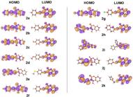
The natural transition orbitals (NTOs) related to the lowest energy electronic transitions of 2a-2k are shown in Figure 4. It is clear from this figure that the highest occupied molecular orbital and (HOMO) lowest unoccupied molecular orbital (LUMO) are in the opposite sides of the molecules, showing electronic transitions with an internal charge transfer (ICT) character in all molecules. It is possible to note that the HOMO orbitals are populated in the S-substituted portion of the derivatives or throughout the molecule, while the population of the LUMO orbitals has a greater contribution from the coumarin unit of the molecules studied. The charge density differences between the first excited and ground states for compounds 2a-2k are shown in Figure S32 in the SI section. They do not reflect the charge transfer character expressed by the NTOs, but instead appear localized on the coumarin unit. This can be explained by the fact that the charge density differences show the charge rearrangements of the whole electronic system that accompany the electronic transition, while the NTO analysis show the dominant hole-particle pairs created by the electronic excitation.
Preliminary analysis of aggregation-induced emission (AIE) properties
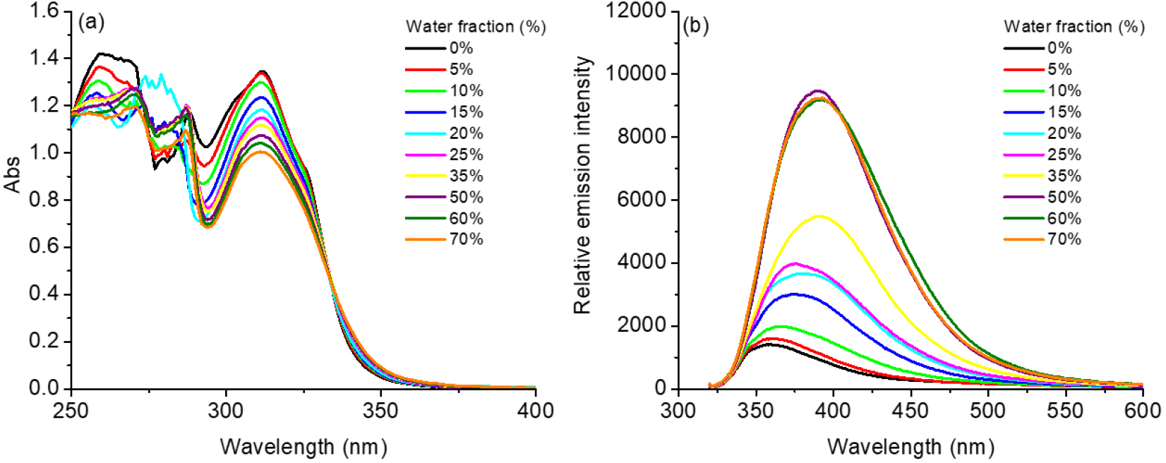
Aggregation-induced emission (AIE) phenomena describe the behavior of a molecule that exhibits low or no luminescence in solution, although an increase in emission is observed in aggregates or in the solid state.24 In this sense, the detection of species in solution can be evaluated by the formation of fluorescent aggregates that are formed by self-organization favored by hydrogen bond interactions or via π-π staking. In the preliminary way, compound 2a was chosen, and its photophysical behavior in the DMSO/H2O mixture (0 to 70% water fraction) was examined to confirm the possibility of the AIE behavior (Figure 5). In the UV-Vis analysis (Figure 5a), a decrease in transition intensities was observed with increasing water fraction (0-70%), likely attributed to possible π-stacking and secondary H-bonding interactions due to the presence of the water protic solvent. In contrast, the steady-state fluorescence emission spectra (Figure 5b) showed the opposite behavior, where an increase in the water fraction enhanced emission peak in the studied derivative 2a, likely by aggregation-induced emission (AIE) phenomenon. This could be explained by the potential formation of aggregates by π-stacking interactions and/or by hydrogen interactions, likely due to the increase in the water fraction, which may favor the formation of fluorescent aggregates in this mixture. Also, shift in the emission peak from 358 to 389 nm was observed as the water fraction increased in derivative 2a, with exhibited an AIE factor (aAIE = F70/F0) of 6.7, suggesting it may display fluorescence emission in the solid state.
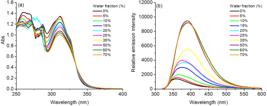
(a) Absorption spectra of compound 2a in DMSO/H2O mixture with different water fractions (0-70%) and (b) steady-state fluorescence emission spectra of compound 2a in DMSO/H2O mixture with different water fractions (0-70%).
Electrochemical analysis
Coumarin derivatives contain rich redox properties and the information about their reduction and oxidation processes is interesting from a biological application, such as redox probes or antioxidant properties.25 Cyclic voltammetry (CV) experiments of 2a-2k were recorded in 0.1 M TBAPF6 with DCM solution, within a range of -2.00 to +2.00 V. Redox parameters are listed in Table 3 and all CV plots of derivatives are shown in the SI section (Figure S33).
Redox potential data of compounds 2a-2k in dichloromethane (DCM) solution (E versus standard hydrogen electrode)
In general, the electrochemical behavior of compounds 2a-2k showed, in all cases, two irreversible reduction and two irreversible oxidation processes, respectively. Cathodic peaks at -0.90 to -1.70 V range can be attributed due to the π-anion and coumarin-phenolate-type species in solution.26 In the positive range, oxidation peaks were observed at the +0.40 to +1.50 V potential range, and this can be attributed to the π-cation and sulfur oxidation (S=O) species stabilized by support electrolyte in solution. Additionally, these species are generated according to the electronic properties of the substituent. For example, derivatives containing electron donor or acceptor groups such as 2c, 2d, and 2g have lower oxidation potentials compared to the derivative 2a (phenyl only), a fact attributed to the change in electronic conjugation in the molecule, favoring these oxidations (Table 3).
Exploring cation optical sensor properties
Preliminary qualitative analyses
Due to their emissive properties and the presence of several coordinating heteroatoms in the coumarin skeleton, which may provide a chelation mode for certain metal ions, the potential of this class of compounds for detecting metal cations in solution was investigated. For this study, compound 2k was selected because it presented the best and most intense emission peak and the possible chelation mode between hard/soft atom species in the same molecule.
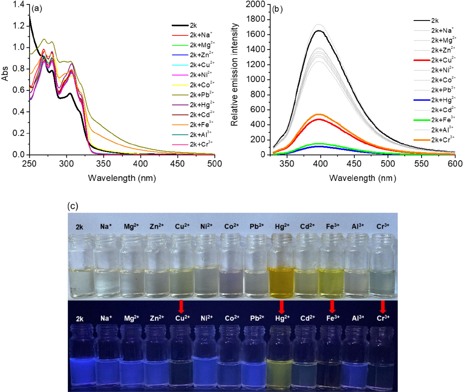
In this way, UV-Vis spectra of 2k (1.0 equiv.) in the absence and presence of 12 different cations (5.0 equiv., independent assays) were recorded using an DMSO/H2O (9:1, v/v) solvent mixture. Thus, derivative 2k showed intense absorption between 280 to 320 nm range, resulting from π→π* transitions, and a change in the color of the solutions in the presence of some cation species such as Cu2+, Hg2+, Fe3+ and Cr3+ ions (Figure 6). The addition of these cations caused a broadening in the transitions, but nothing significant, making it necessary to use a more sensitive technique such as fluorescence emission (Figure 6a). The transitions observed in the presence of the selected ions can be attributed to the presence of ligand-tometal charge transfer transitions (LMCT) from coumarin 2k to ions in solution.
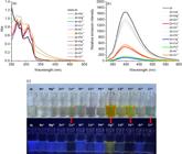
(a) Absorption spectra of compound 2k (2.0 µM; 1.0 equiv.) in the absence or in the presence of selected cation specie (100 µM; 5.0 equiv.) in DMSO/H2O (9:1; v/v). (b) Steady-state fluorescence emission spectra of compound 2k (1.0 equiv.) in the absence or in the presence of selected cation species (5.0 equiv.) in DMSO/H2O (9:1; v/v) and (c) photography of compound 2k in the presence of selected cations, at daylight and UV365nm light conditions.
Analyzing the steady-state fluorescence emission spectra in the absence and presence of the same cation species tested, the blue fluorescence of the 2k solution (1.0 equiv.) became non-emissive only with the addition of the selected ion solution (5.0 equiv.) (Figures 6b-6c). The emission band of the 2k solution was located at approximately 400 nm, with an excitation wavelength of 310 nm. After the addition of the these mentioned ions, we noticed a significant decrease in the fluorescence intensity of the molecular probe (Figure 6b). The observed quenching was predominantly due to the development of the nonfluorescent 2k-cation complex, which can produce static quenching and this fact is due to the paramagnetic/heavy atom effects of the transition metal species.
To gain a deeper understanding of the interaction between the coumarin derivative 2k and the selected ions, further quantitative evaluations were conducted with the compound via fluorescent emission technique. For this purpose, different amounts of a solution of the ion (1.0 to 5.0 equiv.) were added individually to DMSO/H2O (9:1; v/v) mixture solutions of the selected 2k (Figure 7). To this end, the interactive parameters were calculated via KSV, Ka and Kb constants for the selected ions, and thus the Hg2+ species was selected as having the highest values obtained (see Table 4 and Figures S23-S25, SI section).
Quantitative titration parameters between derivative 2k (1.0 equiv.) and selected cations (Hg2+, Cu2+, Fe3+ and Cr3+; 0 to 5.0 equiv.) by steady-state fluorescence emission assays at 298 K
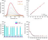
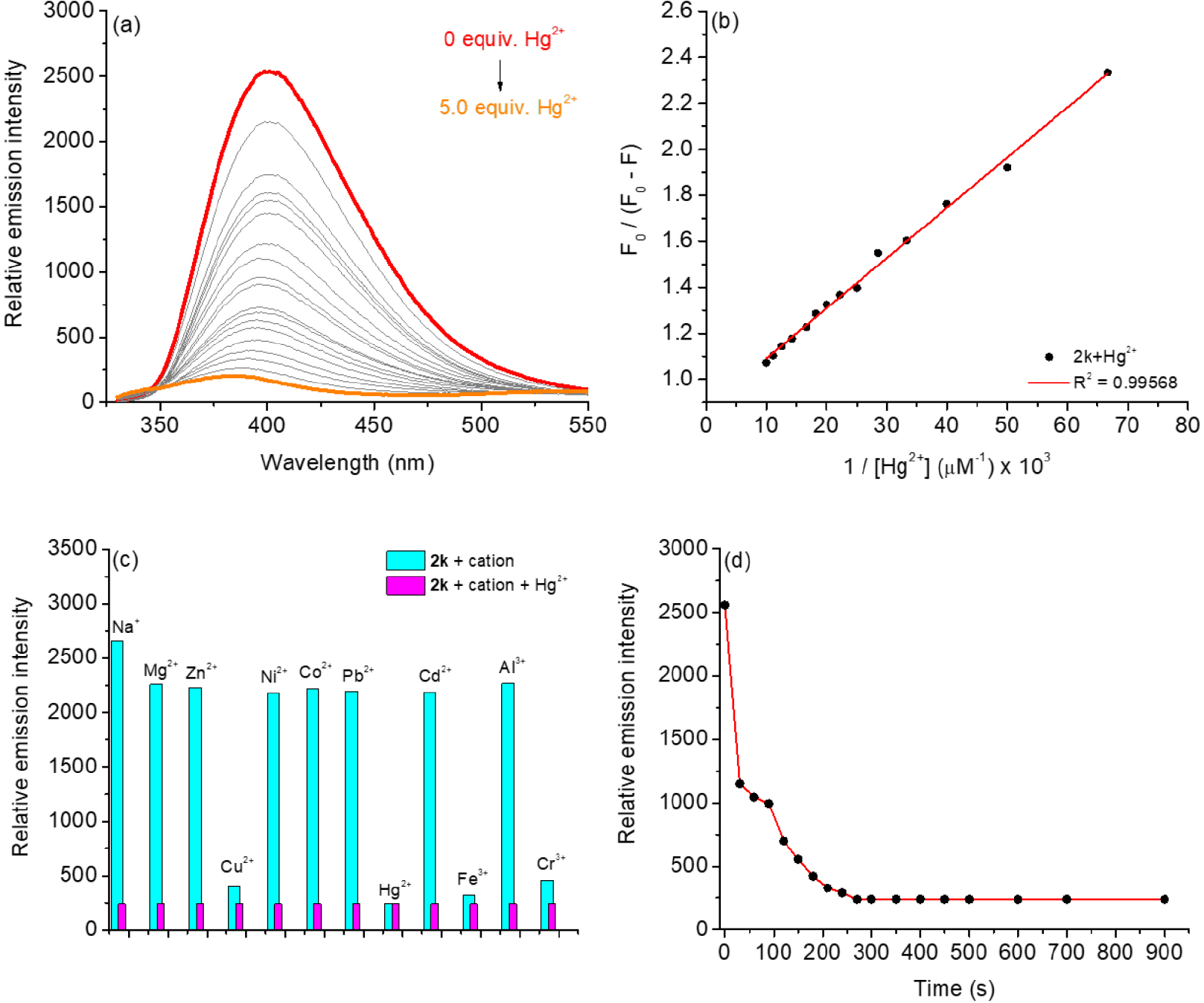
(a) Steady-state fluorescence emission of coumarin 2k (1.0 equiv.) in DMSO/H2O (9:1, v/v) solution in the presence of a different concentration of Hg2+ ions (0 to 5.0 equiv.) with λexc = 310 nm. (b) The linear relationship of probe 2k and Hg2+ ions. (c) Interference study by fluorescence analysis of 2k (1.0 equiv.) and Hg2+ ions (5.0 equiv.) at 400 nm in the presence of different metal cations (5.0 equiv.) with λexc = 310 nm, and (d) kinect profile by fluorescence analysis after addition of 5.0 equivalents of Hg2+ ions in 2k solution.
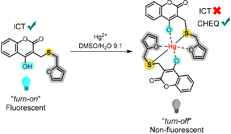
Initially, in the absence of Hg2+ ions, the solution of 2k exhibited fluorescence emission in the blue region. However, with the addition of Hg2+ ions, a non-emissive complex was formed between thio(furyl)coumarin and Hg2+, leading to a significant reduction in its fluorescence intensity (Figure 7a). Figure 7b also presents the relationship between the area under the emission curve and the amount of Hg2+ added, where it can be observed that the most significant quenching occurs in the range of 0 to 30 μM. Based on the literature, the Hg2+ ions detection probably occur through Charge Transfer Quenching (CHEQ) or Charge Exchange Fluorescence (CHEF) phenomena, involving electronic charge transfer transitions from the LMCT.27 Regardless of the specific process, the fluorescence quenching in the presence of a concentration of Hg2+ ions is the result of the CHEQ mechanism, which occurs due to the heavy atom effect of Hg2+ species.
Using the Stern-Volmer quenching equation (see Experimental section), it was possible to determine the KSV quenching constant for the formation of the 2k: Hg2+ complex, with a value in the range of 104 M-1, evidencing a higher fluorescence quenching emission of the compound in the presence of the Hg2+ ions (Table 4). The Ka and Kb constants using the modified Stern-Volmer and the doublelogarithm equations (see Experimental section) were obtained and the values presented are in the same range of 104 M-1, evidencing a strong interaction of the Hg2+ ions with the imidazole skeleton that is probably favoring chelation due to the bidentate nature (S atom of the thiosubstituent unit and O donor atom of the furyl tail and coumarin core) (Table 4). Finally, DGº of derivative 2k with Hg2+ species was calculated from the obtained Kb value. The negative value of DGº energy support the hypothesis that the binding process is spontaneous in the thio-coumarin 2k with Hg2+ ions, favored by the presence of the softer S atom.
In order to further evaluate the possible interference of other cations on the analysis of Hg2+ ions by compound 2k, interference experiments were performed with the other metal ions used. As shown in Figure 7c, when Hg2+ ions were added to the mixture solution with other metal ions and compound 2k, the fluorescence intensity was significantly reduced compared with that in the absence of Hg2+ ions, even in the presence of other ions that also had emission suppression. It was demonstrated that 5.0-fold concentration of other coexisting metal cations had very little effect on the fluorescence response of the 2k-Hg2+ complex. Therefore, the derivative 2k showed good anti-interference ability for the detection of Hg2+ ions, indicating that none of the other metal ions tested prevented the binding of the 2k probe to Hg2+ ions. In addition, the response time is another necessary condition for the coumarin probe 2k to be able to dynamically detect possible real samples in real time. The variations in fluorescence intensity over time, before and after the addition of Hg2+ ions (5.0 equiv.), were recorded to discuss the response time of compound 2k in the presence of Hg2+ ions (Figure 7d). After the observation, the response time indicates that the interaction between derivative 2k and the addition of Hg2+ ions at maximum equivalence (5.0 equiv.) would be from 300 s, which represents a good response time for the detection of these ions in solution.
Lastly, using equations 5 and 6, LOD and LOQ (n = 3) by fluorescence emission of compound 2k for Hg2+ ions were estimated to be 3.66 and 11.10 μM, respectively. A comparative table of LOD values of previously published Hg2+ probes containing coumarin cores is listed in the SI section (Table S1), demonstrating that compound 2k exhibits relatively good sensitivity.
In order to investigate the ratio between 2k and the coordination of Hg2+ ions, the Job’s plot data were analyzed by steady-state fluorescence emission spectroscopy using molar fractions containing 2k and Hg2+ ions. The plot is presented in the SI section (Figure S37). The results indicated that the maximum emission intensity value was obtained when the mole fraction of Hg2+ was set to 0.2. Therefore, 2:1 was the most likely binding stoichiometry for the coordination of 2k and Hg2+ ions, with a proposal represented in Scheme 1.
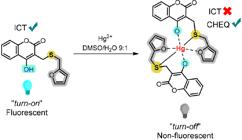
Three possible configurations for the coordination between compound 2k and Hg2+ ions were proposed and theoretically studied, in which a 2:1 stoichiometry is maintained. In the first, the Hg2+ ions are initially coordinated only to the S atoms. In the second, the Hg2+ ion is initially coordinated to the S atoms and to the two O atoms from the coumarin moiety. In the third, the Hg2+ ion is initially coordinated to the S atoms and to the two O atoms from the furyl moieties.
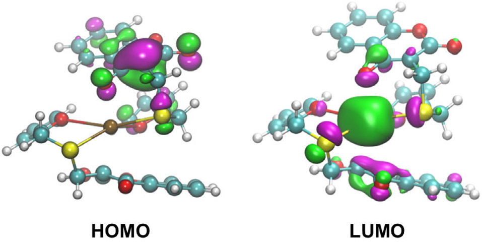
Geometry optimizations and total energy calculations show that the most stable configuration is that in which Hg2+ ions is coordinated to the S atoms and to the O atoms of the furyl group, followed by the structure in which Hg2+ is coordinated only to the S atoms. The optimized structures are shown in Figure S38 (SI section). The absorption spectrum of the most stable configuration is presented in Figure S39, and the NTOs of the lowest-energy electronic transition are shown in Figure 8. The NTOs confirm the LMCT character of the lowest-energy electronic transition.
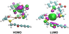
Natural transition orbitals associated to the lowest electronic transition of the Hg2+-2k complex. The left and right panels correspond to the HOMO and LUMO orbitals.
Real sample test - cosmetics
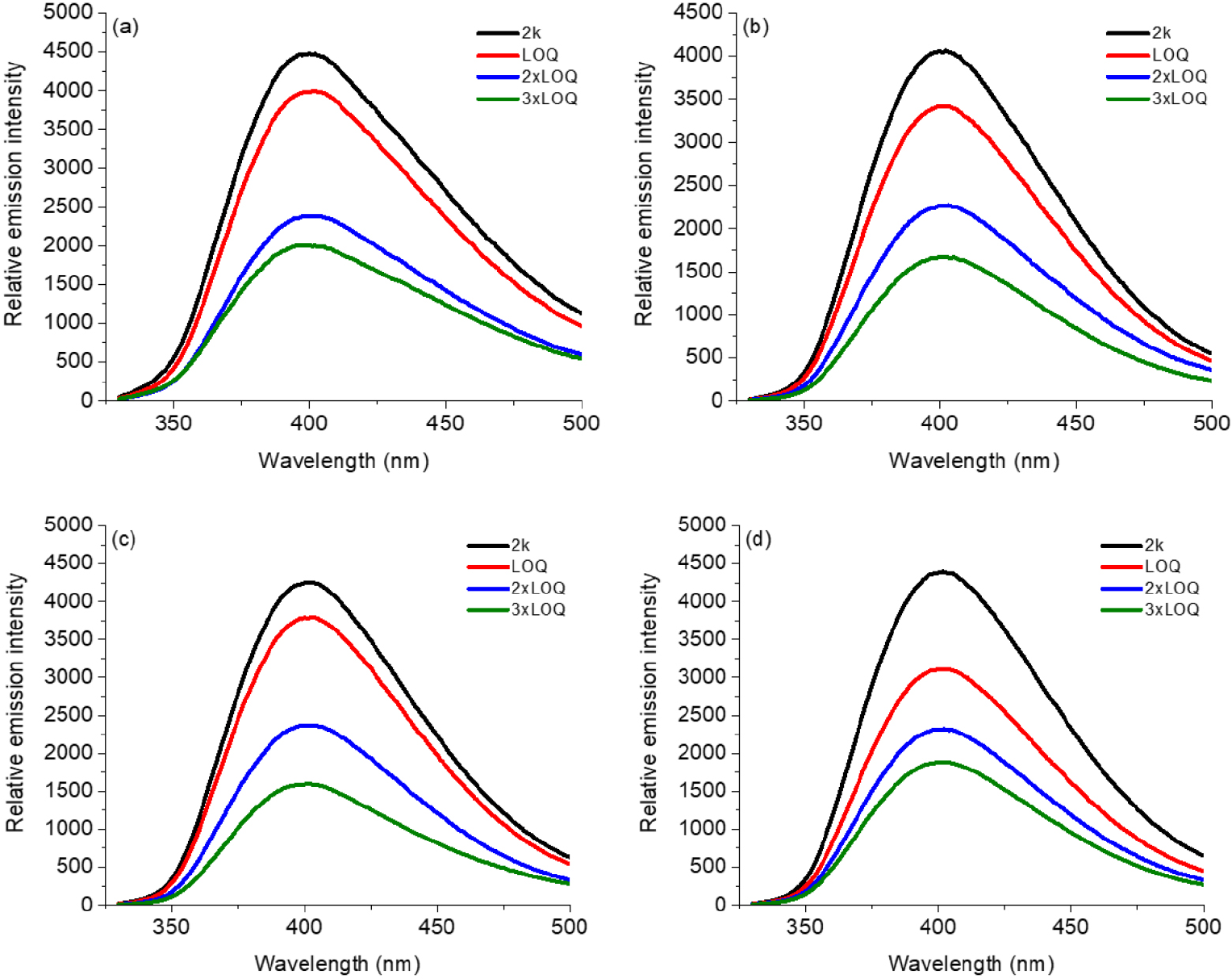
For the tests to determine Hg2+ ions in real samples, four samples were selected (two lipsticks - samples A and B and two face whitening foundations, samples C and D). First, 20 mg of the cosmetic sample was dissolved in 10 mL of isopropanol to prepare a stock solution, and no further sample pre-treatment was required, according to the literature.28 The contents of Hg2+ in two types of cosmetics were measured by steady-state fluorescence emission analysis in Figures 9a-9d. Subsequently, different concentrations based on the LOQ values of Hg2+ (11.10, 22.20 and 33.30 μM) were added to the above samples, respectively. As shown in Figure 8, upon adding LOQ (11.10 μM), 2×LOQ (22.20 μM) and 3×LOQ (33.30 μM) pre-concentrations of Hg2+ ions, the lipstick and face whitening foundation samples shows a significant emission quenching, expressed as Emission Efficiency Quenching (EEQ), in lipsticks (EEQ value ca. 55.4 to 58.7%, Figures 9a-9b) and in face whitening foundation C and D (EEQ value ca. 57.0 to 62.5%, Figures 9c-9d) at 400 nm. These results convincingly demonstrate that 2k could serve as a practical tool to detect the Hg2+ ions content in common beauty products, with little interference from the background.
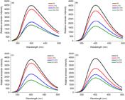
Real tests of derivative 2k in cosmetics: steady-state fluorescence emission spectra at 400 nm of compound 2k in (a) lipstick sample A, (b) lipstick sample B, (c) face whitening foundation sample C and (d) face whitening foundation sample D, with different Hg2+ concentrations (LOQ values). Excitations at λexc = 310 nm in DMSO/H2O 9:1 (v/v) solutions.
Conclusions
Sulfur-substituted 4-hydroxycoumarins 2a-2k were developed and characterized, exhibiting notable photophysical properties. All compounds showed UV absorption and violet-region fluorescence emission (370-465 nm), with modest shifts depending on solvent polarity and substituent nature. Quantum yields were low to moderate (2-18%), with significant Stokes shifts indicating ICT states, supported by DFT calculations and NTO analysis. AIE behavior was observed for 2a, suggesting potential solid-state applications. Electrochemical studies revealed multiple irreversible oxidation and reduction processes, modulated by substituents, relevant for redoxbased applications. 2k emerged as an effective optical sensor for cations, particularly Hg2+, showing strong fluorescence quenching through heavy atom effects and LMCT. It displayed high selectivity, low limits of detection (3.66 µM) and quantification (11.10 µM), rapid response, and successful Hg2+ detection in cosmetic samples. These findings demonstrate that sulfur-substituted 4-hydroxycoumarins are promising fluorescent sensors for heavy metals, with potential applications in environmental, biological, and consumer product analysis.
-
This publication is part of the special issue “Organic Synthesis - BMOS”
Supplementary Information
Supplementary information is available free of charge at http://jbcs.sbq.org.br as PDF file.
Supplementary PDF
Data Availability Statement
The data supporting the findings of this study are available within the article and its SI section.
Acknowledgments
Fellowships from CNPq, CAPES, and FAPERJ are gratefully acknowledged. This work was supported by FAPERJ (E26/210.333/2022, E-26/201.369/2021, E-26/204.583/2024) and CNPq (308006/2022-4, 404587/2021-6). We also thank FIOCRUZ for HRMS measurements. B.A. Iglesias also to thanks the CNPq (PQ grant - 303287/2024-1 and Universal grant 404968/2025-2), CAPES (Finance code 001) and FAPERGS (PqG - 24/2551-0001561-2 and FAPERGS/FAPESP 24/2551-0001939-1).
References
-
1 Suryapratap, J. S.; Nagaiyan, S.; Dyes Pigm. 2022, 202, 110306. [Crossref]
» Crossref -
2 Sontisiri, P.; Promrug, D.; Arthan, D.; Choengchan, N.; Thongyoo. P.; Spectrochim. Acta, Part A 2025, 326, 125170. [Crossref]
» Crossref -
3 Chen, Y.; Tang, S.; Hameed, M. S.; Wang, Q.; Xu, X.; Bao, J.; Wei, S.; Yan, J.; Chen, Q.; Gao, Q.; Liu, H.; Zhang, K.; Han, X.; Spectrochim. Acta, Part A 2025, 325, 125075. [Crossref]
» Crossref -
4 Valmiki, P. A.; Naik, L.; Thippeswamy, M. S.; Maridevarmath, C. V.; Metre, T. V.; Kamble, R. R.; Malimath, G. H.; J. Mol. Struct. 2025, 1322, 140427. [Crossref]
» Crossref -
5 Niranjan, R.; Prasad, G. D.; Arockiaraj, M.; Rajeshkumar, V.; Mahadevegowda, S. H.; J. Mol. Struct. 2025, 1321, 139929. [Crossref]
» Crossref -
6 Jiang, S.; Chen, Y.; Li, Y.; Han, L.; J. Photochem. Photobiol., A 2019, 384, 112031. [Crossref]
» Crossref -
7 Rajeshirke, M.; Tathe, A. B.; Sekar, N.; J. Mol. Liq. 2018, 264, 358. [Crossref]
» Crossref -
8 Torres, F. C.; Gonçalves, G. A.; Vanzolini, K. L.; Merlo, A. A.; Gauer, B.; Holzschuh, M.; Andrade, S.; Piedade, M.; Garcia, S. C.; Carvalho, I.; Poser, G. L.; Kawano, D. F.; Eifler-Lima, V. L.; Cass, Q. B.; J. Braz. Chem. Soc. 2016, 27, 1541. [Crossref]
» Crossref -
9 Gupta, D.; Guliani, E.; Bajaj, K.; Top. Curr. Chem. 2024, 382, 16. [Crossref]
» Crossref -
10 Forezi, L. S. M.; Borba-Santos, L. P.; Cardoso, M. F. C.; Ferreira, V. F.; Rozental, S.; Silva, F. C.; Curr. Top. Med. Chem. 2018, 18, 164. [Crossref]
» Crossref -
11 Forezi, L. S. M.; Froes, T. Q.; Cardoso, M. F. C.; Maciel, C. A. O.; Nicastro, G. G.; Baldini, R. L.; Costa, D. C.; Ferreira, V. F.; Castilho, M. S.; Silva, F. C.; Curr. Top. Med. Chem. 2018, 18, 149. [Crossref]
» Crossref -
12 Chai, J.-D.; Head-Gordon, M.; Phys. Chem. Chem. Phys. 2008, 10, 6615. [Crossref]
» Crossref -
13 Heinrich, G.; Schoof, S.; Gusten, H.; J. Photochem. 1974, 3, 315. [Crossref]
» Crossref -
14 Tomasi, J.; Mennucci, B.; Cammi, R.; Chem. Rev. 2005, 105, 2999. [Crossref]
» Crossref -
15 Gagne, R. R.; Koval, C. A.; Lisensky, G. C.; Inorg. Chem. 1980, 19, 2854. [Crossref]
» Crossref -
16 Cardoso, M. F. C.; Forezi, L. S. M.; Cavalcante, V. G. S.; Juliani, C. S. R.; Resende, J. A. L. C.; Rocha, D. R.; Silva, F. C.; Ferreira, V. F.; J. Braz. Chem. Soc. 2017, 28, 1926. [Crossref]
» Crossref -
17 Rosa, W. C.; Rocha, I. O.; Schmitz, B. F.; Martins, M. A. P.; Zanatta, N.; Tisoco, I.; Iglesias, B. A.; Bonacorso, H. G.; J. Fluor. Chem. 2021, 248, 109822. [Crossref]
» Crossref -
18 Hemdan, S. S.; J. Solut. Chem. 2024, 53, 552. [Crossref]
» Crossref -
19 Li, Y.; Li, G.; Zhang, Q.; Li, Y.; Jia, Q.; Zhang, W.; Feng, X.; Xu, W.; Liu, J.; Chem. Phys. Lett. 2021, 784, 139091. [Crossref]
» Crossref -
20 Lanke, S. K.; Sekar, N.; J. Fluoresc. 2016, 26, 497. [Crossref]
» Crossref -
21 Ríos, M.-C.; Bravo, N.-F.; Macías, M.; Iglesias, B. A.; Portilla, J.; ChemPhotoChem 2025, 9, e202400389. [Crossref]
» Crossref -
22 Gonzaga, D. T.; da Rocha, V. N.; de Souza, A. S.; Piquini, P.; Ferreira, V. F.; Iglesias, B. A.; Dyes Pigm. 2026, 246, 113445. [Crossref]
» Crossref -
23 Bi, T.-J.; Xu, L.-K.; Wang, F.; Ming, M.-J.; Li, X.-Y.; Phys. Chem. Chem. Phys. 2017, 19, 32242. [Crossref]
» Crossref -
24 Zhao, Z.; Zhang, H.; Lam, J. W. Y.; Tang, B. Z.; Angew. Chem., Int. Ed. 2020, 59, 9888. [Crossref]
» Crossref -
25 Huang, C.-N.; Kuo, P.-Y.; Lin, C.-H.; Yang, D.-Y.; Tetrahedron 2007, 63, 10025. [Crossref]
» Crossref -
26 Kim, S.-H.; Jung, E.-J.; So, E.-M.; Shen, C.-Z.; Chun, H.-J.; Kim, Y.-M.; Kim, I.-K.; Bull. Kor. Chem. Soc. 2006, 27, 1329. [Crossref]
» Crossref -
27 Lee, H.; Lee, H.-S.; Reibenspies, J. H.; Hancock, R. D.; Inorg. Chem. 2012, 51, 10904. [Crossref]
» Crossref -
28 Cheng, H.; Yi, F.; Sun, J.; Li, A.; Zhang, X.; Guan, D.; Qu, Y.; Cao, J.; Dyes Pigm. 2024, 226, 112135. [Crossref]
» Crossref
Edited by
-
Editor handled this article:
Emílio Carlos de Lucca Júnior (Guest)