Metabolomics Reveals How Seasonal Variation in the Essential Oils of Ocotea odorifera (Vell.) Rohwer Impacts its Anti-Inflammatory Activity
Abstract
Ocotea odorifera (“canela-sassafras”) has a rich history in the traditional medicine of Brazil for treating inflammatory conditions such as edema, arthritis, and fever. Ethnobotanical records indicate that leaves are commonly prepared by decoction to relieve inflammatory conditions. This research applied metabolomics strategies to investigate how seasonal variation influences the chemical composition and anti-inflammatory activity of O. odorifera leaf essential oils (EOs). Leaves were collected monthly over one year, and EOs were extracted via hydrodistillation. Gas chromatography-mass spectrometry (GC-MS) was applied for EOs chemical composition identification. The EOs were assessed using an ex vivo anti-inflammatory assay, measuring prostaglandin E2 (PGE2) levels in lipopolysaccharide-stimulated human whole blood. Ultra-performance liquid chromatography coupled with tandem mass spectrometry (UPLC-MS/MS) was employed to determine PGE2 inhibition levels. Untargeted metabolomics demonstrated that EOs harvested in autumn and spring exhibited fluctuations in the chemical composition, which favoured greater PGE2 inhibition levels, with pronounced seasonal shifts among the minor compounds. Orthogonal partial least squares discriminant analysis (PLS-DA) revealed terpinen-4-ol as statistically different and positively correlated with the increase in the anti-inflammatory activity. Safrole, a major compound, demonstrated no relevant seasonal variation. These findings highlight and confirm how seasonal chemical dynamics modulate the specialized metabolism and the anti-inflammatory activity profile of O. odorifera EOs.
Keywords:
Ocotea odorifera; “canela-sassafras”; seasonal variation; GC-MS; UPLC-MS/MS; prostaglandin E2
Introduction
Essential oils (EOs) are volatile compounds characterised by a strong odor, known to be antiseptic, and for their medicinal properties, such as analgesic, spasmolytic, bactericidal, virucidal, fungicidal, sedative and anti-inflammatory effects. They can be synthesised by all parts of the plant (buds, flowers, leaves, stems, branches, seeds, fruits, roots, wood or bark) and have an important function in protecting plants due to their biological effects.1-4 Lauraceae is a family of aromatic plants widely distributed across tropical and subtropical regions, encompassing approximately 67 genera and over 2500 species. Brazil, in particular, is home to around 25 genera and 400 species of Lauraceae.5,6 Within this family, the genus Ocotea is notable for its remarkable taxonomic complexity and chemical diversity, consisting of approximately 350 species distributed across the Americas and Africa.6-8 Plants belonging to the Lauraceae, particularly in the Ocotea genus, are renowned for producing a wide array of specialised metabolites, including alkaloids, lignoids, flavonoids, monoterpenes, sesquiterpenes, saponins, and coumarins.8-14
Ocotea odorifera, commonly known as the “sassafras tree”, “canela-sassafras”, or “canela-do-mato”, due to its characteristic odor, is particularly noteworthy within this genus. Traditionally, it has been employed to treat various ailments such as edemas, stomach disorders, arthritis, syphilis, and fever.8,15 Ethnopharmacological reports indicate that O. odorifera leaves are traditionally prepared as a decoction and used in southern Brazil for inflammatory-related conditions, such as rheumatism and painful inflammatory conditions.16,17 Modern scientific investigations have substantiated several of these traditional uses, revealing that O. odorifera possesses significant antioxidant, antimicrobial, antimutagenic, and anti-inflammatory properties.8,15-19 The EOs of O. odorifera are rich in bioactive compounds, particularly safrole, which is often the major constituent, along with other sesquiterpenes and monoterpenes such as α-pinene, β-pinene, and limonene.20-23 The high safrole content has been associated with the antimicrobial and insecticidal properties of the plant, although it also raises concerns due to the known hepatotoxic of safrole and potentially carcinogenic effects when consumed in high quantities or over prolonged periods.24,25
Moreover, inflammation is a complex biological response that serves as the primary defence mechanism of the body against infections, injuries, and any other potentially harmful stimuli. This physiological process involves the activation of a cascade of immune cells and the release of various signalling molecules that orchestrate the inflammatory response through multiple metabolic pathways.26-28 Among these, the cyclooxygenase (COX) pathway leads to the synthesis of prostacyclins (PCI), prostaglandins (PGs), and thromboxanes (TXs), which are key mediators of inflammation, pain, and vascular homeostasis. Simultaneously, the lipoxygenase (LOX) pathway generates lipoxins (LXs), leukotrienes (LTs), and hydroxyeicosatetraenoic acids (HETEs), compounds primarily involved in inflammatory leukocyte recruitment and the regulation of immune responses.29-31
Anti-inflammatory drugs are broadly classified into steroidal anti-inflammatory drugs (SAIDs), known as corticosteroids, and non-steroidal anti-inflammatory drugs (NSAIDs), which are more commonly used as therapeutic agents.32 SAIDs primarily inhibit the phospholipase A2 at the beginning of the cascade, while NSAIDs target it further in the COX pathway. Non-selective NSAIDs inhibit both COX-1 and COX-2; however, COX-1 is responsible for various physiological functions, and its inhibition can lead to adverse effects such as epigastric pain and an increased risk of ulcers.33,34 Given the limitations and side effects associated with current anti-inflammatory drugs, there is a pressing need to explore new substances with anti-inflammatory activity. O. odorifera emerges as a promising natural source, as evidenced by studies demonstrating its anti-inflammatory potential through dual inhibition of edema and neutrophil recruitment.16 While the anti-inflammatory properties of O. odorifera, along with other species in this genus, have been previously documented, these studies have primarily focused on non-volatile compounds prior to isolation of the compounds.15-19,35-37 To our knowledge, no comprehensive investigation has been conducted on the anti-inflammatory activity of its EOs, nor on how seasonal variations might influence their chemical composition and potential activity.
This study aimed to investigate the seasonal variations in the chemical composition of O. odorifera EOs and assess how these variations influence their potential anti-inflammatory activity using untargeted metabolomics strategies. By analysing the EOs extracted from leaves collected monthly over a year, we aimed to identify specific seasonal changes in chemical constituents and draw a parallel between these changes and alterations in the anti-inflammatory profile. The findings also provide insights into the optimal harvesting periods for O. odorifera, contributing to the knowledge of plant-based anti-inflammatory agents of EOs.
Experimental
Solvents, drugs and reactants
The solvents used were of high performance liquid chromatography (HPLC) grade (J.T. Baker, Radnor, PA, USA). Ultrapure water (Milli-Q system, Merck Millipore, Darmstadt, Germany) was used for the preparation of solutions in the anti-inflammatory assay. Reagents and materials for the bioassay, including lipopolysaccharide (LPS) from Escherichia coli O26:B6, dexamethasone, PGE2 standard, chloramphenicol (CAP), polytetrafluoroethylene (PTFE) syringe filters (0.22 μm pore size), Supelclean™ LC-18 SPE cartridges (100 mg sorbent, 1 mL), and the Supelco Manifold System, were all purchased from Sigma-Aldrich (St. Louis, MO, USA). For gas chromatography-mass spectrometry (GC-MS) analyses, helium was used. A homologous series of n-alkanes (C8-C40) used for retention index determination was purchased from Sigma-Aldrich (St. Louis, MO, USA).
Plant material
Leaves of the O. odorifera tree were collected in the urban area of Alfenas-MG, Brazil (21º42’52.2”S, 45º94’97.9”W). A voucher specimen was deposited at the herbarium of the Federal University of Alfenas (UALF 1907). The collection was registered with the National System for the Management of Genetic Heritage and Associated Traditional Knowledge (SisGen) under protocol number A5A8F67, in full compliance with Brazilian regulations governing access to biodiversity. Sampling was carried out monthly for 12 consecutive months, always from the same individual tree and at the same collection site. To minimize diurnal effects on essential oil profiles, collections were performed at a consistent time of day (± a narrow window) and on comparable days each month. Environmental metadata, including exact collection time, temperature, and relative humidity recorded at each event, were documented to support further analysis (Table S1, Supplementary Information (SI) section).
Essential oil extraction
Fresh leaves (130 g) were blended with distilled water (2.0 L) and subjected to hydro-distillation for 2 h following the onset of boiling, using a Clevenger-type apparatus according to previously described procedures in literature.16,38-40 The EOs were collected from the aqueous distillate, separated based on their immiscibility, and subsequently dried over anhydrous sodium sulfate to remove residual moisture. The dried oil was then filtered, weighed to determine yield, and stored in sealed amber glass vials at −20 °C until further analysis.
GC-MS analyses
The EOs, blanks (hexane), quality control (QC) samples and a standard solution containing a mixture of n-alkanes (homologous series from C5 to C29) were separately analysed by gas chromatography (GC-17A, Shimadzu Corporation, Kyoto, Japan) coupled with mass spectrometry (MSQP-5050 A, Shimadzu Corporation, Kyoto, Japan) operating with electron ionization (EI) at 70 eV. All EOs samples were prepared by dissolving in ultrapure hexane (1:100, v/v). A volume of 1.0 μL of each sample was injected in a split ratio of 1:50 at 225 °C and detected at 240 °C. The oven temperature was programmed at a rate of 3 °C min-1 from 60 to 240 °C, during 60 min, and kept for 5 min at 240 °C. Helium was used as a carrier gas with an injection pressure of 99.3 kPa at a linear velocity of 46 cm s-1 and a column constant flow rate of 1.6 mL min-1. Chromatographic separations were carried out using an RtX-5MS capillary chromatographic column (Rtx® Crossbond® 5% diphenyl 95% dimethylpolysiloxane, 30 m × 0.25 mm × 0.25 μm film thickness, Restek, Bellefonte, PA, USA). The MS detector was set to the ion source and interface temperature of 200 and 250 °C, respectively, and the m/z scan range from 40 to 500.
GC-MS data handling and analyses
The GC-MS spectra of O. odorifera EOs were processed by matching the obtained mass spectra against the National Institute of Standards and Technology (NIST08 library, 2008) mass spectral library to automatically calculate the similarity index (SI) with the equipment built-in database. The identification of metabolites was further supported by comparison of the experimentally determined arithmetic retention indices (AI). The AI values were calculated using a homologous series of n-alkanes analyzed under identical chromatographic conditions. These values were then manually compared with literature data, including the Adams database, in conjunction with manual MS spectral matching to support metabolite annotation.41,42 Semi-quantitative analysis was based on the relative abundance of each component, expressed as a percentage of the total peak area in the based peak intensity (BPI) chromatogram.
Experimental design and ex vivo anti-inflammatory assay
The anti-inflammatory assay was conducted following the protocol developed and validated by our research group.18,42-47 Briefly, peripheral venous blood was obtained and pooled from donors who reported not having used anti-inflammatory drugs or CAP (internal standard) at least 15 days before the blood collection. The donors were both male and female healthy volunteers from 20 to 40 years of age. The volunteers signed their informed consent for participation in the research, authorised by the Research Ethics Committee of the Federal University of Alfenas (No. 89325818.1.0000.5142). Randomised experiments were evaluated in triplicate.
The production of the inflammatory mediator prostaglandin E2 (PGE2) was induced in heparinised human whole blood (200 μL) using LPS from E. coli O26:B6 at a final concentration of 10 μg mL-1, in 96-well plates. All test samples were prepared at a stock concentration of 1 mg mL-1, dissolved in a water:dimethyl sulfoxide (1:1, v/v) mixture, and filtered through PTFE membranes with a pore size of 0.45 μm. For the assay, samples were diluted to a final concentration of 10 μg mL-1 in phosphate-buffered saline (PBS; pH 7.2, 0.15 M NaCl, 0.01 M phosphate). Dexamethasone (1 μg mL-1 in PBS) was used as a positive control. Plates were incubated at 37 °C in a humidified atmosphere containing 5% CO2 for 24 h. Following incubation, plates were centrifuged at 1000 rpm for 5 min at 4 °C. For protein precipitation, 500 μL of chilled methanol:acetonitrile (1:1, v/v) were added to 100 μL of plasma. The mixture was centrifuged at 6000 rpm for 10 min at 4 °C. The resulting supernatant was diluted to a final volume of 5 mL with ultrapure water (4.5 mL) and loaded onto solid-phase extraction (SPE) cartridges (Supelclean™ LC-18, 100 mg sorbent, 1 mL capacity). SPE was performed using sequential washes with acidified water (0.1% acetic acid, 1 mL) and methanol (0.1% acetic acid, 1 mL). The methanol fractions were collected into microcentrifuge tubes and evaporated to dryness.
Dried extracts were reconstituted in 100 μL of acetonitrile containing the internal standard (CAP, 25 ng mL-1) before analysis by ultra-performance liquid chromatography in tandem mass spectrometry (UPLC-MS/MS).
UPLC-MS/MS analysis
Quantification of PGE2 was performed using UPLC-MS/MS, a Shimadzu Prominence system equipped with a DGU-20A3 degasser, two LC-20AD pumps, a SIL-20A HT autosampler, a CTO-20A column oven, and a CBM-20A system controller. The system was coupled to a Shimadzu LC-8030 triple quadrupole mass spectrometer operating in electrospray ionisation (ESI) mode, in both positive and negative polarity.
Chromatographic separation was achieved using a reversed-phase Eclipse Plus pre-column (1.8 μm, 2.1 × 5 mm; Agilent, USA) coupled to a Poroshell 120 EC-C18 analytical column (2.7 μm, 3 × 100 mm; Agilent, USA). The mobile phase consisted of (A) ultrapure water with 0.1% formic acid and (B) acetonitrile. The gradient elution was programmed as follows: starting at 40% B, linearly increased to 100% B over 3 min, maintained at 100% B for 1 min, then returned to 40% B over 0.5 min and equilibrated at 40% B for 4 min. The flow rate was set at 0.3 mL min-1, and the injection volume was 20 μL. MS settings were as follows: drying gas (N2) at 15 L min-1 at 450 °C, nebulising gas (N2) at 2 L min-1, collision gas (argon) at 230 kPa, desolvation line (DL) temperature at 250 °C, interface voltage at 3.5 kV, and detector voltage at 2.44 kV. Data acquisition was performed in selected reaction monitoring (SRM) mode using transition m/z 351.2 to m/z 271.2 in the negative ionisation mode [M – H]– for PGE2, according to a previously validated method following the guidelines of Brazilian Health Regulatory Agency (ANVISA) Resolution RDC No. 27/2012.48
Data acquisition and processing were carried out using LabSolutions software (Shimadzu, Japan, 2017). The PGE2 levels were measured based on the peak area ratio related to the internal standard (CAP). Statistical analyses were performed using GraphPad Prism version 9.1.2 (GraphPad Software Inc., La Jolla, CA, USA). Data were analysed by one-way analysis of variance (ANOVA) followed by multiple comparison test of Dunnett. Results are expressed as mean ± standard deviation (SD), with differences considered statistically significant at p < 0.05. To check the validity of the parametric tests, including the paired t-test applied for seasonal comparisons, the normality of the data distribution was assessed,49 using Q-Q (quantile-quantile) plots, a visual method that compares the quantiles of the dataset against a theoretical normal distribution (Figure S1, SI section).
Multivariate statistical analyses
Multivariate statistical analyses were performed online using MetaboAnalyst 6.0.50 Additional analyses (bar graphs and heatmaps) were performed in Python (Anaconda ecosystem) executed in a Jupyter Notebook environment (Python scripts .txt file51), including libraries: pandas, numpy, scikit-learn, matplotlib, seaborn, and statsmodels. Before analysis, data were sum normalised and scaled using Pareto scaling to reduce the influence of large-magnitude variables while preserving data structure and variability. Principal component analysis (PCA) was applied for unsupervised dimensionality reduction, outlier observation and exploratory data analysis.18,52 Partial least squares projections to latent structures with variable importance in projection (PLS-VIP) scores were computed to identify the variables most associated with ex vivo anti-inflammatory activity.18,53,54 PCA and orthogonal partial least squares discriminant analysis (PLS-DA) 2D and 3D score and loading plots, bar graphs, and heatmaps were generated for data visualization of QC and EOs samples. Statistical significance was assessed using p-values, with correction for multiple testing performed using the adjusted false discovery rate (FDR; p < 0.05) and variance important in projection (VIP) scores greater than 1 were considered statistically significant. Additionally, only variables positively correlated with the spring-winter-fall (SWF) sample group (most active seasons) were selected as potential biomarkers related to the observed PGE2 inhibition levels.
Results and Discussion
Ex vivo anti-inflammatory activity
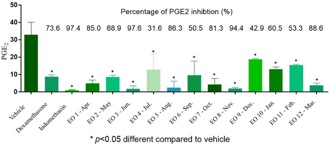
The anti-inflammatory potential of O. odorifera EOs was assessed by measuring their ability to inhibit PGE2 production in LPS-stimulated human whole blood. Positive and negative controls were statistically different (p < 0.05) (Figure 1), supporting the reliability of the anti-inflammatory assay. PGE2 levels were significantly reduced by several EOs compared to the vehicle control (p < 0.05). To validate the reliability of the parametric statistical tests applied, the data distribution was evaluated using Q-Q plots (Figure S1, SI section). The plots indicated that the distribution of PGE2 inhibition data was approximately normal, supporting the use of parametric methods.
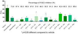
Ex vivo anti-inflammatory results of the O. odorifera EOs (EO1-EO12, obtained from April/2021 to March/2022) and statistical comparison to a negative control (vehicle, PBS + blood + LPS). EOs samples are shown in the x-axis and PGE2 inhibition in the y-axis. Dexamethasone and indomethacin were used as positive controls. The results were analysed by one-way ANOVA, followed by multiple comparison test of Dunnett. Bars represent mean ± standard deviation (SD); *statistical difference to NEG (vehicle) control where p ≤ 0.05.
By analyzing the results, it was evident that the EOs of O. odorifera can be considered statistically anti-inflammatory, though a clear seasonal trend was observed. EOs obtained during summer months, particularly through December and January in the southern hemisphere, showed reduced inhibitory activity, reaching only ca. 50% PGE2 inhibition, which was statistically lower than both the positive controls (dexamethasone and indomethacin) and most other months (p < 0.05). Thus, in contrast, the EOs from non-summer months demonstrated consistently higher PGE2 inhibition, with a mean percentage of ca. 75%. Thus, as the mean PGE2 inhibition of the summer group was statistically lower than that of the autumn, winter, and spring groups (p < 0.05), it suggests that the anti-inflammatory efficacy of O. odorifera essential oils might be season-modulated. In addition, the EOs collected in June, representing the late autumn to early winter transition, exhibited the highest activity (ca. 97%), with no statistical difference from the positive controls. This pattern indicates that summer conditions may negatively impact the biosynthesis of key bioactive compounds responsible for the observed ex vivo anti-inflammatory effects. Also, even though samples were collected always late afternoon, approximately at 5 pm (Table S1, SI section), these obtained results align with the hypothesis that environmental factors, not only temperature, but potentially rainfall, humidity, and other climate conditions, influence the chemical composition and bioactivity of plant-specialized metabolites.55
GC-MS analyses and chemical profile
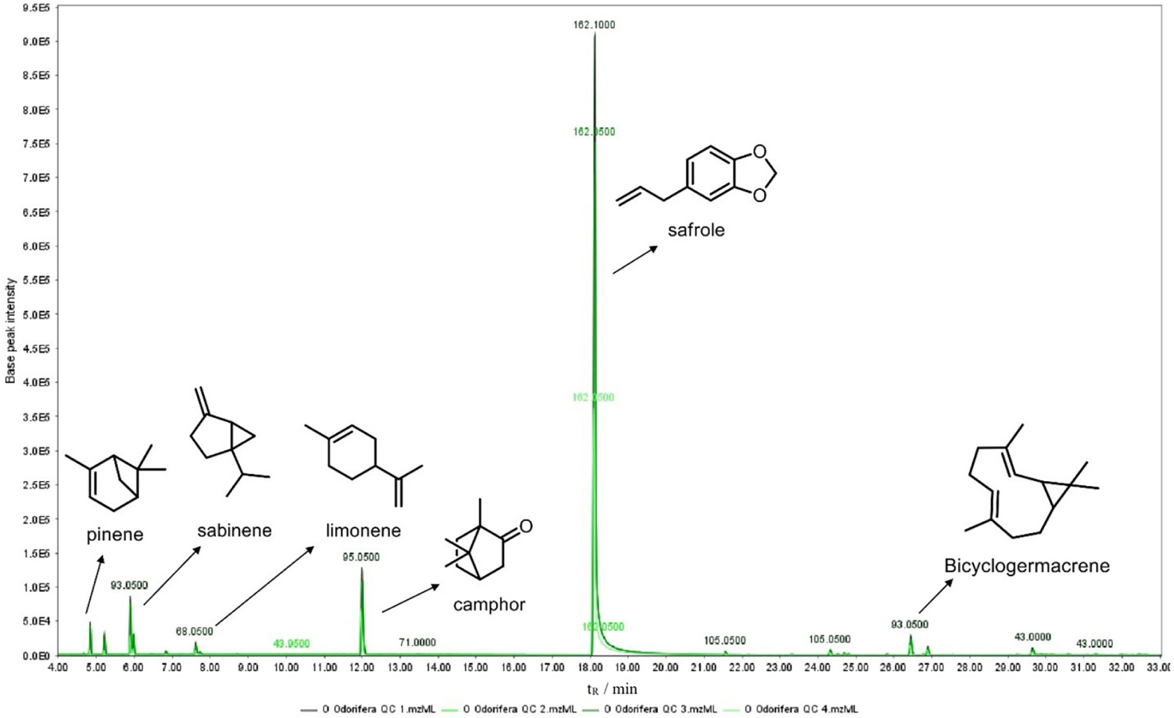
This study reports, for the first time, the seasonal chemical composition of O. odorifera EOs. Compound annotation was carried out by comparing mass spectra with the NIST08 library, supported by Kovats retention index (KI) values for reliable confirmation against literature data. The chemical profile was dominated by phenylpropanoids and monoterpenoids, with a smaller fraction of sesquiterpenes. Seven major compounds safrole (74.1 ± 6.4%), camphor (7.4 ± 0.9%), bicyclogermacrene (3.76 ± 1.7%), spathulenol (2.3 ± 1.1%), cadina-1,4-diene (2.1 ± 1.0%), (+)-sabinene (1.9 ± 0.3%), and limonene (1.3 ± 0.25%) accounted for approximately 90.35% of the total oil composition. In addition to these, minor constituents such as α-pinene (0.8 ± 0.2%), camphene (0.80 ± 0.1%), (−)-β-pinene (0.8 ± 0.4%) and eucalyptol (0.4 ± 0.1%) were also annotated (Figure 2, Table 1). These findings are in line with previous studies reporting O. odorifera as a natural source of safrole and camphor.54 Furthermore, the presence of monoterpenes and sesquiterpenes like eucalyptol, (−)-β-pinene, and camphene is consistent with the typical chemical profiles found in EOs of other species within the Ocotea genus.56,57
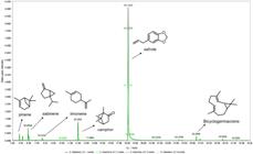
GC-MS BPI chromatogram of O. odorifera leaf EOs. The chromatogram corresponds to the pooled quality control (QC) sample, prepared by combining equal aliquots of the monthly EOs samples collected over the 12-month period (January-December). Peaks correspond to the major volatile compounds annotated based on mass spectral matching (NIST08 library). Highlighted constituents: α-pinene, sabinene, limonene, camphor, safrole and bicyclogermacrene.
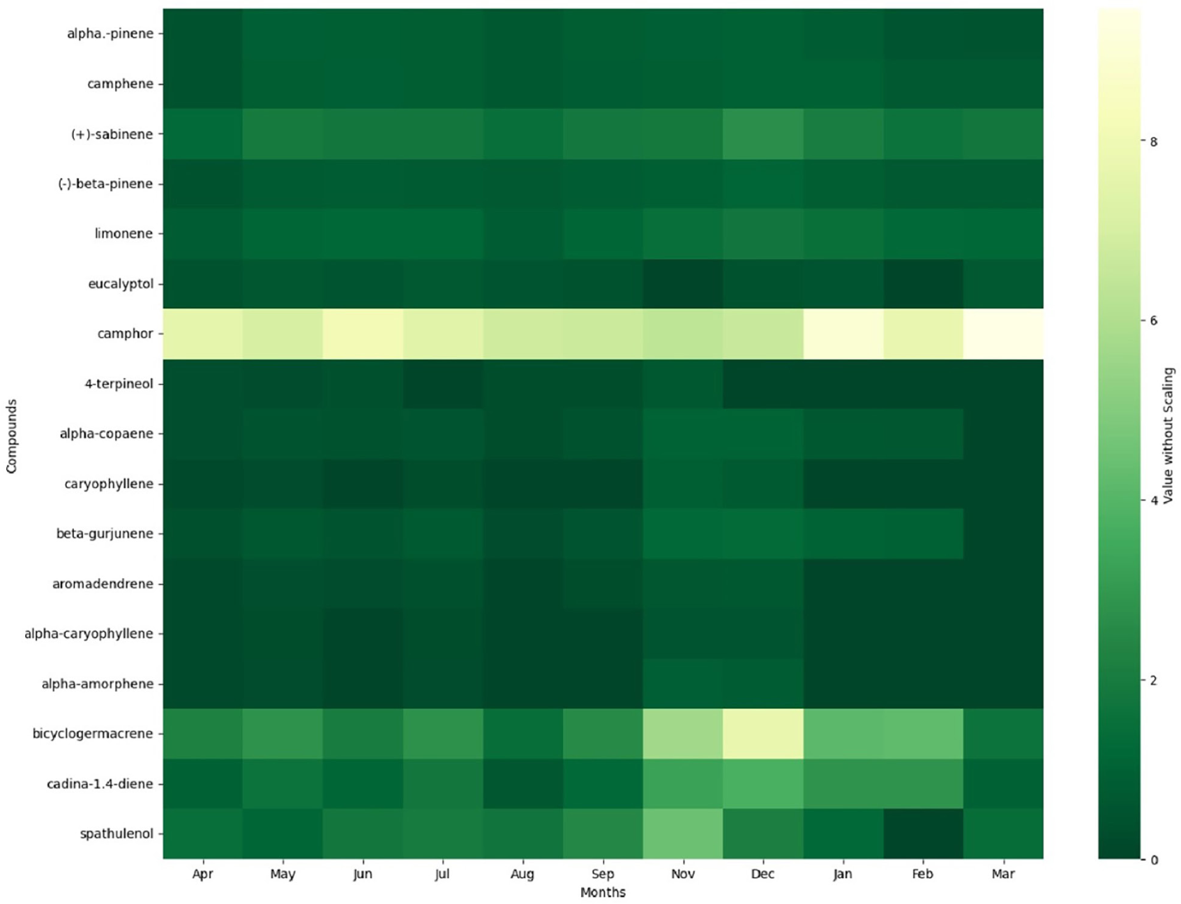
Furthermore, the heatmap provides a clear visual representation of the seasonal variation in the metabolite profile of O. odorifera EOs, highlighting distinct patterns in compound distribution throughout the year (Figure 3). Notably, the major constituent was safrole, ranging from 83% (August, late winter) to 55% (September, medium fall). Due to its overwhelming abundance, safrole was excluded from the heatmap to improve the visualization of the other metabolites, whose relative intensities were otherwise too low to observe clearly. However, the full heatmap including safrole is available in the SI section (Figure S2). Camphor, the second most abundant compound, also displayed relatively high and stable levels, ranging around 9%.
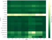
Heatmap of the major volatile compounds in O. odorifera EOs collected monthly over a year. The green colour gradient represents the relative peak area, where lighter colours indicate higher abundance and darker colours represent lower abundance. Safrole, the primary component (ranging from 55 to 83%), was excluded to improve visualization of minor and mid-level compounds. The complete heatmap, including safrole is provided in Figure S2 (SI section).
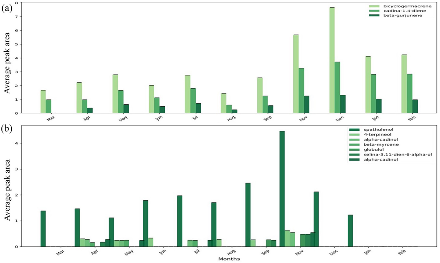
Beyond these two dominant constituents, several compounds exhibited notable seasonal fluctuations, including bicyclogermacrene, cadina-1,4-diene, sabinene, and spathulenol, which showed clear peaks during the late spring and summer months (November-February). These fluctuations are relevant since variations in bioactive compounds may directly impact the biological activity.55 This pattern underscores that the chemical composition of O. odorifera EOs is highly dynamic and closely influenced by seasonal environmental factors, reinforcing the importance of harvest timing for both research, medicinal and industrial applications using it.
Multivariate statistical analyses
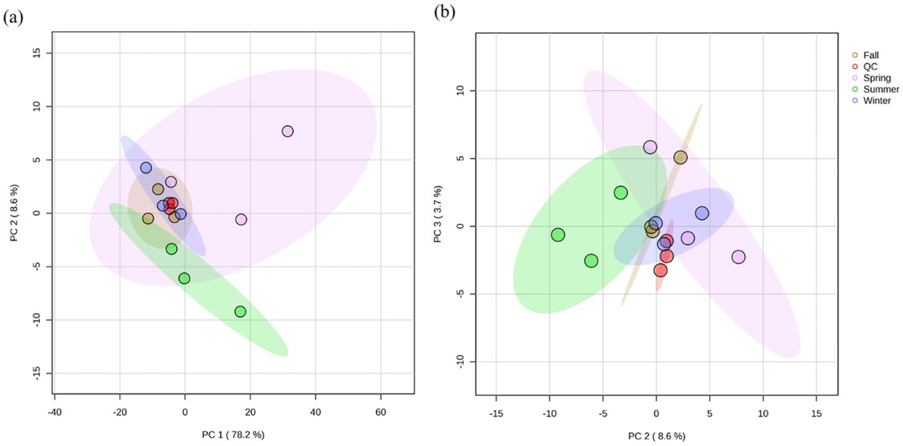
Prior PCA modeling, sum normalization and Pareto scaling were used to reduce the dominance of highly abundant metabolites while retaining contributions from moderate and low-abundance variables, improving the interpretability of seasonal trends.52,58 The PCA model showed strong overall performance (R2 = 0.90), with the first three components explaining 90.5% of the variance (PC1 = 78.2%, PC2 = 8.6%, PC3 = 3.7%), indicating that the dataset is largely governed by a dominant compositional gradient (PC1) with additional, biologically meaningful refinements captured by PC2 and PC3 (Figure 4). A critical feature of the score plots is the behaviour of the QC samples, which form a tight cluster close to the center of the PCA space. This compact grouping indicates instrumental stability and repeatability throughout the analytical sequence, supporting that the observed seasonal dispersion and separation are driven predominantly by true chemical differences among months, rather than batch drift or inconsistent data acquisition.
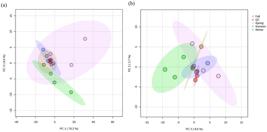
PCA score plots (R² = 0.905) showing seasonal clustering of O. odorifera EOs. (a) PC1 × PC2 and (b) PC2 × PC3.
In the PC1 × PC2 score plot (Figure 4a), most non-summer samples lie relatively close to the centroid, consistent with broadly comparable overall EOs profiles for much of the year. In contrast, the summer samples form the most displaced seasonal group, characterized by a clear shift toward negative PC2 values and a larger internal spread, suggesting that summer oils undergo not only a directional compositional change but also greater month-to-month variability within the season. Winter samples also show a tendency to separate from the central cluster, generally shifting toward more positive PC2 values, reinforcing that PC2 captures a secondary seasonal axis that distinguishes summer from winter beyond the dominant variance described by PC1.52 Additionally, this seasonal differentiation becomes even clearer in the PC2 × PC3 scatter plot (Figure 4b). Here, the summer group remains separated primarily along negative PC2, while winter trends toward positive PC2, indicating that PC2 consistently encodes a key compositional contrast between these seasons. Figure S3 (SI section) combines pairwise PCA score scatterplots (PC1-PC2, PC1-PC3, PC2-PC3), kernel density distributions of scores on each PC (diagonal panels), and a 3D PCA score plot (PC1-PC2-PC3); together they show tight QC clustering near the centroid and seasonal score shifts (notably summer toward negative PC2), with significant separation across PC planes (p = 0.022-0.012). These observations sharpen the distinction of summer oils as chemically different from most other samples, consistent with the heatmap evidence for shifts in the most abundant constituents.
Partial least squares discriminant analysis
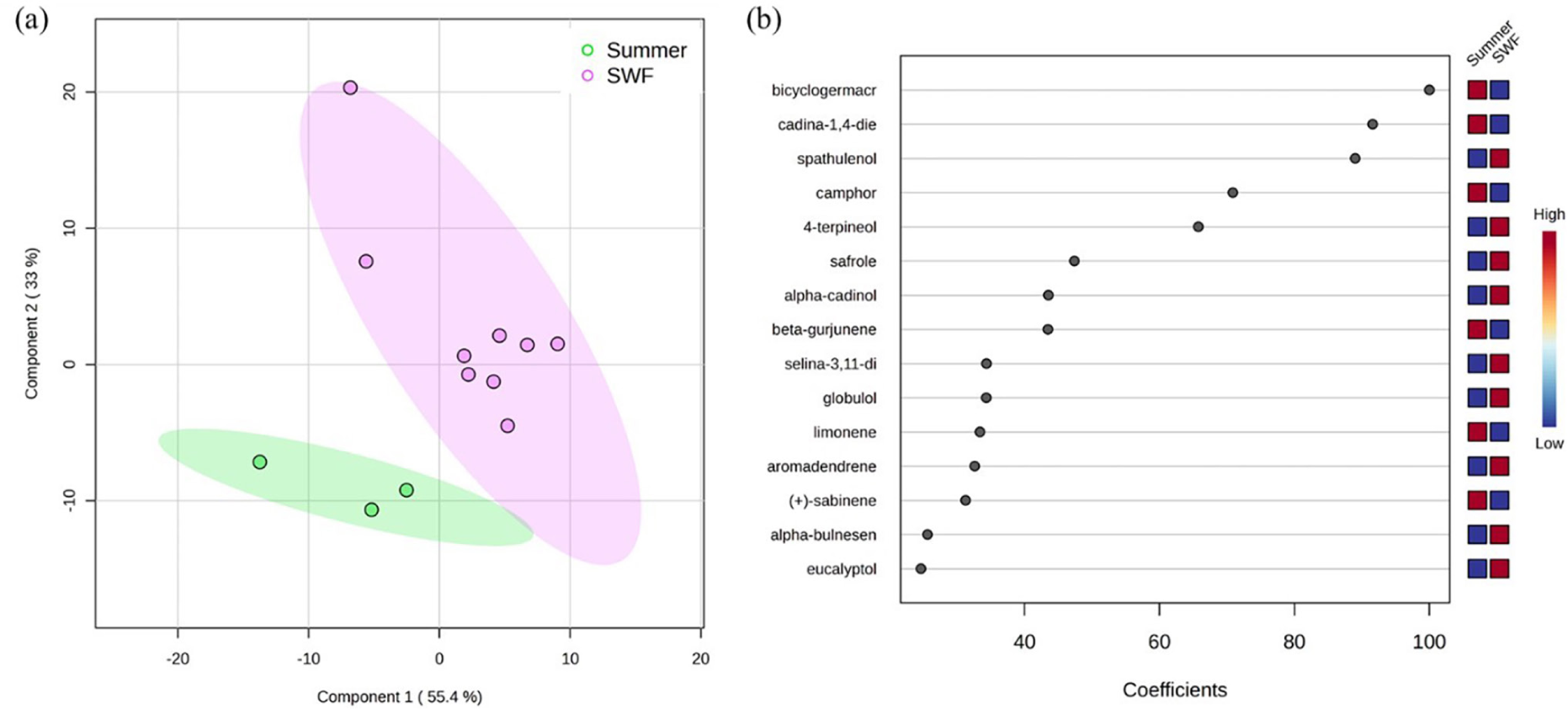
PLS-DA enhanced the separation between samples classified as spring, winter, fall (SWF), representing periods of high anti-inflammatory activity, and summer, representing periods of low anti-inflammatory activity. To ensure a robust model, sum normalisation and Pareto scaling were again applied to reduce the dominance of high-variance metabolites while preserving contributions from other variables.58 The resulting three-component model demonstrated an excellent fit (R2 = 0.97) and satisfactory predictive power (Q2 = 0.74). The 2D score plot (Figure 5a) clearly distinguished the summer months (December, January, and February) from the others SWF group (March to November), with no overlap in their 95% confidence intervals.59 To identify the specific metabolites driving this seasonal discrimination, variable importance in projection (VIP) scores and correlation coefficients were analyzed. Metabolites with a VIP score > 1 were considered significant drivers of the separation of the model16,18 (Figure S4 and Table S2, SI section). The correlation plot (Figure 5b) ranked the top features based on their relationship with the seasonal groups.
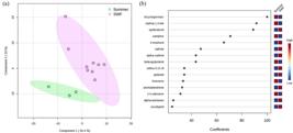
(a) PLS-DA score plot (2D) (5-fold CV, R2 = 0.97, Q2 = 0.74), demonstrating the separation between samples collected in summer (green) and SWF (purple), with shaded areas representing the 95% confidence intervals; (b) PLS-DA ranked by their correlation coefficients. The coloured boxes on the right indicate the relative abundance (red: high; blue: low) of each compound in the respective groups.
The anti-inflammatory profile suggests a significant seasonal trend in the bioactivity of O. odorifera EOs. EOs collected during the summer exhibited significantly lower PGE2 inhibition percentages (42.9% December, 60.5% January, and 53.3% February) compared to non-summer periods, which often exceeded 80-90% inhibition (e.g., April 85.0%, June 97.6%, and November 94.4%). This discrimination highlights the influence of seasonality and environmental factors on the chemical profile and, consequently, the anti-inflammatory profile of the oil. Specifically, the summer group clustered on the negative side of the PLS1 axis, correlating with reduced anti-inflammatory activity, while the SWF group aligned on the positive side of PLS1, associated with higher anti-inflammatory activity. These corroborate that the potential seasonal suppression of the anti-inflammatory activity during summer is likely linked to variations in metabolite abundance of minor compounds, as safrole kept the major compound throughout the year.
In this context, the compounds such as bicyclogermacrene, cadina-1,4-diene, β-gurjunene, and α-copaene showed strong positive correlations with the summer group, which corresponds to periods of lower anti-inflammatory activity (Table S3, SI section). These metabolites exhibited strong positive correlations with the predictive component OPLS1, indicating that these compounds might be characteristic of the summer period, when the anti-inflammatory potential is reduced. Conversely, less statistically significant metabolites are more associated with the SWF months, which correspond to periods of higher anti-inflammatory activity.
Most of these metabolites did not reach the conventional thresholds for statistical significance (p < 0.05) under univariate analysis. However, the terpinen-4-ol was statistically different (VIP > 1 and p value < 0.05) and positively correlated with the SWF group. Despite this, their consistent distribution pattern suggests biological relevance. Supporting this observation, the seasonality behavior of the two most statistically promising compounds (e.g., spathulenol and terpinen-4-ol) exhibited markedly higher concentrations during bioactive months and were markedly reduced during the summer months (Figure 6). This striking seasonal fluctuation reinforces the hypothesis that these compounds may contribute to the anti-inflammatory potential of O. odorifera EOs. Nevertheless, further investigations with larger sample sizes and complementary bioassays are essential to validate their role in contributing to the anti-inflammatory properties of the O. odorifera EOs.
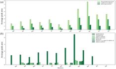
(a) Monthly variation in the average peak area (%) of compounds associated with the summer period, including bicyclogermacrene, cadina-1,4-diene, and β-gurjunene; (b) monthly distribution of oxygenated metabolites, such as spathulenol and terpinen-4-ol, associated with the high-bioactivity months (e.g., June and November).
In LPS-stimulated human blood, PGE2 is generated predominantly through the inducible COX-2 pathway, and essential-oil constituents capable of modulating COX-2 expression or upstream inflammatory signaling may plausibly reduce PGE2 production.60,61 In this context, terpinen-4-ol, the only feature meeting both VIP and univariate significance thresholds in our dataset, has been reported to exhibit anti-inflammatory activity in experimental models, including attenuation of pro-inflammatory signalling and, in some systems, reduction of interleukins and COX-2/iNOX/PGE2-related responses.60,61 Likewise, spathulenol has been described as an anti-inflammatory sesquiterpenoid in macrophage-based and immunomodulatory models, where reductions in NO and down-regulation of inflammatory mediators, including COX-2/iNOS-related endpoints, have been also reported.62-64 Importantly, the marked seasonal reduction of these oxygenated terpenes during summer mirrors the lower PGE2 inhibition, supporting the hypothesis that they contribute possibly in combination with other minor constituents, to the higher anti-inflammatory efficacy observed in non-summer months.61-64 Taken together, our results suggest that seasonal changes in the minor oxygenated fraction, rather than fluctuations in the dominant safrole content, may better explain the observed modulation of PGE2 inhibition in our human whole blood ex vivo assay.
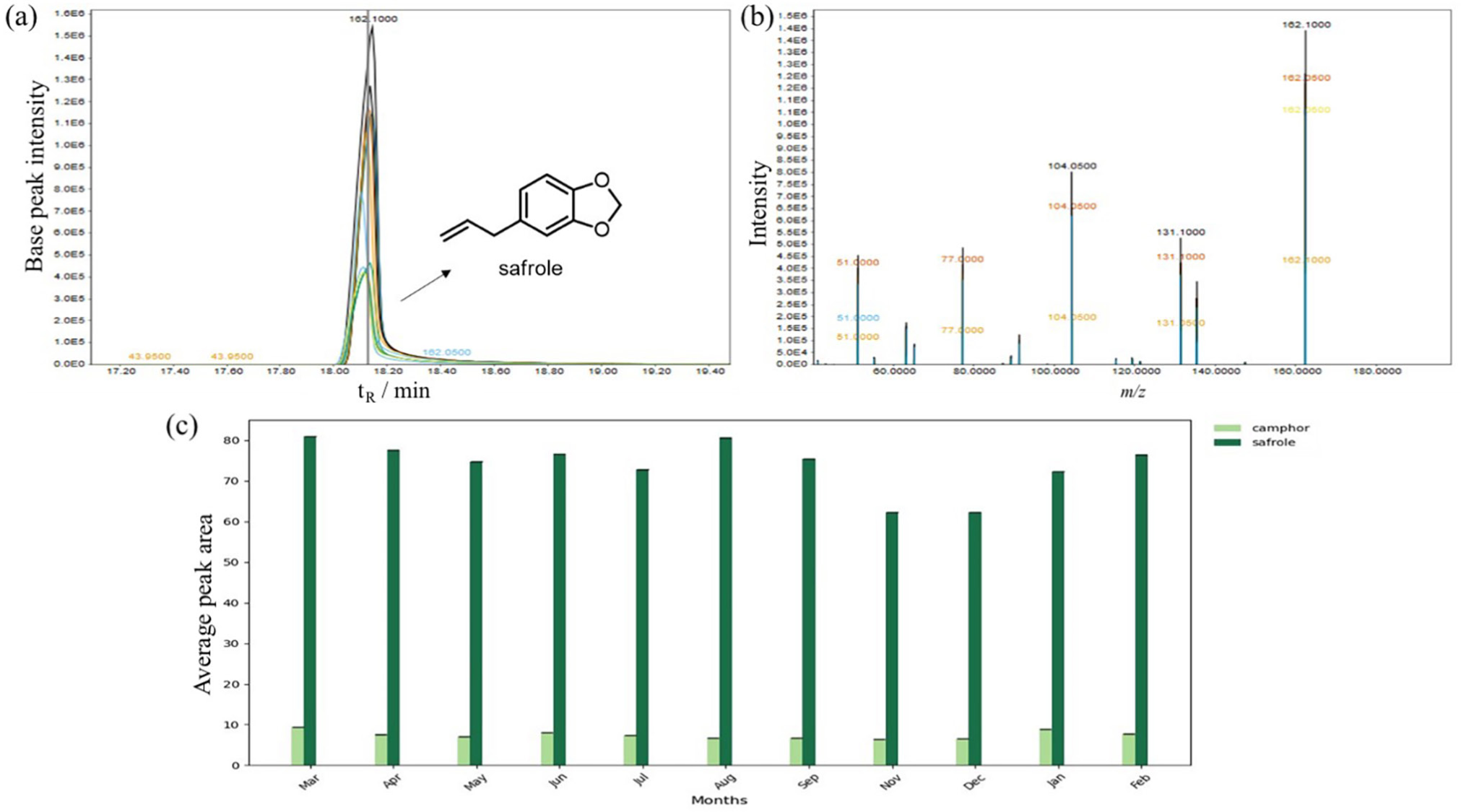
Therefore, a particularly noteworthy finding of this research concerns the chemical stability of safrole (Figure 7). Unlike the other metabolites associated with peak bioactivity, which fluctuated significantly, safrole remained the dominant constituent throughout the study. It exhibited a consistently high concentration (mean of 74.14 ± 6.4%), with the highest peaks in August, February, and March, and relatively lower in November and December, followed by camphor (mean of 7.4 ± 0.9%), which also remained stable during the year. Thus, although safrole dominated the EOs composition throughout the year, its relative stability and weak alignment with the seasonal bioactivity profile indicate that the major constituent alone is unlikely to explain the observed modulation of PGE2 inhibition.

(a) Total ion chromatogram (TIC) extracted peak at tR = 18.10 min that corresponds to safrole; (b) mass spectrum of the safrole peak showing the molecular ion M+ at m/z 162 and characteristic fragmentation patterns (m/z 104, m/z 131 and m/z 135); (c) comparative analysis of major constituents (safrole: 74.1%; camphor: 7.4%) across sampling periods of the year.
Moreover, as safrole is the major compound, and the biomarker of the O. odorifera EOs, its abundance also suggests a highly stable biosynthetic pathway for phenylpropanoids in the leaf EOs of O. odorifera species. This pattern further emphasizes the importance of understanding not only the seasonal dynamics of beneficial compounds but also the consistent presence of potentially toxic constituents like safrole, which has documented hepatotoxic and carcinogenic potential, and thus must be carefully managed in any industrial or pharmacological application derived from its EOs.
Conclusions
This study provides the first comprehensive characterization of the seasonal dynamics of O. odorifera EOs, integrating chemical profiling with ex vivo anti-inflammatory activity. Our findings demonstrate that while the oil maintains a remarkably stable presence of safrole and camphor throughout the year, its biological efficacy is highly modulated by seasonality. The anti-inflammatory potential, measured via PGE2 inhibition levels in human whole blood, significantly decreased during the summer months, compared to the rest of the year. PCA and PLS-DA confirmed that summer EOs are chemically distinct, characterized by an increased abundance of sesquiterpenes such as bicyclogermacrene and cadina-1,4-diene, which inversely correlated with bioactivity. While oxygenated terpenes such as spathulenol, terpinen-4-ol and α-cadinol suggest that these minor constituents may play a synergistic role in the therapeutic effects of the oil. These results emphasize that for O. odorifera, the major compound does not dictate the seasonal shift in the anti-inflammatory activity profile; rather, the minor volatile fraction seems to be more sensitive to environmental conditions, and might drive the medicinal quality. Consequently, harvest timing is a critical factor for the pharmaceutical and industrial standardization of O. odorifera EOs.
Supplementary Information
Supplementary information is available free of charge at http://jbcs.sbq.org.br as PDF file.
Supplementary Information
Data Availability Statement
All data is online available at DOI: https://zenodo.org/records/19153319 and https://zenodo.org/records/18281195
Acknowledgments
The authors acknowledge the following funding agencies for the fellowships and financial support for this research project: CNPq (Grant Nos. 408115/2023-8, 408229/2025-0, 309500/2025-7, 304916/2025-0); FAPEMIG (Grant Nos. APQ-05218-23, APQ-00544-23, APQ02882-24, APQ-05607-24), FAPESP (Grant No. 2024/04606-5) and CAPES (Finance Code 001).
References
-
1 Bakkali, F.; Averbeck, S.; Averbeck, D.; Idaomar, M.; Food Chem. Toxicol. 2008, 46, 446. [Crossref]
» Crossref -
2 Anthony, J. P.; Fyfe, L.; Smith, H.; Trends Parasitol. 2005, 21, 462. [Crossref]
» Crossref -
3 André, W. P. P.; Ribeiro, W. L. C.; de Oliveira, L. M. B.; Macedo, I. T. F.; Rondon, F. C. M. E.; Bevilaqua, C. M. L.; Acta Sci. Vet. 2018, 46, 14. [Crossref]
» Crossref -
4 Sadgrove, N. J.; Padilla-González, G. F.; Phumthum, M.; Plants 2022, 11, 789. [Crossref]
» Crossref -
5 Custódio, D. L.; Veiga Jr., V. F.; RSC Adv. 2014, 4, 21864. [Crossref]
» Crossref -
6 Antonio, A. S.; Veiga Jr., V. F.; Wiedemann, L. S. M.; Phytochemistry 2020, 173, 112314. [Crossref]
» Crossref -
7 Zuluaga, J. C. P.; van der Werff, H.; Park, B.; Eaton, D. A. R.; Comita, L. S.; Queenborough, S. A.; Donoghue, M. J.; Am. J. Bot. 2021, 108, 664. [Crossref]
» Crossref -
8 Salleh, W. M. N. H. W.; Ahmad, F.; J. Appl. Pharm. Sci. 2017, 7, 204. [Crossref]
» Crossref -
9 Camargo, M. J.; Miranda, M. L. D.; Kagamida, C. M.; Rodrigues, E. D.; Garcez, F. R.; Garcez, W. S.; Quim. Nova 2013, 36, 1008. [Crossref]
» Crossref -
10 Alves, M. F.; Katchborian-Neto, A.; Bueno, P. C. P.; Carnevale-Neto, F.; Casoti, R.; Ferreira, M. S.; Murgu, M.; Paula, A. C. C.; Dias, D. F.; Soares, M. G.; Chagas-Paula, D. A.; RSC Adv. 2024, 14, 10481. [Crossref]
» Crossref -
11 Katchborian-Neto, A.; Alves, M. F.; Bueno, P. C. P.; Nicácio, K. J.; Ferreira, M. S.; Oliveira, T. B.; Barbosa, H.; Murgu, M.; Ladvocat, A. C. C. P.; Dias, D. F.; Soares, M. G.; Lago, J. H. G.; Chagas-Paula, D. A.; Brief. Bioinform. 2024, 25, bbae013. [Crossref]
» Crossref -
12 Santos, M. F. C.; Feliciano, C. R.; Neto, A. K.; Paula, D. A. C.; Soares, M. G.; Nat. Prod. Res. 2021, 37, 1217. [Crossref]
» Crossref -
13 Silva, A. F.; Santos, M. F. C.; Maiolini, T. S. C.; Salem, P. P. O.; Murgu, M.; de Paula, A. C. C.; Silva, E. O.; Nicácio, K. J.; Ferreira, A. G.; Dias, D. F.; Soares, M. G.; Chagas-Paula, D. A.; Phytochem. Lett. 2021, 42, 52. [Crossref]
» Crossref -
14 Alves, M. F.; Katchborian-Neto, A.; Casoti, R.; Leite, F. B.; de Paula, A. C. C.; Dias, D. F.; Soares, M. G.; Arruda Sanchez, T.; de Paula, D. A. C.; Chem. Biodiversity 2025, 22, e202402227. [Crossref]
» Crossref -
15 Yamaguchi, M. U.; Garcia, F. P.; Cortez, D. A. G.; Ueda-Nakamura, T.; Dias Filho, B. P.; Nakamura, C. V.; Antonie van Leeuwenhoek, Int. J. Gen. Mol. Microbiol. 2011, 99, 507. [Crossref]
» Crossref -
16 de Alcântara, B. G. V.; Oliveira, F. P.; Katchborian-Neto, A.; Casoti, R.; Domingos, O. S.; Santos, M. F. C.; Oliveira, R. B.; de Paula, A. C. C.; Dias, D. F.; Soares, M. G.; Chagas-Paula, D. A.; J. Ethnopharmacol. 2021, 264, 133378. [Crossref]
» Crossref -
17 Alcântara, B. G. V.; Neto, A. K.; Garcia, D. A.; Casoti, R.; Oliveira, T. B.; Ladvocat, A. C. C. P.; Edrada-Ebel, R.; Soares, M. G.; Dias, D. F.; Chagas-Paula, D.; Chem. Biodiversity 2023, 20, e202300650. [Crossref]
» Crossref -
18 Katchborian-Neto, A.; Jesus, K. J.; Cruz, J. C.; Bueno, P. C. P.; Murgu, M.; Dias, D. F.; Soares, M. G.; Paula, A. C. C.; Chagas-Paula, D. A.; Phytomedicine 2023, 120, 155060. [Crossref]
» Crossref -
19 Gontijo, D. C.; Brandão, G. C.; Gontijo, P. C.; de Oliveira, A. B.; Diaz, M. A. N.; Fietto, L. G.; Leite, J. P. V.; Food Chem. 2017, 230, 618. [Crossref]
» Crossref -
20 Simić, A.; Soković, M. D.; Ristić, M.; Grujić-Jovanović, S.; Vukojević, J.; Marin, P. D.; Phytother. Res. 2004, 18, 713. [Crossref]
» Crossref -
21 Mossi, A. J.; Zanella, C. A.; Kubiak, G.; Lerin, L. A.; Cansian, R. L.; Frandoloso, F. S.; Prá, V. D.; Mazutti, M. A.; Costa, J. A. V.; Treichel, H.; Renew. Agric. Food Syst. 2014, 29, 161. [Crossref]
» Crossref -
22 Almeida, R. S.; Ribeiro-Filho, J.; Freitas, P. R.; de Araújo, A. C. J.; dos Santos, E. L.; Tintino, S. R.; Moura, T. F.; Ferreira, V. A.; Ferreira, B. A.; Fonseca, V. J. A.; Leite, P. I. P.; Silva, A. C. A.; Silva, L. E.; do Amaral, W.; Deschamps, C.; Siyadatpanah, A.; Wilairatana, P.; Coutinho, H. D. M.; J. Infect. Public Health 2022, 15, 373. [Crossref]
» Crossref -
23 Passos, B. G.; Albuquerque, R. D. D. G.; Muñoz-Acevedo, A.; Echeverria, J.; Llaure-Mora, A. M.; Ganoza-Yupanqui, M. L.; Rocha, L.; Fitoterapia 2022, 156, 10565. [Crossref]
» Crossref -
24 Ni, Y. L.; Shen, H. T.; Lee, M. W.; Yeh, K. L.; Chiang, C. Y.; Kuan, Y. H.; Tzu Chi Med. J. 2020, 32, 344. [Crossref]
» Crossref -
25 Yang, A. H.; Zhang, L.; Zhi, D. X.; Liu, W. L.; Gao, X.; He, X.; Xenobiotica 2018, 48, 1164. [Crossref]
» Crossref -
26 Keyel, P. A.; Cytokine 2014, 69, 136. [Crossref]
» Crossref -
27 Mahesh, G.; Kumar, K. A.; Reddanna, P.; J. Inflamm. Res. 2021, 14, 253. [Crossref]
» Crossref -
28 Chen, L.; Deng, H.; Cui, H.; Fang, J.; Zuo, Z.; Deng, J.; Li, Y.; Wang, X.; Zhao, L.; Oncotarget 2018, 9, 7204. [Crossref]
» Crossref -
29 Hwang, S. H.; Wecksler, A. T.; Wagner, K.; Hammock, B. D.; Curr. Med. Chem. 2013, 20, 1783. [Crossref]
» Crossref -
30 Funk, C. D.; Science 2001, 294, 1871. [Crossref]
» Crossref -
31 Rudrapal, M.; Eltayeb, W. A.; Rakshit, G.; El-Arabey, A. A.; Khan, J.; Aldosari, S. M.; Alshehri, B.; Abdalla, M.; Sci. Rep. 2023, 13, 8656. [Crossref]
» Crossref -
32 Steele, V. E.; Hawk, E. T.; Viner, J. L.; Lubet, R. A.; Mutat. Res., Fundam. Mol. Mech. Mutagen. 2003, 523, 137. [Crossref]
» Crossref -
33 Timoshenko, A. V.; Xu, G.; Chakrabarti, S.; Lala, P. K.; Chakraborty, C.; Exp. Cell Res. 2003, 289, 265. [Crossref]
» Crossref - 34 Rang, H. P.; Dale, M. M.; Ritter, J. M.; Flower, R. J.; Henderson, G.; Farmacologia, 7th ed.; Elsevier: Amsterdam, Netherlands, 2011.
-
35 Cabral, M. M. O.; Barbosa-Filho, J. M.; Maia, G. L. A.; Chaves, M. C. O.; Braga, M. V.; de Souza, W.; Soares, R. O. A.; Exp. Parasitol. 2010, 124, 319. [Crossref]
» Crossref -
36 Garcez, F. R.; da Silva, A. F. G.; Garcez, W. S.; Linck, G.; Matos, M. D. F. C.; Santos, E. C. S.; Queiroz, L. M. M.; Planta Med. 2011, 77, 383. [Crossref]
» Crossref -
37 Barbosa, H.; da Silva, R. L. C. G.; Costa-Silva, T. A.; Tempone, A. G.; Antar, G. M.; Lago, J. H. G.; Caseli, L.; Bioorg. Chem. 2020, 101, 103978. [Crossref]
» Crossref -
38 de Oliveira Jr., R. G.; Ferraz, C. A. A.; Silva, J. C.; de Oliveira, A. P.; Diniz, T. C.; Silva, M. G.; Quintans Jr., L. J.; de Souza, A. V. V.; dos Santos, U. S.; Turatti, I. C. C.; Lopes, N. P.; Lorenzo, V. P.; Almeida, J. R. G. S.; Molecules 2017, 22, 900. [Crossref]
» Crossref -
39 Lima, M. N. N.; Costa, J. S.; Guimarães, B. A.; Freitas, J. J. S.; Setzer, W. N.; Silva, J. K. R.; Maia, J. C. S.; Figueiredo, P. L. B.; Molecules 2023, 28, 3371. [Crossref]
» Crossref -
40 Garcia, D. A.; Costa, L. P. D. M.; Rosa, W.; Ladvocat, A. C. C. P.; Dias, D. F.; Soares, M. G.; Ionta, M.; Chagas-Paula, D. A.; Chem. Biodiversity 2024, 21, e202400692. [Crossref]
» Crossref -
41 Gonzalez, F. R.; Nardillo, A. M.; J. Chromatogr. A 1999, 842, 29. [Crossref]
» Crossref - 42 Adams, R. P.; Identification of Essential Oil Components by Gas Chromatography/Mass Spectrometry, 4th ed.; Allured: Carol Stream, United States, 2007.
-
43 Santos, M. F. C.; Nicácio, K. J.; Katchborian-Neto, A.; Ferreira, M. S.; Miranda, D. O.; Andrade, J. V.; Pereira, H. A.; Jesus, E. G.; Souza, T. B. S.; Morais-Urano, R. P.; Dias, D. F.; Chagas-Paula, D. A.; Soares, M. G.; Nat. Prod. Res. 2024, 38, 393. [Crossref]
» Crossref -
44 Nicácio, K. J.; Ferreira, M. S.; Katchborian-Neto, A.; Costa, M. L.; Murgu, M.; Dias, D. F.; Soares, M. G.; Chagas-Paula, D. A.; Chem. Biodiversity 2022, 19, e202100966. [Crossref]
» Crossref -
45 Leão, L. P. M. O.; Neto, A. K.; Nicácio, K. J.; Lavorato, S. N.; Leite, F. B.; Teixeira, K. C.; Murgu, M.; de Paula, A. C. C.; Soares, M. G.; Chagas-Paula, D. A.; Dias, D. F.; Chem. Biol. Drug Des. 2024, 104, e14634. [Crossref]
» Crossref -
46 Ferreira, M. S.; Katchborian-Neto, A.; Nicácio, K. J.; Santos, M. F. C.; Dias, D. F.; Chagas-Paula, D. A.; Soares, M. G.; Nat. Prod. Res. 2024, 39, 2635. [Crossref]
» Crossref -
47 Silva, D. O.; Santos, M. F. C.; Nicácio, K. J.; Neto, A. K.; Lago, J. H. G.; Chagas-Paula, D. A.; Dias, D. F.; Soares, M. G.; Nat. Prod. Res. 2024, 38, 1054. [Crossref]
» Crossref -
48 Rosa, W.; Domingos, O. S.; Salem, P. P. O.; Caldas, I. S.; Murgu, M.; Lago, J. H. G.; Sartorelli, P.; Dias, D. F.; Chagas-Paula, D. A.; Soares, M. G.; Phytochem. Anal. 2021, 32, 859. [Crossref]
» Crossref -
49 Vinaixa, M.; Samino, S.; Saez, I.; Duran, J.; Guinovart, J. J.; Yanes, O.; Metabolites 2012, 2, 775. [Crossref]
» Crossref -
50 MetaboAnalyst, version 6.0; Xia Lab, McGill University; Montreal, QC, Canada, 2024. [Link] accessed in March 2026
» Link -
51 Nicácio, K. J.; Ferreira, M. S.; Salem, P. P. O.; Oliveira, R. A.; Lago, J. H. G.; Soares, M. G.; Dias, D. F.; Katchborian Neto, A.; Chagas-Paula, D.; Metabolomics reveals how seasonal variation in essential oils of Ocotea odorifera (Vell.) Rohwer impacts its anti-inflammatory activity, version 0.1; Zenodo, Brazil, 2026. [Link] accessed in March 2026
» Link -
52 Worley, B.; Powers, R.; Curr. Metabolomics 2012, 1, 92. [Crossref]
» Crossref -
53 Bylesjö, M.; Rantalainen, M.; Cloarec, O.; Nicholson, J. K.; Holmes, E.; Trygg, J.; J. Chemom. 2006, 20, 341. [Crossref]
» Crossref -
54 Boccard, J.; Rutledge, D. N.; Anal. Chim. Acta 2013, 769, 30. [Crossref]
» Crossref -
55 Gobbo-Neto, L.; Lopes, N. P.; Quim. Nova 2007, 30, 374. [Crossref]
» Crossref -
56 Almeida, R. S.; Freitas, P. R.; Araújo, A. C. J.; Menezes, I. R. A.; Santos, E. L.; Tintino, S. R.; Moura, T. F.; Ribeiro Filho, J.; Ferreira, V. A.; Silva, A. C. A.; Silva, L. E.; do Amaral, W.; Deschamps, C.; Iriti, M.; Coutinho, H. D. M.; Antibiotics 2020, 9, 247. [Crossref]
» Crossref -
57 Alcoba, A. E. T.; Melo, D. C.; de Andrade, P. M.; Dias, H. J.; Pagotti, M. C.; Magalhães, L. G.; Junior, W. G. F.; Crotti, A. E. M.; Miranda, M. L. D.; Nat. Prod. Res. 2018, 32, 2865. [Crossref]
» Crossref -
58 Saccenti, E.; Hoefsloot, H. C. J.; Smilde, A. K.; Westerhuis, J. A.; Hendriks, M. M. W. B.; Metabolomics 2014, 10, 361. [Crossref]
» Crossref -
59 Zanatta, A. C.; Vieira, N. C.; Dantas-Medeiros, R.; Vilegas, W.; Edrada-Ebel, R. A.; Metabolites 2023, 13, 349. [Crossref]
» Crossref -
60 Matera, R.; Lucchi, E.; Valgimigli, L.; Molecules 2023, 28, 901. [Crossref]
» Crossref -
61 Grazul, M.; Kwiatkowski, P.; Hartman, K.; Kilanowicz, A.; Biomedicines 2023, 11, 2381. [Crossref]
» Crossref -
62 Costa, I. F. J. B.; Simão, T. L. B. V.; Calixto, S. D.; Pereira, R. V.; Konno, T. U. P.; Pinto, S. C.; Tinoco, L. W.; Lasunskaia, E.; Leal, I. C. R.; Muzitano, M. F.; Rodriguésia 2021, 72, 1. [Crossref]
» Crossref -
63 Mohanta, O.; Ray, A.; Jena, S.; Sahoo, A.; Panda, S. S.; Das, P. K.; Nayak, S.; Panda, P. C.; Molecules 2023, 28, 5817. [Crossref]
» Crossref -
64 Sá, R. C. S.; Andrade, L. N.; Sousa, D. P.; Nat. Prod. Commun. 2015, 10, 1767. [Crossref]
» Crossref
Edited by
-
Editor handled this article:
Hector Henrique F. Koolen (Associate)