An Integrated Leaf Spray Mass Spectrometry and Feature-Based Molecular Networking Strategy for Glycoside Annotation in Strychnos jobertiana Leaves
Abstract
Glycosides are widely distributed plant metabolites with remarkable structural diversity and broad biological activities. However, comprehensive strategies for their systematic recognition, extraction, and annotation from complex plant matrices remain scarce, especially for chemically underexplored Amazonian species such as Strychnos jobertiana. In this study, we present an integrated analytical strategy combining leaf spray chemical profiling and feature-based molecular networking (FBMN) analysis to guide extract preparation and enable glycoside annotation from S. jobertiana leaves. Rapid chemical profiling by leaf spray mass spectrometry (LS-MS) enabled the early recognition of glycoside-related ions, including alkaloid and flavonoid derivatives, guiding the preparation of an aqueous extract processed by solid-phase extraction (SPE) before liquid chromatography-mass spectrometry (LC-MS) analysis. The resulting data were explored using FBMN supported by spectral search analysis and manual interpretation of tandem mass spectra. In parallel, the isolation of desoxycordifoline (1), harman-3-carboxylic acid (2), kaempferol 3-O-(2”,6”-di-O-rhamnopyranosyl) glucopyranoside (3), and kaempferol-3-O-neohesperidoside (4) provided structural support for the annotation of molecular clusters. Two major glycoside clusters were characterized, one dominated by flavonoid glycosides, and another composed primarily of monoterpene-derived glycosides. Overall, 21 compounds were annotated, including derivatives related to compounds 1-4, such as secoxyloganin (8), kaempferol-3-O-glucoside (5), kaempferol-3-O-rutinoside (6), and kaempferol-3-O-rungioside (7), and diagnostic fragment ions associated with glycoside compounds were highlighted. This integrated workflow demonstrates the value of LS-MS profiling as a decision-making tool for extract preparation and highlights feature-based molecular networking (FBMN) as a powerful approach for glycoside annotation, contributing to the chemical knowledge of Amazonian plant species.
Introduction
Glycosides constitute one of the most widespread and structurally diverse classes of plant secondary metabolites, occurring across multiple taxonomic groups and biosynthetic pathways.1 They encompass a wide range of natural product classes, including alkaloids, flavonoids, terpenes, and organic acids, each contributing distinct physicochemical and biological properties.1-3 These metabolites play essential roles in plant physiology and ecology, such as chemical defense, signaling, and interactions with herbivores and microorganisms.4 From a pharmacological perspective, glycosides are associated with a broad spectrum of biological activities, including antioxidant, antimicrobial, anti-inflammatory, and cytotoxic effects, making them recurrent targets in natural product research.1-4 Their remarkable diversity arises from the combination of structurally distinct aglycones with one or more sugar units, which modulate solubility, stability, and bioactivity.5
Despite their importance, comprehensive strategies capable of rapidly surveying glycoside-rich profiles and guiding downstream analytical decisions are still scarce, largely due to the limitations of currently available preliminary screening approaches, such as classical phytochemical assays. These tests are typically non-specific and may yield positive results for multiple classes of glycosylated metabolites. In addition, they generally require relatively large amounts of plant material and provide no structural information, making them unsuitable for guiding downstream analytical workflows. These limitations are particularly evident in the context of Amazonian plant species, which represent one of the richest reservoirs of biodiversity worldwide but remain largely underexplored.6 Many taxa from this biome lack detailed chemical investigations, and available studies are often restricted to isolated compounds or targeted classes of metabolites. Within this context, species belonging to the genus Strychnos are of special interest due to their well-documented capacity to biosynthesize structurally diverse alkaloids, flavonoids, and terpenoid derivatives, including glycosylated forms.7 However, while some Strychnos species have been studied extensively, others, such as Strychnos jobertiana, remain poorly characterized from a metabolomic perspective, with limited information regarding their glycoside composition.
Recent advances in mass spectrometry-based approaches have provided new opportunities to overcome these challenges by enabling faster and more informative chemical profiling of plant materials.8 Ambient ionization methods, in particular, allow the direct analysis of samples with minimal or no preparation, preserving chemical information that might otherwise be altered during extraction.9-11 Among these approaches, leaf spray mass spectrometry (LS-MS) stands out as a powerful tool for the rapid assessment of plant chemical profiles. By enabling the direct ionization of metabolites from intact leaves, this technique offers a snapshot of the chemical composition of plant tissues, facilitating the early recognition of dominant metabolite classes.12 Importantly, such preliminary information can serve as a decision-making tool, guiding the selection of extraction solvents and sample preparation strategies tailored to the chemical features observed, addressing a critical step in metabolomic studies where extract preparation is frequently performed without prior knowledge of the sample’s chemical profile.13
In parallel, advances in computational metabolomics have transformed the way complex mass spectrometry data are interpreted. As an example, feature-based molecular networking (FBMN), implemented within the Global Natural Products Social Molecular Networking (GNPS) platform, has emerged as a robust approach for organizing tandem mass spectrometry data according to spectral similarity, allowing structurally related metabolites to be visualized as molecular clusters.14,15 This strategy has proven particularly advantageous in plant metabolomics, where chemical diversity is high and reference standards are often unavailable. By grouping related features, molecular networking facilitates the propagation of structural information within clusters and supports the annotation of compound families, including glycosides, based on shared fragmentation patterns.15,16 When combined with manual interpretation of tandem mass spectra and complementary experimental evidence, such as the isolation of reference standards, this approach enhances confidence in metabolite annotation while reducing reliance on exhaustive isolation procedures.17
In this context, the present study aims to develop and apply an integrated analytical strategy combining leaf spray chemical profiling and FBMN to investigate the glycoside composition of S. jobertiana leaves. Initial leaf spray analysis was employed to rapidly assess the chemical profile and recognize glycoside-related ions, including alkaloid and flavonoid derivatives, guiding the preparation of an aqueous extract. This extract was subsequently processed by solid-phase extraction (SPE) and analyzed by ultra-high-performance liquid chromatography coupled to high-resolution tandem mass spectrometry (UHPLC-HRMS/MS), and the resulting data were explored through FBMN. In parallel, the isolation of reference compounds provided structural support for the annotation of molecular clusters.
Experimental
General experimental procedures
An analytical micro knife mill model 80374 (Hamilton Beach, SP, Brazil) was used to grind the material. A freeze-dryer model Alpha 1-2 LDplus (Martin Christ, Osterode am Harz, Germany) was used for lyophilization. The mass spectrometry (MS) data were acquired with an LCQ Fleet ion trap and an Orbitrap Exploris 240 mass spectrometer (Thermo Scientific, Waltham, MA, USA). Semi-preparative high-performance liquid chromatography (HPLC) analyses were performed on a UFLC system (LC-6 AD pump; DGU-20A5 degasser; SPD-20AV UV detector; Rheodyne injector; CBM-20A communication module) (Shimadzu, Columbia, MD, USA). The nuclear magnetic resonance (NMR) data were acquired on an AVANCE III HD NMR spectrometer (Bruker, Karlsruhe, Germany). Whatman grade 1 filter paper (Sigma-Aldrich, St. Louis, MO, USA) was used for filtration. All the solvents used for extraction, chromatography and the low-resolution MS experiments were HPLC grade, purchased from Tedia (Fairfield, OH, USA), and the water was purified via a Milli-Q system (Merck KGaA, Darmstadt, Germany).
Plant material
The leaves of S. jobertiana were collected in May 2018 at the Reserva Florestal Adolpho Ducke, located in the city of Manaus, Amazonas State, Brazil (2°56’30” S, 59°57’0.6” W). The botanical material was identified by the parataxonomist José Ferreira Ramos (Instituto Nacional de Pesquisas da Amazônia, INPA), based on dichotomous identification keys and comparison with authenticated herbarium specimens. A voucher specimen of S. jobertiana has been deposited in the herbarium of the INPA under the accession number 282887. Access to the genetic heritage was registered in the Sistema Nacional de Gestão do Patrimônio Genético e do Conhecimento Tradicional Associado (SisGen) under the registration code AF4CFA7. Immediately after collection, the material was air-dried at ambient temperature (approximately 20 °C) for 7 days and properly stored.
Leaf spray mass spectrometry (LS-MS) analysis
LS-MS analysis was performed using an LCQ Fleet ion trap mass spectrometer, prepared according to a previously described methodology.13 A representative leaf was cut into triangular pieces (10 mm base × 10 mm height) and fixed with a metal clip positioned approximately 10 mm from the mass spectrometer inlet. A high voltage was applied directly to the plant tissue after wetting it with approximately 10 µL of HPLC-grade methanol, promoting the formation of a spray of charged droplets that transported endogenous metabolites into the mass spectrometer inlet.13 Mass spectra were acquired in continuous monitoring mode. MS analytical conditions were: capillary voltage, 20 V; capillary temperature, 275 °C; tube lens, 115 V; mass range: 200-1000 m/z. Helium (He) was used as a collision gas with collision energies ranging from 18 to 20%.
Extraction and isolation
Powdered plant material (20 g) was extracted with distilled water (1,000 mL) by infusion for 15 min, as previously described.7,18,19 The resulting aqueous extract was filtered through filter paper and subsequently freeze-dried, affording the leaf aqueous extract (3.16 g, 15.8% yield). An aliquot of the leaf aqueous extract (1 g) was subjected to a cleanup procedure following a previously reported method.18 Briefly, the sample was dissolved in distilled water (2 mL) and loaded onto a cartridge packed with C18 phase (10 g), previously activated with HPLC-grade methanol (100 mL) and conditioned with distilled water (100 mL). The cartridge was washed with distilled water (100 mL) and subsequently eluted with HPLC-grade methanol (100 mL). The methanolic eluate was evaporated under a nitrogen gas stream, yielding the methanol fraction (0.72 g).
An aliquot of the methanol fraction (240 mg) was further fractionated by semi-preparative HPLC using water (A) and methanol (B) as mobile phases. The gradient elution program was 20-80% B (v/v) over 24 min, at a flow rate of 3.5 mL min-1. Separations were performed on a C18 column (250 mm × 10 mm, 5 μm; Phenomenex, Torrance, CA, USA), with UV detection at 280 and 316 nm. Eight injections (30 mg each), prepared in a water-methanol mixture (80:20, v/v; 100 μL), were performed, yielding 15 fractions (SJSPE1-15). Fractions SJSPE8-10 (44.5 mg) were pooled and subjected to re-fractionation under the same chromatographic conditions, affording compound 1 (18.4 mg), compound 2 (3.2 mg), compound 3 (6.7 mg), and compound 4 (2.4 mg).
Compounds 1, 3, and 4 were solubilized in 600 μL of methanol-d4 (CD3OD), whereas compound 2 was solubilized in 600 μL of dimethyl sulfoxide-d6 (DMSO-d6). All samples were subjected to one-dimensional (1D) and two-dimensional (2D) NMR experiments on an AVANCE III HD NMR spectrometer operating at 11.75 T, observing 1H and 13C at 500 and 125 MHz, respectively. All 1H and 13C NMR chemical shifts (δ) are given in ppm relative to tetramethylsilane (TMS) at 0.00 ppm as an internal reference, and the coupling constants (J) are given in hertz.
UHPLC-HRMS/MS analysis
UHPLC-HRMS/MS analysis was performed using a Vanquish Flex UHPLC system coupled to an Orbitrap Exploris 240 mass spectrometer, equipped with an electrospray ionization (ESI) source operating in negative ionization mode. The analysis was performed as a single experimental run. The methanol fraction was dissolved in water-methanol (80:20, v/v) at a concentration of 500 ppm, centrifuged, and filtered through a 0.22 μm nylon membrane (Allcrom, São Paulo, Brazil). An aliquot (2 μL) was injected into a Hypersil GOLD column (50 × 3 mm, 1.9 μm; Thermo Scientific). The mobile phase consisted of 0.1% formic acid in water (A) and methanol (B), using the following gradient program: 0-15 min, 20-80% B; 15-25 min, 80% B, at a flow rate of 0.4 mL min-1. The ion source parameters were set as follows: spray voltage, −3.0 kV; sheath gas, 35; auxiliary gas, 7; sweep gas, 2; ion transfer tube temperature, 275 °C; vaporizer temperature, 320 °C; and radio frequency (RF) lens, 70%. Data-dependent acquisition (DDA) mode was employed, targeting the five most intense precursor ions. A normalized collision energy of 35% was applied for MS/MS experiments.
Feature-based molecular networking (FBMN) and annotation
The MS/MS data were converted to the .mzML format using MSConvert (ProteoWizard)20 and processed in MZmine 4.8.3021 using the easy workflow configuration of MZwizard, optimized for UHPLC-Orbitrap data-dependent acquisition (DDA) experiments. The resulting feature list and corresponding MS/MS spectra files were uploaded to the GNPS2 web platform for the construction of FBMN.15 Molecular network parameters were set as follows: precursor and fragment ion mass tolerances of 0.02 Da, a minimum cosine score of 0.6, at least 3 matched fragment ions, a maximum of 10 neighbors per node, and a maximum of 100 molecular families. For spectral library matching, a minimum cosine score of 0.7 and at least 3 matched fragment ions were required. Network visualization was performed using Cytoscape v. 3.10.4 (Cytoscape Consortium, San Diego, CA, USA, 2023).
Results and Discussion
LS-MS chemical profiling as a decision-making tool
The use of leaf spray mass spectrometry was initially justified as a rapid and minimally invasive approach to obtain preliminary chemical information directly from S. jobertiana leaves, allowing an informed assessment of dominant metabolite classes prior to extraction. Such early-stage chemical insight is particularly valuable in metabolomic investigations, where extract preparation represents a critical step and is often performed without prior knowledge of the chemical composition of the sample.
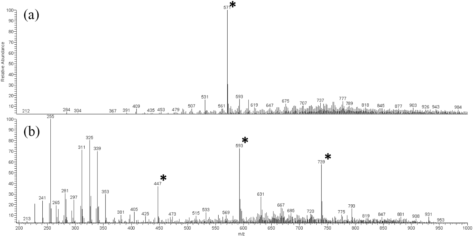
Full scan analysis in positive ion mode revealed a relatively simple spectral profile, dominated by a base peak at m/z 571 (Figure 1a). In contrast, negative ion mode provided a substantially more informative chemical fingerprint (Figure 1b), with multiple ions detected across a broad mass range between m/z 200 and 800. Although notable signals attributable to fatty acids and common contaminants were observed between m/z 200 and 340, prominent ions at m/z 447, 593, and 741 stood out as chemically relevant features.
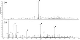
LS-MS chemical profiling of S. jobertiana leaves acquired in positive (a) and negative (b) ionization modes with * denoting ions selected for MS/MS experiments.
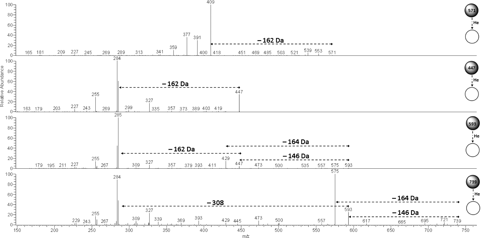
The MS/MS spectrum of the ion at m/z 571 (Figure 2) revealed an initial neutral loss of 162 Da (m/z 571 → 409), characteristic of glycosylated compounds.18,22 Subsequent neutral losses of 18 Da (m/z 409 → 391), 32 Da (m/z 409 → 377), and 70 Da (m/z 409 → 359) were observed, forming a fragmentation pattern consistent with the monoterpene indole alkaloid (MIA) desoxycordifoline, previously reported from the leaves of S. peckii.18
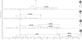
MS/MS spectra of the ions at m/z 571 (positive ionization mode) and 447, 593, and 739 (negative ionization mode), obtained by collision-induced dissociation (CID).
The ion at m/z 447 exhibited a distinct MS/MS spectrum, characterized by an initial loss of 162 Da (m/z 447 → 285) and a base peak at m/z 284, indicative of a radical cleavage process. This fragmentation behavior is characteristic of flavonoid glycosides and is consistent with a kaempferol-derived structure, also previously reported in Strychnos species.7,18 Similarly, the MS/MS spectrum of the ion at m/z 593 displayed sequential neutral losses of 146 Da (m/z 593 → 447) and 162 Da (m/z 447 → 285), corresponding to rhamnose and glucose units, respectively.22 An additional loss of 164 Da (m/z 593 → 429) supported the presence of a neohesperidoside moiety, indicating a kaempferol glycoside bearing a rhamnopyranosyl-(1→2)-glucopyranoside disaccharide.22
The MS/MS spectrum of the ion at m/z 741 further reinforced this interpretation, showing competitive neutral losses of 146 Da (m/z 741 → 593) and 164 Da (m/z 741 → 575), as well as a combined loss of 308 Da (m/z 593 → 285), consistent with a kaempferol derivative containing an additional rhamnose unit linked to a neohesperidoside core. Collectively, these observations revealed a strong presence of both alkaloid and flavonoid glycosides in the leaves of S. jobertiana.
Based on this preliminary chemical assessment, the decision was made to prepare an aqueous extract, followed SPE to remove free sugars, salts, and other interfering constituents while enriching the sample in glycosylated metabolites. This strategy has previously proven to be effective for the enrichment of glycosylated alkaloids from S. peckii18 and flavonoid glycosides from S. subcordata,23 supporting its applicability to S. jobertiana. Furthermore, considering the structural diversity commonly reported within the genus Strychnos, including variations in sugar type (e.g., glucose and galactose)23-25 and substitution patterns on flavonoid scaffolds, chromatographic fractionation was undertaken to isolate representative compounds capable of supporting subsequent liquid chromatography-mass spectrometry data annotation.23
In this context, the alkaloids desoxycordifoline (1) and harman-3-carboxylic acid (2), as well as the flavonoid glycosides kaempferol 3-O-(2”,6”-di-O-rhamnopyranosyl)glucopyranoside, also known as clitorin (3), and kaempferol-3-O-neohesperidoside (4) were successfully isolated from S. jobertiana leaves. These compounds were fully characterized by one- and two-dimensional nuclear magnetic resonance (1D/2D) spectroscopy and confirmed through comparison with literature data,26-29 providing essential structural anchors for the subsequent FBMN analysis. Clitorin (3) and kaempferol-3-O-neohesperidoside (4) are reported for the first time in the genus Strychnos.
LC-MS profiling and FBMN annotation
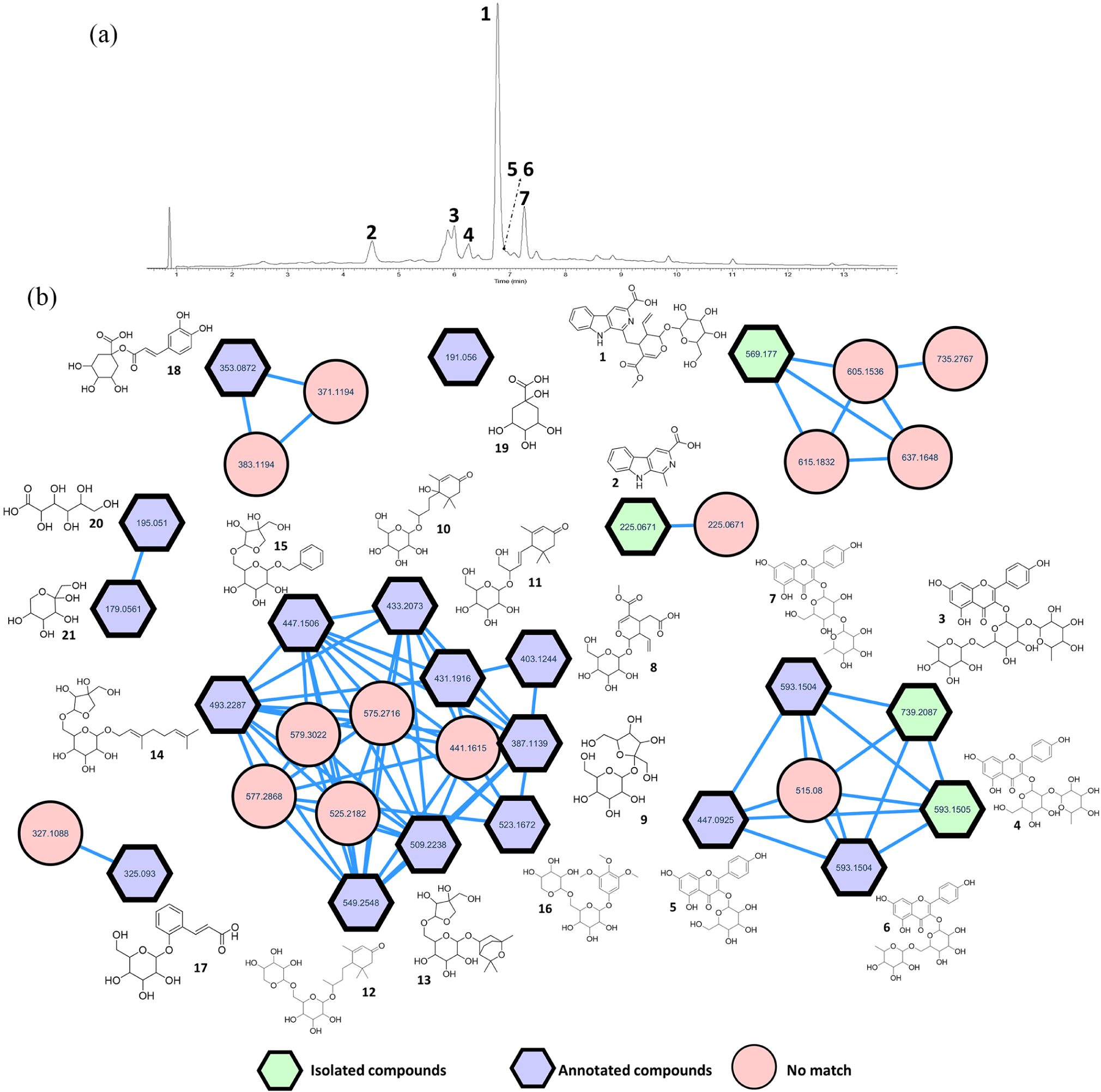
Liquid chromatography with diode-array detection (LC-DAD) provided an initial overview of the chemical profile of the extract (Figure 3a). The chromatogram was characterized by a limited number of well-resolved peaks, indicating a relatively low chromatographic complexity. The major peak, eluting at 6.84 min, corresponded to the isolated compound desoxycordifoline (1), while the peaks observed at retention times of 4.60, 6.06, and 6.33 min were assigned to harman-3-carboxylic acid (2), clitorin (3), and kaempferol 3-O-neohesperidoside (4), respectively. Overall, the LC-DAD profile suggested that a small number of metabolites dominated the extract composition.
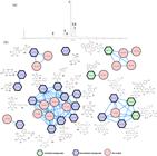
LC-DAD chromatogram highlighting the peaks corresponding to desoxycordifoline (1), harman-3-carboxylic acid (2), clitorin (3), kaempferol 3-O-neohesperidoside (4), and kaempferol glycoside derivatives (5-7) (a), and FBMN overview generated from data acquired in negative ionization mode, showing the organization and structures of the annotated compounds (b).
In contrast to the apparent simplicity observed by LC-DAD, FBMN analysis of the LC-MS data revealed a considerably richer chemical landscape. A large number of features were detected and organized into molecular clusters, with the majority of glycoside-related features grouped into two main clusters (Figure 3b). Notably, the cluster associated with clitorin (3) derivatives contained the ions at m/z 447 and 593 previously observed during LS-MS analysis, confirming the consistency between the preliminary ambient profiling and the LC-MS dataset. Within this cluster, at least three chromatographically resolved isomers corresponding to the ion at m/z 593 were detected. These features were tentatively annotated using the library of the GNPS2 platform as kaempferol 3-O-neohesperidoside (4) (cosine similarity = 0.9974), kaempferol-3-O-glucoside (5) (cosine similarity = 0.9976), kaempferol-3-O-rutinoside (6) (cosine similarity = 0.8553), and kaempferol 3-O-rungioside (7) (cosine similarity = 0.9979). Remarkably, clitorin (3) itself was also correctly annotated with a cosine similarity score of 0.9168. For all these annotations, mass errors equal to or lower than 1 ppm were observed (Table 1) for the corresponding molecular formulae, supporting their assignment as structurally related compounds.
Annotated compounds in S. jobertiana leaves based on feature-based molecular networking (FBMN) analysis in negative ionization mode
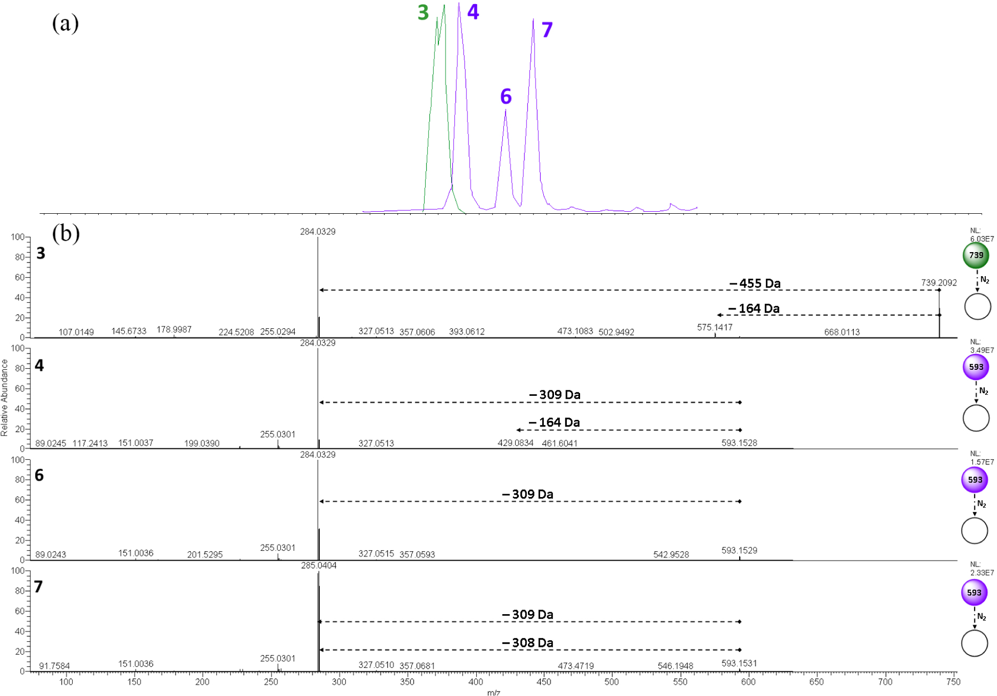
A more detailed inspection of the extracted ion chromatograms (EIC) (Figure 4a) revealed that the isomeric compounds 4, 6, and 7 eluted within a narrow retention time window between 6.33 and 7.33 min. Analysis of the MS/MS spectrum of compound 3 (Figure 4b), acquired using higher-energy collisional dissociation (HCD), revealed a dominant base peak at m/z 284 and a partial suppression of fragment ions previously observed under collision-induced dissociation (CID) during leaf spray analysis. This behavior reduced the amount of structurally informative fragmentation available for manual interpretation under HCD conditions. Nevertheless, a low-intensity neutral loss of 164 Da (m/z 739 → 575) could still be detected, supporting the presence of a rhamnopyranosyl-(1→2)-glucopyranoside disaccharide moiety.
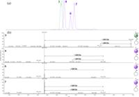
Extracted ion chromatogram (EIC) for the ions at m/z 593 and 739 (a) and higher-energy collisional dissociation (HCD) MS/MS spectra of compounds 3, 4, 6, and 7 (b), all acquired in negative ionization mode.
Similarly, the MS/MS spectrum of compound 4 exhibited a neutral loss of 164 Da (m/z 593 → 429), indicating a structure closely related to compound 3 but lacking one rhamnose unit, in agreement with the annotation proposed by the GNPS2 library and the structural elucidation. In contrast, compound 6 displayed a direct radical cleavage of the disaccharide unit, with a loss of 309 Da (m/z 593 → 284), a fragmentation pathway consistent with flavonoid glycosides containing a rhamnopyranosyl-(1→6)-glucopyranoside disaccharide. It is important to note that, although this compound was annotated here as kaempferol-3-O-rutinoside, considering its close structural relationship with compound 3 and its previous annotation/isolation in S. peckii,7 S. spinosa,25 S. nux-vomica,30 and S. pseudoquina,31 its isomer kaempferol 3-O-robinobioside, bearing galactose instead of glucose, has been isolated from S. variabilis.24 This observation highlights the structural complexity of flavonoid glycosides within the genus Strychnos and underscores the potential need for compound isolation to achieve unequivocal confirmation of sugar stereochemistry. Finally, the MS/MS spectrum of compound 7 showed a distinctive fragmentation pattern, characterized by nearly equivalent radical and non-radical competitive losses of 308 Da (m/z 593 → 285) and 309 Da (m/z 593 → 284), respectively. Unlike the other isomers, its base peak was observed at m/z 285. The rhamnopyranosyl-(1→3)-glucopyranoside disaccharide pattern proposed by the spectral library may explain this distinct fragmentation behavior relative to the other isomeric kaempferol glycosides.
The major glycoside cluster was anchored by the annotation of secoxyloganin (8) (cosine similarity = 0.9902), a secoiridoid (monoterpene derivative) glycoside structurally related to the isolated compound desoxycordifoline (1). Directly connected to this node in the molecular network, sucrose (9) was also annotated (cosine similarity = 0.9871), suggesting that this cluster represents a group of relatively simple glycosides. Thus, the organization of this cluster appears to comprise monoterpene-derived glycosides bearing glucose or structurally related sugars as the carbohydrate moiety.
Spectral library annotations obtained through the GNPS2 platform followed this pattern, proposing several monoterpene-derived glycosides, including icariside B5 (10), NCGC00380268-01 (11), NCGC00380271-01 (12), NCGC00385425-01 (13), and geranyl 6-O-β-D-apiofuranosyl-β-D-glucopyranoside (14). Among these, compound 14 exhibited the longest retention time on the C18 column, which is consistent with the increased hydrophobicity imparted by its geranyl side chain. In addition, benzenoid glycoside derivatives were annotated within this cluster, such as icariside F2 (15) and NCGC00180666-01 (16). Glycosides of this type have previously been isolated from the dried bark and wood of S. axillaris,32 including 3,4,5-trimethoxyphenol 1-O-β-D-apiofuranosyl-(1→6)-β-D-glucopyranoside, tachioside, isotachioside, calleryanin, and vanilloloside, and from the dried branches of S. spinosa,25 including 2,4,6-trimethoxyphenol 1-O-α-D-glucopyranoside, benzyl alcohol O-α-L-arabinopyranosyl-(1→6)-α-D-glucopyranoside, supporting the plausibility of these annotations within the genus.
Interestingly, most compounds within this cluster displayed recurrent diagnostic fragment ions at m/z 59.0139, 71.0139, 85.0295, 89.0243, 101.0243, 113.0245, and 119.0351, which are characteristic of glycosides such as sucrose (Table 1). These fragment ions suggest that the final fragmentation step in the monoterpene-derived glycosides by HCD predominantly involves cleavage of the sugar moiety, yielding negatively charged carbohydrate-derived ions. This observation is chemically consistent with the fact that, with the exception of secoxyloganin (8), all compounds annotated within this cluster were detected as formate adducts, favoring fragmentation pathways centered on the carbohydrate portion.
Other phenolic glycosides were also annotated in other clusters based on high cosine similarity scores in the spectral library search, including melilotoside (17) and 1-caffeoylquinic acid (18). Furthermore, simpler metabolites such as quinic acid (19), gluconic acid (20), and fructose (21) followed similar annotation pathways within the network. Finally, the isolated compound harman-3-carboxylic acid (2) was found to be connected to a neighboring node displaying high spectral similarity, suggesting the presence of a closely related isomer. However, despite the strong spectral resemblance, confident annotation of this feature was not possible based on the available data.
Conclusions
This study addressed the challenge of systematically recognizing and annotating glycosides in complex plant matrices, particularly in chemically underexplored Amazonian species such as S. jobertiana. By integrating leaf spray chemical profiling with LC-MS and FBMN, we established a coherent analytical strategy that connects early chemical insight with comprehensive metabolite annotation. LS-MS proved to be an effective decision-making tool, enabling the rapid recognition of glycoside-rich profiles directly from plant material and guiding the selection of extraction and sample preparation strategies. This preliminary step ensured that subsequent chromatographic and mass spectrometric analyses were tailored to capture the chemical features most relevant to the metabolome of the plant. The application of FBMN further allowed structurally related metabolites to be into coherent clusters, facilitating the annotation of both flavonoid and monoterpene-derived glycosides. Structural support provided by isolated reference compounds strengthened annotation confidence and enabled detailed interpretation of fragmentation behavior. Overall, this integrated workflow led to the annotation of 21 compounds and the identification of diagnostic fragment ions characteristic of glycosides, expanding the chemical knowledge of S. jobertiana and reinforcing the relevance of combining complementary analytical approaches. However, it is important to note that MS/MS-based annotation of glycosides does not always provide unequivocal structural information, particularly regarding sugar identity and glycosylation position, which may lead to ambiguity in distinguishing isomeric compounds. This limitation highlights the importance of complementary approaches, such as compound isolation and full spectroscopic characterization. Notably, desoxycordifoline (1), harman-3-carboxylic acid (2), clitorin (3), and kaempferol-3-O-neohesperidoside (4) were isolated and reported for the first time in S. jobertiana, with compounds 3 and 4 being described for the first time in the genus Strychnos.
Supplementary Information
Supplementary information (NMR spectra of the isolated compounds, the corresponding NMR data table, and MS/MS spectra of the annotated compounds with their mirror plots) is available free of charge at http://jbcs.sbq.org.br as PDF file.
Supplementary Information
Data Availability Statement
The FBMN dataset is publicly available in the link https://gnps2.org/status?task=4213d78e755846efa42917076defedbd
Acknowledgments
The authors are grateful to CNPq (grant number 315040/2021-1, 443823/2024-3; 302141/2025-1), FAPEAM, FINEP, and CAPES (Finance Code 001) for their financial support. The authors are also grateful to Central Analítica (CA/UFAM) and Centro Multiusuário para Análise de Fenômenos Biomédicos (CMABio/UEA) for analysis. The authors acknowledge the use of ChatGPT (OpenAI, GPT-5.3) for assistance with language editing (grammar and spelling) and for providing an initial visual background for the graphical abstract.
References
-
1 Kytidou, K.; Artola, M.; Overkleeft, H. S.; Aerts, J. M. F. G.; Front. Plant Sci 2020, 11, 357. [Crossref]
» Crossref -
2 Delbrouck, J. A.; Desgagné, M.; Comeau, C.; Bouarab, K.; Malouin, F.; Boudreault, P. L.; Molecules 2023, 28, 4957. [Crossref]
» Crossref -
3 Kowsalya, K.; Vidya, N.; Halka, J.; Preetha, J. S. Y.; Saradhadevi, M.; Sahayarayan, J. J.; Gurusaravanan, J., Arun, M.; Glycoconjugate J. 2025, 42, 107. [Crossref]
» Crossref - 4 Jadhav, R.; Kumar, S.; Ansari, Z. G.; Chauhan, P.; Chouhan, V. In Plant Secondary Metabolites; CRC Press: Boca Raton, USA, 2025, p. 153-176.
-
5 Vasudevan, U. M.; Lee, E. Y.; Biotechnol. Adv. 2020, 41, 107550. [Crossref]
» Crossref -
6 de Freitas, P. D.; Galetti Jr., P. M. In Population Genetics in the Neotropics; Bohrer Monteiro Siqueira, M. V.; Konzen, E. R.; Galetti Jr., P. M., eds.; Springer: Cham, Switzerland, 2025. [Crossref]
» Crossref -
7 Cassas, F.; Santos, C. L. G.; Silva, F. M. A.; Cass, Q. B.; Anal. Bioanal. Chem. 2025, 417, 4557. [Crossref]
» Crossref -
8 Ma, X.; Molecules 2022, 27, 6466. [Crossref]
» Crossref -
9 Turner, S. R.; Reynolds, J. C.; Turner, M. A.; Heaney, L. M.; Anal. Sci. Adv 2022, 3, 67. [Crossref]
» Crossref -
10 Turner, S. R.; Sears, P.; Heaney, L. M.; Anal. Sci. Adv 2023, 4, 133. [Crossref]
» Crossref -
11 Shi, L.; Habib, A.; Bi, L.; Hong, H.; Begum, R.; Wen, L.; Crit Rev. Anal. Chem. 2024, 54, 1584. [Crossref]
» Crossref -
12 Liu, J.; Wang, H.; Cooks, R. G.; Ouyang, Z.; Anal. Chem 2011, 83, 7608. [Crossref]
» Crossref -
13 de Lima, B. R.; da Silva, F. M. A.; Soares, E. R.; de Almeida, R. A.; da Silva-Filho, F. A.; Barison, A.; Costa, E. V.; Koolen, H. H. F.; de Souza, A. D. L.; Pinheiro, M. L. B.; J. Braz. Chem. Soc. 2020, 31, 79. [Crossref]
» Crossref -
14 Nothias, L.-F.; Petras, D.; Schmid, R.; Dührkop, K.; Rainer, J.; Sarvepalli, A.; Protsyuk, I.; Ernst, M.; Tsugawa, H.; Fleischauer, M.; Aicheler, F.; Aksenov, A. A.; Alka, O.; Allard, P.-M.; Barsch, A.; Cachet, X.; Caraballo-Rodriguez, A. M.; da Silva, R. R.; Dang, T.; Garg, N.; Gauglitz, J. M.; Gurevich, A.; Isaac, G.; Jarmusch, A. K.; Kameník, Z.; Kang, K. B.; Kessler, N.; Koester, I.; Korf, A.; Le Gouellec, A.; Ludwig, M.; Martin H. C.; McCall, L.-I.; McSayles, J.; Meyer, S. W.; Mohimani, H.; Morsy, M.; Moyne, O.; Neumann, S.; Neuweger, H.; Nguyen, N. H.; Nothias-Esposito, M.; Paolini, J.; Phelan, V. V.; Pluskal, T.; Quinn, R. A.; Rogers, S.; Shrestha, B.; Tripathi, A.; van der Hooft, J. J. J.; Vargas, F.; Weldon, K. C.; Witting, M.; Yang, H.; Zhang, Z.; Zubeil, F.; Kohlbacher, O.; Böcker, S.; Alexandrov, T.; Bandeira, N.; Wang, M.; Dorrestein, P. C.; Nat. Methods 2020, 17, 905. [Crossref]
» Crossref -
15 Wang, M.; Carver, J. J.; Phelan, V. V.; Sanchez, L. M.; Garg, N.; Peng, Y.; Nguyen, D. D.; Watrous, J.; Kapono, C. A.; Luzzatto-Knaan, T.; Porto, C.; Bouslimani, A.; Melnik, A. V.; Meehan, M. J.; Liu, W.-T.; Crüsemann, M.; Boudreau, P. D.; Esquenazi, E.; Sandoval-Calderón, M.; Kersten, R. D.; Pace, L. A.; Quinn, R. A.; Duncan, K. R.; Hsu, C.-C.; Floros, D. J.; Gavilan, R. G.; Kleigrewe, K.; Northen, T.; Dutton, R. J.; Parrot, D.; Carlson, E. E.; Aigle, B.; Michelsen, C. F.; Jelsbak, L.; Sohlenkamp, C.; Pevzner, P.; Edlund, A.; McLean, J.; Piel, J.; Murphy, B. T.; Gerwick, L.; Liaw, C.-C.; Yang, Y.-L.; Humpf, H.-U.; Maansson, M.; Keyzers, R. A.; Sims, A. C.; Johnson, A. R.; Sidebottom, A. M.; Sedio, B. E.; Klitgaard, A.; Larson, C. B.; P, C. A. B.; Torres-Mendoza, D.; Gonzalez, D. J.; Silva, D. B.; Marques, L. M.; Demarque, D. P.; Pociute, E.; O’Neill, E. C.; Briand, E.; Helfrich, E. J. N.; Granatosky, E. A.; Glukhov, E.; Ryffel, F.; Houson, H.; Mohimani, H.; Kharbush, J. J.; Zeng, Y.; Vorholt, J. A.; Kurita, K. L.; Charusanti, P.; McPhail, K. L.; Nielsen, K. F.; Vuong, L.; Elfeki, M.; Traxler, M. F.; Engene, N.; Koyama, N.; Vining, O. B.; Baric, R.; Silva, R. R.; Mascuch, S. J.; Tomasi, S.; Jenkins, S.; Macherla, V.; Hoffman, T.; Agarwal, V.; Williams, P. G.; Dai, J.; Neupane, R.; Gurr, J.; Rodríguez, A. M. C.; Lamsa, A.; Zhang, C.; Dorrestein, K.; Duggan, B. M.; Almaliti, J.; Allard, P.-M.; Phapale, P.; Nothias, L.-F.; Alexandrov, T.; Litaudon, M.; Wolfender, J.-L.; Kyle, J. E.; Metz, T. O.; Peryea, T.; Nguyen, D.-T.; VanLeer, D.; Shinn, P.; Jadhav, A.; Müller, R.; Waters, K. M.; Shi, W.; Liu, X.; Zhang, L.; Knight, R.; Jensen, P. R.; Palsson, B. O.; Pogliano, K.; Linington, R. G.; Gutiérrez, M.; Lopes, N. P.; Gerwick, W. H.; Moore, B. S.; Dorrestein, P. C.; Bandeira, N.; Nat. Biotechnol 2016, 34, 828. [Crossref]; Global Natural Product Social Molecular Networking (GNPS). [Link] accessed in January. 2026
» Crossref» Link -
16 Yang, J. Y.; Sanchez, L. M.; Rath, C. M.; Liu, X.; Boudreau, P. D.; Bruns, N.; Glukhov, E.; Wodtke, A.; Felicio, R.; Fenner, A.; Wong, W. R.; Linington, R. G.; Zhang, L.; Debonsi, H. M.; Gerwick, W. H.; Dorrestein, P. C.; J. Nat. Prod 2013, 76, 1686. [Crossref]
» Crossref -
17 Phinney, K. W.; Ballihaut, G.; Bedner, M.; Benford, B. S.; Camara, J. E.; Christopher, S. J.; Davis, W. C.; Dodder, N. G.; Eppe, G.; Lang, B. E.; Long, S. E.; Lowenthal, M. S.; Nelson, B. C.; Prendergast, J. L.; Reiner, J. L.; Rimmer, C. A.; Sander, L. C.; Schantz, M. M.; Sharpless, K. E.; Vocke, R. D.; Wood, L. J.; Anal. Chem. 2013, 85, 11732. [Crossref]
» Crossref -
18 Santos, C. L. G.; Neves, K. O. G.; Silva-Filho, F. A.; Lima, B. R.; Costa, E. V.; Souza, A. D. L.; Koolen, H. H. F.; Pinheiro, M. L. B.; Silva, F. M. A.; Front. Nat. Prod. 2023, 2, 1189619. [Crossref]
» Crossref -
19 Santos, C. L. G.; Angolini, C. F. F.; Neves, K. O. G.; Costa, E. V.; Souza, A. D. L.; Pinheiro, M. L. B.; Silva, F. M. A.; Rapid Commun. Mass Spectrom 2020, 34, e8683. [Crossref]
» Crossref -
20 Kessner, D.; Chambers, M.; Burke, R.; Agus, D.; Mallick, P.; Bioinformatics 2008, 24, 2534. [Crossref]
» Crossref -
21 Schmid, R.; Heuckeroth, S.; Korf, A.; Smirnov, A.; Myers, O.; Dyrlund, T. S.; Bushuiev, R.; Murray, K. J.; Hoffmann, N.; Lu, M.; Sarvepalli, A.; Zhang, Z.; Fleischauer, M.; Dührkop, K.; Wesner, M.; Hoogstra, S. J.; Rudt, E.; Mokshyna, O.; Brungs, C.; Ponomarov, K.; Mutabdžija, L.; Damiani, T.; Pudney, C. J.; Earll, M.; Helmer, P. O.; Fallon, T. R.; Schulze, T.; Rivas-Ubach, A.; Bilbao, A.; Richter, H.; Nothias, L.-F.; Wang, M.; Orešič, M.; Weng, J.-K.; Böcker, S.; Jeibmann, A.; Hayen, H.; Karst, U.; Dorrestein, P. C.; Petras, D.; Du, X.; Pluskal, T.; Nat. Biotechnol 2023, 41, 447. [Crossref]
» Crossref -
22 Castro, A. P. A.; Lima, B. R.; França, R. S.; Leocadio, B. R. C.; Silva-Filho, F. A.; Fernandes, C. C.; Souza, A. D. L.; Koolen, H. H. F.; Pinheiro, M. L. B.; Silva, F. M. A.; Rapid Commun. Mass Spectrom 2026, 40, e10163. [Crossref]
» Crossref -
23 França, R. S.; Bataglion, G. A.; Castro, A. P. A.; Leocadio, B. L. C.; Neves, K. O. G.; da Silva-Filho, F. A.; Lima, B. R.; Cassas, F.; Koolen, H. H. F.; Souza, A. D. L.; Pinheiro, M. L. B.; Cass, Q. B.; Silva, F. M. A.; J. Chromatogr. A 2026, 1766, 466598. [Crossref]
» Crossref -
24 Brasseur, T.; Angenot, L.; Phytochemistry 1986, 25, 1171. [Crossref]
» Crossref -
25 Itoh, A.; Oya, N.; Kawaguchi, E.; Nishio, S.; Tanaka, Y.; Kawachi, E.; Akita, T.; Nishi, T.; Tanahashi, T.; J. Nat. Prod 2005, 68, 1434. [Crossref]
» Crossref -
26 Kazuma, K.; Noda, N.; Suzuki, M.; Phytochemistry 2003, 62, 229. [Crossref]
» Crossref -
27 Pereira, M. M.; Souza Jr., S. N.; Alcântara, A. F. C.; Piló-Veloso, D.; Alves, R. B.; Machado, P. O.; Azevedo, A. O.; Moreira, F. H.; Castro, M. S. A.; Raslan, D. S.; Rev. Bras. Plantas Med 2006, 8, 1. [Crossref]
» Crossref -
28 Brandt, V.; Tits, M.; Geerlings, A.; Frédérich, M.; Penelle, J.; Delaude, C.; Verpoorte, R.; Angenot, L.; Phytochemistry 1999, 51, 1171. [Crossref]
» Crossref -
29 Wu, H.; Dushenkov, S.; Ho, C. T.; Sang, S.; Food Chem 2009, 115, 592. [Crossref]
» Crossref -
30 Eldahshan, O. A.; Abdel-Daim, M. M.; Cytotechnology 2015, 67, 831. [Crossref]
» Crossref -
31 da Silva, M. A.; Rafacho, B. P. M.; Hiruma-Lima, C. A.; Rocha, L. R. M.; dos Santos, L. C.; Sannomiya, M.; Souza-Brito, A. R. M.; Vilegas, W.; Chem. Pharm. Bull 2005, 53, 881. [Crossref]
» Crossref -
32 Itoh, A.; Tanaka, Y.; Nagakura, N.; Akita, T.; Nishi, T.; Tanahashi, T.; Phytochemistry 2008, 69, 1208. [Crossref]
» Crossref
Edited by
-
Editor handled this article:
Paulo Wender P. Gomes (Guest Editor)