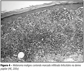
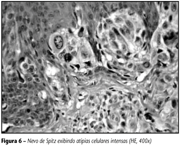
The challenge and the diagnostic difficulty imposed by some melanocytic lesions are well known by the pathologists. The lack of uniform diagnostic criteria is, without a doubt, one of the main causes for the high rates of disagreement described in literature. The aim of this report is to review twelve selected criteria in order to reduce diagnostic disagreement. These are: 1) size; 2) symmetry; 3) lateral delimitation; 4) maturation; 5) pagetoid spreading; 6) necrosis/ulceration; 7) inflammatory reaction; 8) regression; 9) cellular atypias; 10) mitosis; 11) melanization; 12) isolated cells proliferation. The characteristics of each one and their application in the diagnosis of melanomas were shown, always remembering possible exceptions in which these criteria might present in benign melanocytic lesions. Farther more, tables of differential diagnostic features are offered, for benign and malignant melanocytic lesions, using these histopathological criteria. As any criterion must not be considered by itself, the rigorous application of this data set here provided can substantially help the generalist surgical pathologist (non-specialist in melanocytic lesions) to solve some of the problems at his daily routine.
Melanoma; Differential diagnosis; Spitz nevus; Dysplastic nevus


