Abstracts
OBJECTIVE: Primary benign tumors of the trachea and main bronchi are uncommon. Interventional bronchoscopy allows the diagnosis and the treatment of some of these lesions. METHODS: We reviewed four cases endoscopically treated at our institution. RESULTS: Two patients had hamartoma, and two patients had endobronchial lipoma. In all of the cases, the interventional technique for the resection was the use of a polypectomy snare and electrocautery. The only complication reported was one episode of bronchospasm. CONCLUSIONS: Minimally invasive bronchoscopic resection is a safe, effective method for treating selected benign tumors of the main airway and has a low complication rate.
Bronchoscopy; Bronchial neoplasms; Hamartoma; Lipoma
OBJETIVO: Tumores benignos primários da traqueia e dos brônquios principais são incomuns. A broncoscopia intervencionista permite o diagnóstico e o tratamento de algumas dessas lesões. MÉTODOS: Revisamos quatro casos tratados endoscopicamente em nossa instituição. RESULTADOS: Dois pacientes tinham hamartoma, e dois pacientes apresentaram lipoma endobrônquico. Em todos os casos, a técnica de intervenção para a ressecção foi o uso de alça de polipectomia e eletrocautério. A única complicação relatada foi um episódio de broncoespasmo. CONCLUSÕES: O tratamento broncoscópico minimamente invasivo é um método seguro e efetivo para o tratamento bem-sucedido de alguns tumores benignos da via aérea principal, com um baixo índice de complicações.
Broncoscopia; Neoplasias brônquicas; Hamartoma; Lipoma
CASE SERIES
Minimally invasive bronchoscopic resection of benign tumors of the bronchi* * Study carried out at the Respiratory Endoscopy Department, University of São Paulo School of Medicine Hospital das Clínicas, São Paulo, Brazil. ,** ** A versão completa em português deste artigo está disponível em www.jornaldepneumologia.com.br
Ascedio Jose RodriguesI; David CoelhoII; Sérvulo Azevedo Dias JúniorIII; Márcia JacomelliIV; Paulo Rogério ScordamaglioV; Viviane Rossi FigueiredoVI
IAttending Physician. Respiratory Endoscopy Department, University of São Paulo School of Medicine Hospital das Clínicas, São Paulo, Brazil
IIResident. Respiratory Endoscopy Department, University of São Paulo School of Medicine Hospital das Clínicas, São Paulo, Brazil
IIIResident. Respiratory Endoscopy Department, University of São Paulo School of Medicine Hospital das Clínicas, São Paulo, Brazil
IVAttending Physician. Respiratory Endoscopy Department, University of São Paulo School of Medicine Hospital das Clínicas, São Paulo, Brazil
VAttending Physician. Respiratory Endoscopy Department, University of São Paulo School of Medicine Hospital das Clínicas, São Paulo, Brazil
VITechnical Director. Respiratory Endoscopy Department, University of São Paulo School of Medicine Hospital das Clínicas, São Paulo, Brazil
Correspondence to Correspondence to: Ascedio Jose Rodrigues Serviço de Endoscopia Respiratória, HC-FMUSP, Prédio dos Ambulatórios, 6º Andar, Bloco 03 Avenida Dr. Enéas de Carvalho Aguiar, 255, Cerqueira César CEP 05017-000, São Paulo, SP, Brasil Tel. 55 11 2661-5612 E-mail: ascedio@gmail.com
ABSTRACT
OBJECTIVE: Primary benign tumors of the trachea and main bronchi are uncommon. Interventional bronchoscopy allows the diagnosis and the treatment of some of these lesions.
METHODS: We reviewed four cases endoscopically treated at our institution.
RESULTS: Two patients had hamartoma, and two patients had endobronchial lipoma. In all of the cases, the interventional technique for the resection was the use of a polypectomy snare and electrocautery. The only complication reported was one episode of bronchospasm.
CONCLUSIONS: Minimally invasive bronchoscopic resection is a safe, effective method for treating selected benign tumors of the main airway and has a low complication rate.
Keywords: Bronchoscopy; Bronchial neoplasms; Hamartoma; Lipoma
Introduction
Primary tumors of the trachea and bronchi account for 1-2% of all types of cancer of the respiratory tract. In adults, benign lesions (mostly lipomas, leiomyomas, hamartomas, and inflammatory polyps) account for only 20% of all tumors of the main airways.(1)
Hamartomas, despite being the most common benign tumors of the lung, have a low prevalence in the general population, and 90% are located within the lung parenchyma, rarely causing symptoms.(2) Endobronchial lipomas are even rarer, accounting for only 0.1% of lung tumors.(3)
Benign endobronchial lesions usually occur in large bronchi and grow into the lumen, causing progressive airflow obstruction. The clinical presentation of these lesions is variable, and the most common signs and symptoms are stridor, wheezing, hemoptysis, chronic cough, recurrent pulmonary infections, and dyspnea (in varying degrees, depending on the location and degree of the obstruction). Such lesions are usually detected by conventional X-rays when they cause atelectasis or obstructive pneumonia.
In recent years, endoscopic treatment has been increasingly used to treat benign endobronchial lesions. In selected cases, therapeutic bronchoscopy is effective, safe, and minimally invasive, offering the possibility of same-day discharge after the procedure.(4) Resection by electrocautery and argon plasma coagulation has been described, as has the use of laser and cryotherapy. In the present study, we present the clinical features and results of the bronchoscopic treatment of four cases of benign tumors of the bronchial tree.
Methods
We reviewed the cases of four patients with benign tumors of the bronchial tree that were treated endoscopically. A summary of the characteristics of the patients is shown in Table 1. The Research Ethics Committee of the University of São Paulo School of Medicine Hospital das Clínicas approved the study.
Results
Case 1
A 51-year-old male patient presented with a primary complaint of nocturnal cough, which had worsened over the previous three months. The physical examination was unremarkable. There were no abnormal findings on the chest X-ray. Simple spirometry, methacholine challenge test, and CT scans of the paranasal sinuses showed no abnormalities. Chest CT scans revealed an irregular lesion of low attenuation, which partially obstructed the left main bronchus.
Flexible bronchoscopy revealed that the tumor had a non-friable, lobulated surface, and that it obstructed 80% of the left main bronchus. The biopsy revealed a lipomatous hamartoma. Argon plasma coagulation associated with cup forceps resection via bronchoscopy was performed in three sessions due to the size of the tumor and its wide base on the airway wall. In the first session, rigid bronchoscopy was performed, which allowed the removal of a large amount of tissue. At six months after the procedures, control chest CT and bronchoscopy showed no further damage, and the patient was asymptomatic.
Case 2
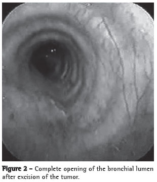
A 32-year-old female patient presented with recurrent pneumonia in the left lower lobe. During the clinical investigation, a chest X-ray showed atelectasis of the left lower lobe. Chest CT scans showed a lesion that partially obstructed the left main bronchus. Flexible bronchoscopy showed an elevated lesion, with a bosselated pink surface and a pedicle on the medial wall of the left main bronchus, blocking 90% of its lumen, as well as a large amount of purulent secretion distal to the lesion (Figure 1). The histological examination of the biopsy specimen was compatible with hamartoma. Systemic antibiotics were administered for one week prior to endoscopic intervention. The lesion was uneventfully excised using a flexible bronchoscope, a polypectomy snare, and electrocautery. There was complete opening of the bronchial lumen, as well as good clinical and radiological evolution (Figure 2). There were no clinical signs of recurrence 15 months after the procedure.
Case 3
A 39-year-old male patient presented with recurrent pneumonia. A chest X-ray showed partial atelectasis of the left lower lobe. Chest CT scans confirmed partial atelectasis. Flexible bronchoscopy showed a lesion, with a smooth, yellowish surface, that totally obstructed the posterior segment of the left lower lobe. A biopsy fragment was consistent with lipoma. The lesion was uneventfully excised with the use of a flexible bronchoscope, a polypectomy snare, and electrocautery. There was complete opening of the bronchial lumen. After a 12-month follow-up period, the patient remained asymptomatic, with good clinical and radiological evolution.
Case 4
A 78-year-old female patient presented with recurrent pneumonia in the right lower lobe. Chest X-ray was normal. Chest CT scans showed a protruding lesion into the bronchial lumen of the right lower lobe bronchus, with fat attenuation. Bronchoscopy revealed a bilobulated tumor, with a smooth, yellowish surface, that obstructed 80% of the lumen of the right lower lobe bronchus. Microscopic examination revealed a lipoma. The lesion was excised with the use of a flexible bronchoscope, a polypectomy snare, and electrocautery. There was complete opening of the bronchial lumen. The patient had significant bronchospasm immediately after the procedure and had to be hospitalized for 24 h. The patient was asymptomatic at 30 days after the procedure. As of this writing, the patient has yet to return for the medical follow-up.
Discussion
In this case series, the endobronchial lesions were lipomas and hamartomas. Clinically, the patients presented with chronic cough or a history of recurrent respiratory infections, and the initial tests failed to help define the diagnosis. In all cases, despite the benign appearance of the lesions at bronchoscopy, a diagnosis of malignant lesion was excluded only after the histological analysis of tumor fragments.
Pulmonary hamartomas are benign lesions that consist of pulmonary and bronchial elements, which are usually combined with cartilaginous tissue, fat, and smooth muscle.(2) Endobronchial hamartomas account for only 10% of intrathoracic hamartomas. This subtype is often associated with symptoms secondary to obstruction and irritation of the airways.(5) Lipomas are less common, with a prevalence of only 0.1-0.5% of all lung tumors.(6)
Most benign lesions are asymptomatic and are typically discovered by chance during routine radiological examinations. These lesions show some typical features, such as a "popcorn" pattern of calcification, which can be seen on conventional chest X-rays or chest CT scans in patients with hamartomas,(7,8) as well as solid lesions with fat attenuation, which is the main characteristic of lipomas.(9)
Most patients with endobronchial hamartoma or lipoma who are referred for bronchoscopy are symptomatic. The most common symptoms in patients with endobronchial hamartomas or lipomas are recurrent respiratory infections, cough, hemoptysis, dyspnea, and chest pain. In asymptomatic patients, the diagnosis is typically made on the basis of abnormal findings on chest X-rays requested for other reasons.(2,3) The main radiological changes found in these patients are signs of volume loss (such as atelectasis or complete lobar collapse), signs of alveolar filling, and pulmonary nodules.(2) The definitive diagnosis is made by bronchoscopy with biopsy. Endoscopic examination typically reveals a lesion with a smooth, regular, soft surface that is non-friable to the touch of the device. Although these features suggest a benign lesion, on some occasions, it might be difficult to distinguish between these tumors and malignant lesions based on macroscopic findings alone; therefore, biopsy should be performed routinely in all patients presenting with such lesions.(2,10)
In the cases presented here, we removed the lesions with bronchoscopic resection, using electrocautery and a polypectomy snare. In one case, three sessions were required in order to complete the resection. Each of the other three patients was treated in a single session. One patient presented with post-procedure bronchospasm, which was resolved within 24 h through clinical measures.
The technique employed for the resection of benign tumors of the airways depends on the tumor location, the presence of lung parenchymal disease reaching the distal airways, the presence of extrabronchial growth, and eventual symptoms. The management of such tumors should be individualized according to the characteristics of each patient and lesion. The standard treatment consists of the removal of the lesion by bronchotomy, thoracotomy, lobectomy, or (in rare instances) pneumonectomy.(5) Due to the benign nature of these lesions, endoscopic resection has become a popular technique for their resection, because it provides satisfactory results without the risks associated with thoracotomy.(11-14) In a recent multicenter study, endoscopic procedures were shown to be quite effective and safe for the diagnosis and treatment of endobronchial lipomas.(15)
Endobronchial resection is an excellent choice given that most endobronchial lesions are intraluminal. Various methods for resecting endobronchial lesions include the use of laser, electrocautery, cryotherapy, and argon plasma coagulation. Care must be taken when using such devices because of the risks of airway perforation and fistula formation. The partial removal of the lesion allows increased permeability of segmental bronchi, as well as allowing the implantation of the tumor within the bronchial wall to be located and assessed. Subsequently, complete excision of the tumor and treatment of the tumor base reduces the chance for local recurrence and maintains the patency of the affected bronchus.(4)
The advantages of the use of bronchoscopy in the management of these tumors include less invasiveness, lesser need for general anesthesia, quicker bronchial opening, shorter hospital stays, and lower costs. Benign tumors of the airway can cause respiratory symptoms, especially when there is severe obstruction of the airway. The diagnosis of these lesions can be challenging, and endoscopic removal, performed by an experienced bronchoscopist, is a safe and effective alternative to surgical resection. However, surgery should be considered for patients with recurrence, extrabronchial extension of the lesions, or destruction of the peripheral lung due to long-term atelectasis, as well as when technical difficulties that could prevent complete bronchoscopic removal are anticipated.
References
1. Baldi BG, Fernandes CJ, Salge JM, Takagaki TY. Tracheal polyp. J Bras Pneumol. 2007;33(5):616-20.
2. Cosío BG, Villena V, Echave-Sustaeta J, de Miguel E, Alfaro J, Hernandez L, et al. Endobronchial hamartoma. Chest. 2002;122(1):202-5.
3. Muraoka M, Oka T, Akamine S, Nagayasu T, Iseki M, Suyama N, et al. Endobronchial lipoma: review of 64 cases reported in Japan. Chest. 2003;123(1):293-6.
4. Ferreira D, Almeida J, Parente B, Moura E Sá J. Complete resection of endobronchial hamartomas via bronchoscopic techniques, electrosurgery by Argon plasma and laser [Article in Portuguese]. Rev Port Pneumol. 2007;13(5):711-9.
5. Kaya S, Karalezli A, Balkan E, Cakiroğlu E, Hasanoğlu HC. Endobronchial hamartoma removed by flexible fiberoptic bronchoscopy via electrocautery. Tuberk Toraks. 2006;54(3):273-6.
6. Bof AM, Rapoport A, Paier LC, Diaz YL, Leiro LC, Pando-Serrano RR, et al. Endobronchial lipoma. J Bras Pneumol. 2005;31(6):555-8
7. Silva VA, Kataguiri P, Trufelli DC, Matos LL, Neves- Pereira JC, Campos JR. Pulmonary hamartoma as a differential diagnosis of breast cancer metastasis: case report. J Bras Pneumol. 2007;33(6):738-42.
8. Yilmaz S, Ekici A, Erdogan S, Ekici M. Endobronchial lipomatous hamartoma: CT and MR imaging features (2004:5b). Eur Radiol. 2004;14(8):1521-4.
9. Basoglu A, Celik B, Akdag AO, Sengul AT. Endobronchial lipoma: a rare cause of bronchial occlusion. Interact Cardiovasc Thorac Surg. 2004;3(2):263-4.
10. Karabulut N, Bir F, Yuncu G, Kiter G. Endobronchial lipomatous hamartoma: an unusual cause of bronchial obstruction (2007: 7b). Eur Radiol. 2007;17(10):2687-90.
11. David O, Beasley MB, Minardi AJ Jr, Malek F, Kovitz KL. Management of endobronchial hamartoma. J La State Med Soc. 2003;155(2):110-2.
12. Sahin AA, Aydiner A, Kalyoncu F, Tokgozoglu L, Baris YI. Endobronchial hamartoma removed by rigid bronchoscope. Eur Respir J. 1989;2(5):479-80.
13. Stey CA, Vogt P, Russi EW. Endobronchial lipomatous hamartoma: a rare cause of bronchial occlusion. Chest. 1998;113(1):254-5.
14. Shah H, Garbe L, Nussbaum E, Dumon JF, Chiodera PL, Cavaliere S. Benign tumors of the tracheobronchial tree. Endoscopic characteristics and role of laser resection. Chest. 1995;107(6):1744-51.
15. Nassiri AH, Dutau H, Breen D, Colchen A, Quiot JJ, Nguyen B, et al. A multicenter retrospective study investigating the role of interventional bronchoscopic techniques in the management of endobronchial lipomas. Respiration. 2008;75(1):79-84.
Submitted: 18 July 2011.
Accepted, after review: 6 September 2011.
Financial support: None.
- 1. Baldi BG, Fernandes CJ, Salge JM, Takagaki TY. Tracheal polyp. J Bras Pneumol. 2007;33(5):616-20.
- 2. Cosío BG, Villena V, Echave-Sustaeta J, de Miguel E, Alfaro J, Hernandez L, et al. Endobronchial hamartoma. Chest. 2002;122(1):202-5.
- 3. Muraoka M, Oka T, Akamine S, Nagayasu T, Iseki M, Suyama N, et al. Endobronchial lipoma: review of 64 cases reported in Japan. Chest. 2003;123(1):293-6.
- 4. Ferreira D, Almeida J, Parente B, Moura E Sá J. Complete resection of endobronchial hamartomas via bronchoscopic techniques, electrosurgery by Argon plasma and laser [Article in Portuguese]. Rev Port Pneumol. 2007;13(5):711-9.
- 6. Bof AM, Rapoport A, Paier LC, Diaz YL, Leiro LC, Pando-Serrano RR, et al. Endobronchial lipoma. J Bras Pneumol. 2005;31(6):555-8
- 7. Silva VA, Kataguiri P, Trufelli DC, Matos LL, Neves- Pereira JC, Campos JR. Pulmonary hamartoma as a differential diagnosis of breast cancer metastasis: case report. J Bras Pneumol. 2007;33(6):738-42.
- 8. Yilmaz S, Ekici A, Erdogan S, Ekici M. Endobronchial lipomatous hamartoma: CT and MR imaging features (2004:5b). Eur Radiol. 2004;14(8):1521-4.
- 9. Basoglu A, Celik B, Akdag AO, Sengul AT. Endobronchial lipoma: a rare cause of bronchial occlusion. Interact Cardiovasc Thorac Surg. 2004;3(2):263-4.
- 10. Karabulut N, Bir F, Yuncu G, Kiter G. Endobronchial lipomatous hamartoma: an unusual cause of bronchial obstruction (2007: 7b). Eur Radiol. 2007;17(10):2687-90.
- 11. David O, Beasley MB, Minardi AJ Jr, Malek F, Kovitz KL. Management of endobronchial hamartoma. J La State Med Soc. 2003;155(2):110-2.
- 12. Sahin AA, Aydiner A, Kalyoncu F, Tokgozoglu L, Baris YI. Endobronchial hamartoma removed by rigid bronchoscope. Eur Respir J. 1989;2(5):479-80.
- 13. Stey CA, Vogt P, Russi EW. Endobronchial lipomatous hamartoma: a rare cause of bronchial occlusion. Chest. 1998;113(1):254-5.
- 14. Shah H, Garbe L, Nussbaum E, Dumon JF, Chiodera PL, Cavaliere S. Benign tumors of the tracheobronchial tree. Endoscopic characteristics and role of laser resection. Chest. 1995;107(6):1744-51.
- 15. Nassiri AH, Dutau H, Breen D, Colchen A, Quiot JJ, Nguyen B, et al. A multicenter retrospective study investigating the role of interventional bronchoscopic techniques in the management of endobronchial lipomas. Respiration. 2008;75(1):79-84.
Publication Dates
-
Publication in this collection
10 Jan 2012 -
Date of issue
Dec 2011
History
-
Received
18 July 2011 -
Accepted
06 Sept 2011