
Abstracts
Introduction:
Endoscopic submucosal dissection (ESD) is an already established procedure in the treatment of gastric and esophageal cancer in its early stages. Colorectal lesions, initially approached by endoscopic mucosal resection en bloc or in fragments, are the current focus for submucosal approach, especially for superficial lateral spreading tumor of 20 mm-diameter. The experience of Japanese centers, which are reference in therapeutic endoscopy, demonstrates reduction in the rate of disease recurrence with this approach and, according to specific histopathological criteria, may avoid colectomy in some cases of malignant neoplasia.11. Fukuzawa M, Gotoda T. History of endoscopic submucosal dissection and role for colorectal endoscopic submucosal dissection: a Japanese perspective. Gastrointest Interv. 2012.
2. Saito Y, Uraoka T, Matsuda T, Emura F, Ikehara H, Mashimo Y, et al. Endoscopic treatment of large superficial colorectal tumors: a case series of 200 endoscopic submucosal dissections (with vídeo). Gastrointest Endosc. 2007.-33. Yoshida N, Wakabayashi N, Kanemasa K, Sumida Y, Hasegawa D, Inoue K, et al. Endoscopic submucosal dissection for colorectal tumors: technical difficulties and rate of perforation. Endoscopy. 2009.
Case report:
The patient was 50-year-old female. She underwent endoscopic submucosal dissection of a rectal lateral spreading tumor measuring 50 mm, located 8 cm from the anal margin. The procedure was performed without major complications, with just two points for muscle layer detachment, without gross perforation and closed with metal clips. However, the patient developed air leakage to the peritoneum, retroperitoneum, mediastinum and subcutaneous tissue, being only treated with clinical procedures and without additional intervention.
Conclusion:
It is vital to know and be able to apply the technique of ESD, in addition to addressing its complications, since despite the numerous benefits compared to surgery, ESD can result in serious outcomes.44. Hotta K, Fuji T, Saito Y, Matsuda T. Local recurrence after endoscopic resection of colorectal tumors. Int J Colorectal Dis. 2009.,55. Saito Y, Fukuzawa M, Matsuda T, Fukunaga S, Sakamoto T, Uraoka T, et al. Clinical outcome of endoscopic submucosal dissection versus endoscopic mucosal resection of large colorectal tumors as determined by curative resection. Surg Endosc. 2010.
Lateral spreading tumor; Pneumoretroperitoneum; Pneumomediastinum; Pneumoperitoneum; Endoscopic submucosal dissection
Introdução:
A dissecção endoscópica da submucosa (ESD) já é procedimento consagrado no tratamento do câncer gástrico e esofagiano em suas fases precoces. As lesões colorre-tais, inicialmente abordadas por mucossectomia, em bloco ou em fragmentos, são o foco atual para a abordagem submucosa, principalmente para os tumores de crescimento lateral superficial a partir de 20 mm de diâmetro. A experiência de centros japoneses, referências em endoscopia terapêutica, demonstram redução no índice de recidiva da doença com esta abordagem e, segundo critérios histopatológicos específicos, podem evitar uma colectomia em alguns casos de neoplasia maligna.11. Fukuzawa M, Gotoda T. History of endoscopic submucosal dissection and role for colorectal endoscopic submucosal dissection: a Japanese perspective. Gastrointest Interv. 2012.
2. Saito Y, Uraoka T, Matsuda T, Emura F, Ikehara H, Mashimo Y, et al. Endoscopic treatment of large superficial colorectal tumors: a case series of 200 endoscopic submucosal dissections (with vídeo). Gastrointest Endosc. 2007.-33. Yoshida N, Wakabayashi N, Kanemasa K, Sumida Y, Hasegawa D, Inoue K, et al. Endoscopic submucosal dissection for colorectal tumors: technical difficulties and rate of perforation. Endoscopy. 2009.
Relato de caso:
Trata-se de paciente de 50 anos, submetida à dissecção endoscópica da submucosa de lesão de crescimento lateral, com 50 mm, localizada no reto, a 8 cm da margem anal. O procedimento foi realizado sem maiores intercorrências, com apenas dois pontos de afastamento da muscular, sem perfuração grosseira, fechados com clipe. Entretanto, a paciente evoluiu com escape aéreo para peritônio, retroperitônio, mediastino e subcútis, sendo tratada sem intervenção adicional, apenas com manejo clínico.
Conclusão:
É de fundamental importância conhecer e saber aplicar a técnica da ESD, além de abordar suas complicações, uma vez que, mesmo repleta de benefícios em relação à cirurgia, ela pode apresentar desfechos graves.44. Hotta K, Fuji T, Saito Y, Matsuda T. Local recurrence after endoscopic resection of colorectal tumors. Int J Colorectal Dis. 2009.,55. Saito Y, Fukuzawa M, Matsuda T, Fukunaga S, Sakamoto T, Uraoka T, et al. Clinical outcome of endoscopic submucosal dissection versus endoscopic mucosal resection of large colorectal tumors as determined by curative resection. Surg Endosc. 2010.
Lesão de crescimento lateral; Pneumorretroperitôneo; Pneumomediastino; Pneumoperitôneo; Dissecção submucosa; endoscópica
Introduction
Colonoscopy is widely used not only as a diagnostic procedure, but also with a therapeutic goal, being much prized by minimally invasive medicine.
ESD of early esophageal and gastric carcinomas is already practiced worldwide. The same technique applied to not-invasive pre-malignant and malignant colorectal lesions is not yet accepted as standard procedure. But this procedure is becoming increasingly feasible, to the extent that the technology extends the capabilities with tools appropriate to this procedure.11. Fukuzawa M, Gotoda T. History of endoscopic submucosal dissection and role for colorectal endoscopic submucosal dissection: a Japanese perspective. Gastrointest Interv. 2012.
2. Saito Y, Uraoka T, Matsuda T, Emura F, Ikehara H, Mashimo Y, et al. Endoscopic treatment of large superficial colorectal tumors: a case series of 200 endoscopic submucosal dissections (with vídeo). Gastrointest Endosc. 2007.-33. Yoshida N, Wakabayashi N, Kanemasa K, Sumida Y, Hasegawa D, Inoue K, et al. Endoscopic submucosal dissection for colorectal tumors: technical difficulties and rate of perforation. Endoscopy. 2009. Thus, ESD allows the required professional training for a proper accomplishment of the method. Despite the prolonged surgical time and long learning curve, this method is superior to the piecemeal mucosal resection and has a lower rate of local recurrence and greater healing potential, besides allowing a histopathologic diagnosis for an accurate disease staging.44. Hotta K, Fuji T, Saito Y, Matsuda T. Local recurrence after endoscopic resection of colorectal tumors. Int J Colorectal Dis. 2009.,55. Saito Y, Fukuzawa M, Matsuda T, Fukunaga S, Sakamoto T, Uraoka T, et al. Clinical outcome of endoscopic submucosal dissection versus endoscopic mucosal resection of large colorectal tumors as determined by curative resection. Surg Endosc. 2010.
The perforations and bleeding are more common in this technique; but thus far the benefits conferred to the patient outweigh the risks. Moreover, the literature shows that conservative treatment of these complications has been possible in most cases.66. Tanaka S, Oka S, Chayama K. Colorectal endoscopic submucosal dissection: presente status and future perspective, including its differentiation from endoscopic mucosal resection. J Gastroenterol. 2008.,77. Fujishiro M, Yahagi M, Kakushima N, Kodashima S, Muraki Y, Ono S, et al. Outcomes of endoscopic submucosal dissection for colorectal epitelial neoplasms in 200 consecutive cases. Clin Gastroenterol Hepatol. 2007.
Case report
A 50-year-old female patient was examined and who reported abdominal cramping pain in hypogastrium, diarrhea alternating with normal bowel habit and hematochezia for about a year. The terminal ileum colonoscopy showed a type-II high
granular flat lesion, located about 8 cm from the anal margin, measuring 50 mm in its greatest diameter. Our hospital did not have the needed equipment for colonoscopy imaging magnification.
The patient was healthy, with criteria for cure of breast adenocarcinoma treated with left mastectomy, ipsilateral axillary lymphadenectomy and adjuvant treatment with radiation and chemotherapy 18 years ago. She reported a family history of colorectal cancer in a first-degree relative (mother, age 70).
The patient was submitted to a colonoscopy with submucosal dissection of the lesion under general anesthesia. Submucosal infiltration with glycerol 12% stained with indigo carmine for submucosal expansion and for better visualization of planes and vessels was performed. During dissection, 0.1% carboxymethylcellulose was used, in order to keep for longer the submucosal expansion. The muscle layer detachment was identified at two points; there was no gross perforation, with the biggest one measuring about 8 mm. Both perforations were closed with metal clips, without subsequent intercurrences (Fig. 1).
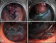
(A) Indigo carmine-stained LST; (B) dissection; (C) totally resected lesion showing points of muscle layer detachment; (D) closure with metal clips.
The patient remained hemodynamically stable, and was successfully extubated at the end of colonoscopy. When the procedure was over, an extensive subcutaneous emphysema throughout the right half of the body was noted, but without clinical consequence. The abdomen was flaccid and painless and the patient did not complain of dyspnea nor chest pain. An immediate workup was performed with chest radiography, which showed pneumomediastinum, besides the extensive subcutaneous emphysema. Still in the operative suite, the abdominal radiograph revealed retropneumoperitoneum and pneumoperitoneum (Fig. 2).

The patient was transferred to the intensive care unit in ambient air for monitoring. At that time, she was alert and oriented. The patient remained NPO during 48 h and with intravenous antibiotic therapy. The computed tomography for control, performed in 48 h, showed pneumomediastinum, pneumoperitoneum, pneumoretroperitoneum, air delamination of rectal walls and minimum amount of perihepatic free fluid and of fluid among bowel loops (Fig. 3).

(A) pneumomediastinum; (B) pneumoretroperitoneum, pneumoperitoneum and subcutaneous emphysema.
The patient remained clinically asymptomatic and stable. Therefore, we proceeded with conservative treatment. The patient tolerated a restricted liquid diet by the third day after the procedure; on the fifth day, the patient received a bland diet. On the fourth day, she produced soft stools without right red blood or mucus, and was discharged on the sixth day of progression. At the outpatient visit, 15 days after the procedure, the patient was clinically well and reported no complications in this period.
The histopathological examination of the sample revealed tubulovillous adenoma with low-grade dysplasia and foci of high-grade dysplasia.
Discussion
The literature reports several cases of pneumoperitoneum during ESD and an increasing number of air leakage into the abdominal retrocavity in rectal endoscopic approaches. Anatomically, this space is contiguous with the mediastinum and the subcutaneous space.88. Sato K, Itoh S, Shigiyama F, Kitagawa T, Maetani I. Pneumoretroperitoneum, pneumomediastinum and subcutaneous emphysema after colorectal endoscopic submucosal dissection (ESD) with air insuflation. J Interv Gastroenterol. 2011.,99. Park NS, Choi JH, Lee DH, Kim YJ, Kim ES, Jung SW, et al. Pneumoretroperitoneum, pneumomediastinum, pneumopericardium, and subcutaneous emphysema after colonoscopic examination. Gut Liver. 2007.
In our case, the non-occurrence of a gross perforation (as in other reports) is interesting, but only the divulsion of circular muscle fibers. Even so, metal clips were applied, but this was enough for the air leak. It is very important to note that the insufflation of carbon dioxide (CO2) is less harmful to the patient, by being an easily absorbed and rapidly eliminated gas, unlike the ambient air.1010. Saito Y, Uraoka T, Matsuda T, Emura F, Ikehara H, Mashimo Y, et al. A pilot study to assess the safety and efficacy of carbon dioxide insufflation during colorectal endoscopic submucosal dissection with the patient under conscious sedation. Gastrointest Endosc. 2007;65:537–42. Unfortunately this technology is not available in our service.
Japanese centers with large series of ESD reported incidences of perforation between 1.4 and 10.4%, without regard to the location of the lesion, but with its size and the presence of fibrosis. Clamps of coagulation are the primary cause of perforation.1111. Yoshida N, Wakabayashi N, Kanemasa K, Sumida Y, Hasegawa D, Inoue K, et al. Endoscopic submucosal dissection for colorectal tumors: technical difficulties and rate of perforation. Endoscopy. 2009;41: 758-61. The perforation site should be clipped once it has been identified; and in presence of abdominal distention, paracentesis decompression should be performed. In our case, this procedure was not necessary, since at the end of the procedure the patient had a flabby and painless abdomen.1212. Zhou PH, Yao LQ, Qin XY Endoscopic submucosal dissection for colorectal epithelial neoplasm. Surg Endosc. 2009;23:1546-51. Some cases of perforation are not detected by endoscopy, but only upon the control radiograph or CT. Most perforations are treated conservatively1313. Yoshida N, Yagi N, Naito Y, Yoshikawa T. Safe procedure in endoscopic submucosal dissection for colorectal tumors focused on preventing complications. World J Gastroenterol. 2010;16:1688-95.
Despite technical difficulties and an extensive learning curve, ESD should be encouraged, because the ultimate goal is the reduction of sequels left by other more vigorous approaches to cancer.
Conclusion
In ESD-treated patients, microperforations can be treated by conservative procedures, provided that the hospital count on a full-time multidisciplinary team and propaedeutic and therapeutic care, including a team for immediate surgical approach in case of adverse developments.
REFERENCES
-
1Fukuzawa M, Gotoda T. History of endoscopic submucosal dissection and role for colorectal endoscopic submucosal dissection: a Japanese perspective. Gastrointest Interv. 2012.
-
2Saito Y, Uraoka T, Matsuda T, Emura F, Ikehara H, Mashimo Y, et al. Endoscopic treatment of large superficial colorectal tumors: a case series of 200 endoscopic submucosal dissections (with vídeo). Gastrointest Endosc. 2007.
-
3Yoshida N, Wakabayashi N, Kanemasa K, Sumida Y, Hasegawa D, Inoue K, et al. Endoscopic submucosal dissection for colorectal tumors: technical difficulties and rate of perforation. Endoscopy. 2009.
-
4Hotta K, Fuji T, Saito Y, Matsuda T. Local recurrence after endoscopic resection of colorectal tumors. Int J Colorectal Dis. 2009.
-
5Saito Y, Fukuzawa M, Matsuda T, Fukunaga S, Sakamoto T, Uraoka T, et al. Clinical outcome of endoscopic submucosal dissection versus endoscopic mucosal resection of large colorectal tumors as determined by curative resection. Surg Endosc. 2010.
-
6Tanaka S, Oka S, Chayama K. Colorectal endoscopic submucosal dissection: presente status and future perspective, including its differentiation from endoscopic mucosal resection. J Gastroenterol. 2008.
-
7Fujishiro M, Yahagi M, Kakushima N, Kodashima S, Muraki Y, Ono S, et al. Outcomes of endoscopic submucosal dissection for colorectal epitelial neoplasms in 200 consecutive cases. Clin Gastroenterol Hepatol. 2007.
-
8Sato K, Itoh S, Shigiyama F, Kitagawa T, Maetani I. Pneumoretroperitoneum, pneumomediastinum and subcutaneous emphysema after colorectal endoscopic submucosal dissection (ESD) with air insuflation. J Interv Gastroenterol. 2011.
-
9Park NS, Choi JH, Lee DH, Kim YJ, Kim ES, Jung SW, et al. Pneumoretroperitoneum, pneumomediastinum, pneumopericardium, and subcutaneous emphysema after colonoscopic examination. Gut Liver. 2007.
-
10Saito Y, Uraoka T, Matsuda T, Emura F, Ikehara H, Mashimo Y, et al. A pilot study to assess the safety and efficacy of carbon dioxide insufflation during colorectal endoscopic submucosal dissection with the patient under conscious sedation. Gastrointest Endosc. 2007;65:537–42.
-
11Yoshida N, Wakabayashi N, Kanemasa K, Sumida Y, Hasegawa D, Inoue K, et al. Endoscopic submucosal dissection for colorectal tumors: technical difficulties and rate of perforation. Endoscopy. 2009;41: 758-61.
-
12Zhou PH, Yao LQ, Qin XY Endoscopic submucosal dissection for colorectal epithelial neoplasm. Surg Endosc. 2009;23:1546-51.
-
13Yoshida N, Yagi N, Naito Y, Yoshikawa T. Safe procedure in endoscopic submucosal dissection for colorectal tumors focused on preventing complications. World J Gastroenterol. 2010;16:1688-95.
Publication Dates
-
Publication in this collection
Oct-Dec 2014
History
-
Received
06 July 2014 -
Accepted
11 Aug 2014


