Abstract
This review shows the structure, mode of action, and actual epidemiological data about EAST1 toxin. It is a particularly intriguing bacterial toxin that may subvert multiple cellular processes to yield intestinal epithelial cell secretion. EAST1 toxin was first described in strains of EAggEC that were associated with persistent diarrhea primarily in developing world countries. Molecular organization, mobility, and data in literature are suggesting that EAST1 could be a transposon. The insertion sequences in Escherichia coli and some of the usual transposition mechanisms as well as regulation are reviewed. This review emphasizes the presence of the gene astA in EPEC, EAggEC, A-EPEC, ETEC, DAEC, EIEC, and in non-diarrheagenic E. coli. It also discusses here the presence of the astA gene in Salmonella spp. and future perspectives for understanding its role in diarrheal disease in both bacterial genera.
EAST1 toxin; astA gene; virulence in Escherichia coli; virulence in Salmonella spp
REVIEW ARTICLE
East1 toxin and its presence in a changing microbial world
C. P. Sousa
Centro de Ciências da Saúde, Universidade Federal da Paraíba Campus Universitário, João Pessoa, Paraíba, Brasil
Address to correspondence Address to correspondence C. P. de SOUZA Centro de Ciências da Saúde, Universidade Federal da Paraíba Campus Universitário, s/n 58 900-740, João Pessoa, Paraíba, Brasil Phone: 55 67 422-7118 prokarya@zipmail.com.br
ABSTRACT
This review shows the structure, mode of action, and actual epidemiological data about EAST1 toxin. It is a particularly intriguing bacterial toxin that may subvert multiple cellular processes to yield intestinal epithelial cell secretion. EAST1 toxin was first described in strains of EAggEC that were associated with persistent diarrhea primarily in developing world countries. Molecular organization, mobility, and data in literature are suggesting that EAST1 could be a transposon. The insertion sequences in Escherichia coli and some of the usual transposition mechanisms as well as regulation are reviewed. This review emphasizes the presence of the gene astA in EPEC, EAggEC, A-EPEC, ETEC, DAEC, EIEC, and in non-diarrheagenic E. coli. It also discusses here the presence of the astA gene in Salmonella spp. and future perspectives for understanding its role in diarrheal disease in both bacterial genera.
Key words: EAST1 toxin; astA gene; virulence in Escherichia coli; virulence in Salmonella spp.
INTRODUCTION
1 Historical introduction
Prolegomenous
The genome of each organism contains not only information for its functioning in the current environment, but its potential to evolve novel functions that will allow it to thrive in alternative environments.
This potential information is subject to a variety of selective constraints that limit the organisms actual ability to create any particular new function.
Today many organisms are evolving rapidly as populations adapt to the huge increase in human populations and associated environmental changes (1).
The world of bacteria represents a large gene pool, which is available for microorganisms to evolve divergently and to adapt rapidly to new niches. Pathogenic bacteria have co-evolved with their hosts. In favor of rapid replication and intergeneric gene exchange, bacteria have developed strategies to resist host defense and to be transmitted efficiently to new hosts (76).
Diarrhea is a major cause of worldwide mortality and morbidity, particularly in young children. For all diarrheal pathogens, there is a sequence of mucosal interactions requiring mucus attachment and penetration followed by interaction with the tissue and elicitation of host damage (103). Virulence properties are produced by pathogens to allow them to breach infection barriers and execute this sequence. Given that pathogens are defined by reference to their hosts, opportunities of living as parasites or saprophytes lie well into the future, since multi-cellular creatures had yet to evolve (112).
Escherichia coli
E. coli maybe the most versatile of human pathogens. This bacterium is not only the dominant Gram-negative facultative anaerobe in human gastrointestinal tract; it is also a potent pathogen capable of causing a variety of diseases by a dizzying array of mechanisms. E. coli can cause no less than six clinical syndromes of diarrhea with overlapping, but distinct symptoms and epidemiology; it can cause urinary tract infections ranging from asymptomatic bacteriuria to urosepsis; and it can cause neonatal meningitis, pneumonia, cholecystitis, and wound infections.
The ability of a single species to co-exist with its host as a symbiont and to cause so many distinct diseases is a property of the organism genetic heterogeneity.
The E. coli population is composed of commensal organisms that live harmlessly in the gut, opportunistic pathogens that occasionally infect normally sterile tissues, and specialized pathogens that possess specific virulence attributes promoting organism ability to cause disease (101).
While much of the E. coli genome is common to all strains of the species, the various clinical syndromes described to E. coli are caused by pathotypes that differ from each other and from commensal strains because they have acquired distinct sets of virulence genes. These genes are carried on plasmids, or lysogenic bacteriophages, or on large chromosomal insertions known as pathogenicity islands. The distinctive clinical syndromes recognized in patients infected with different E. coli pathotypes are a direct result of interactions with the host encoded by the various gene set combinations (54,65,84,108).
Presumably, these combinations of virulence genes were not selected by virtue of their ability to harm humans; these genes, however, endow the organism with the ability to occupy distinctive niches within (or between) hosts. Unfortunately, little is known about the precise selective advantage provided to the organism by the various sets of virulence genes. Despite these attempts at organization, it is important to keep in mind that the free flow of genetic information in nature is a force that complicates our efforts to identify individual E. coli strains as members of one or another pathotype. Strains that share virulence features with more than one group have been identified and will likely be recognized with increasing frequency. The species is not static, but is constantly evolving in leaps and bounds by the acquisition of new genetic determinants (42).
Diarrheagenic Escherichia coli strains are major pathogens associated with enteric disease in many parts of the world. Besides the virulence potential of particular E. coli strains, it is well known nowadays that some E. coli categories have the ability to elaborate and express potential virulence factors, such as adhesins and toxins (66).
Enteroaggregative E. coli (EAggEC) have been implicated as an emerging cause of pediatric diarrhea in the developing world (66) and as the causative organism in a number of outbreaks in the developed world (47,95).
EaggEC-mediated diarrhea is predominantly secretory in nature; stools contain mucus and often blood but generally no polymorphonuclear leukocytes (66).
Although enterotoxins (EAST1, PET, and PIC) have been described in EAggEC (23,24,85), the EAggEC pathogenesis has not yet been completely described.
The first evidence of enterotoxins elaborated by the E. coli pathotype EAggEC was presented by Savarino et al.(85). These authors described a homologue of the ETEC ST toxin (designated EAST1 for enteroaggregative thermostable enterotoxin), which is produced by about 40% of EAggEC strains associated with persistent diarrhea in children in Chile.
What are toxins ?
Toxins and other virulence determinants are mechanisms for gaining access to environments in our bodies and to the nutrients sequestered within them, for releasing these nutrients in usable form, and for moving toxins and nutrients to new hosts when they are expended.
It is important to note that toxins are substances that are able to induce host damage (59). Although many microorganisms secrete toxins that conform to these general criteria, only relatively few have in experiments actually been shown to influence pathogenicity of the producing organism (110).
Probably the definition of toxins could be that they are components or products of microorganisms which, when extracted and introduced alone into host animals, can reproduce disease symptoms normally associated with infection without infestation by their original microbes (110).
Pathogenic bacteria produce many substances that are directly or indirectly toxic to host cells. Secreted microbial proteins, usually enzymes that kill host cells at very low concentrations are exotoxins. Other non-proteinaceous toxic bacterial substances, such as lipopolysaccharide (LPS) or endotoxin, do not use a direct enzymatic mechanism to injure host cells and are biologically active at higher concentrations. Endotoxin is a critical component of the Gram-negative cell wall (30).
A-B toxins have two components: i) the B subunit(s), which mediates binding to the host cell receptor and facilitates delivery of the toxin into the host cell, and ii) the A subunit, which contains the toxic activity.
The A toxic subunit ranges from ADP-ribosylating activity (e.g., Vibrio cholerae, Bordetella pertussis, and Corynebacterium diphtery toxins) to proteolytic activity (e.g., Clostridium botulinum, and C. tetani toxins).
Parts of the A subunit are often conserved, especially the regions that are critical for enzymatic activity, although the B domain often show variations, presumably to provide host and tissue binding specificity (30).
The pore-forming toxins function by insertion into the host cell plasma membrane, which causes the formation of a pore or channel, leading to lysis of the host cell. This RTX family is found in many Gram-negative pathogens (107). Other families of pore-forming toxins have been described, such as the family of sulfhydryl-activated cytolysins, found in Gram-positive microorganisms.
A family of secreted bacterial products contains the immunoglobulin A (IgA) protease-type proteins, found in several Gram-negative pathogens. These bacteria encode their own auto-transporters.
Another family of conserved toxins contains heat-stable toxins (86). These toxins activate host guanylate cyclase and share homology with guanilin, an endogenous activator of intestinal guanylate cyclase (18), which may indicate an evolutionary relationship between these molecules or, alternatively, a convergent evolution of a virulence factor that exploits an existing host cell function.
Mechanisms of action of some important bacterial toxins
Table 1 shows the major toxins involved in classic toxinoses in which toxin production is an important virulence determinant and for which the mode of action is well known.
Microbial protein toxins are generally described as exotoxins to distinguish them from bacterial endotoxin, the lipid A moiety of lipopolysaccharide O antigens.
Endotoxin is an integral component of the Gram-negative outer membrane that plays an essential structural role in cellular survival and division. Its toxic activity is minimal during normal bacterial growth, and there is no significant toxin release into the surrounding environment. The power of this kind of toxin is seen when endotoxin is released by cell lysis, and the lipid A portion stimulates a massive immune response resulting in septic shock that is often fatal to the host (44).
Endotoxin-induced stimulation of the immune system bactericidal activity will exert strong selective pressure for antigenic variation to allow at least low-level survival.
Exotoxins, on the other hand, may be described as secreted proteins whose site of action may be some distance from the site of infection, and which exert their specific effects at low concentrations, often by enzymatic activities of some kind. Exotoxin implies the concept of a protein found outside the bacterial cell, and many are indeed actively exported during growth. The major exotoxins are those that i) cause damage to the cell membrane and ii) enter host cells and exert their toxic activities against intracellular targets (Table 1).
Epidemiology and prevalence of EAST1 sequence
It has been recognized for some years that certain E. coli strains (85,86,88,100,113,115), more recently Salmonella spp. (99,101), are able to produce a low molecular weight heat-stable enterotoxin, active in an Ussing-chamber model.
Using colony hybridization, Savarino et al. (88) have shown that the astA gene occurs in all strains of E. coli O157:H7, in 41% of EAggEC and ETEC, and in 22% of EPEC. Furthermore, the probe detected the EAST1 sequence in 22% E. coli strains isolated from asymptomatic children. These authors have shown that the DNA probe did not react with strains of EIEC, or Yersinia enterocolitica or Vibrio cholerae strains.
Sousa (99) using PCR and colony hybridization has shown the distribution and expression of the astA gene (EAST1 toxin) in 345 strains of the Enterobacteriaceae family including Escherichia coli [205], Salmonella [42], Shigella [13], Edwardsiella [10], Klebsiella [11], Proteus [21], Morganella [13], Citrobacter [2], and Enterobacter [28]. The gene was found in 28.3% and 11.9% in samples of E. coli and Salmonella spp., respectively.
Sousa (99) has shown that most of positive samples of E. coli categories were found in EHEC (88%), EAggEC (86.6%), and A-EPEC (58.3%). The astA gene was also found in 16.6% of E. coli samples without virulence genes.
There was no significant variation in gene astA presence between serotypes of each E. coli category. In 32 strains of EIEC, two serotypes (O144:H- and O164:H-) were found that possessed and expressed the gene in Ussing chambers.
Five Salmonella spp. strains possessing the gene were isolated from foods. S. agona had biological activity in Ussing chambers (101). The results of nucleotide sequencing of the PCR product of strain O144:H- belonging to EIEC category, showed that the toxin EAST1 was 100% identical to the enterotoxin of ETEC H10707 and 98% identical to the astA gene of EAggEC. In the amino acid sequence analysis deduced from the nucleotide sequence was 100% identical to the enterotoxin 1 of ETEC H10707, and 96% identical to EAST1 of EaggEC. The difference was only in one amino acid, where Alanin was replaced by Tyrosin (98).
A total of 476 E. coli samples isolated from weaned pigs with diarrhea and/or edema disease were tested for the presence of EAST1 gene by PCR; 31.3% of all isolates carried the EAST1 sequence. Isolation rates of EAST1 positive strains that were ETEC were 35.6% (ETEC being enterotoxin and fimbrial positive strains) or 49.7% (ETEC being enterotoxin positive strains. The results of Choi et al al. (13) may represent an additional determinant in the pathogenesis of E. coli diarrhea in weaned pigs.
It is of concern that strains producing EAST1 and causing human disease belong to a very broad range of O: H serotypes. Sousa (99) and Sousa and Dubreuil (101) described the presence of this gene in S. agona and S. london strains isolated from foods and in S. thyphi isolated from clinical sources. S. agona induces an increase in potential difference in rabbit mucosal tissue mounted in Ussing chambers.
In three separate outbreaks of diarrhea linked to EAST1, it has been reported that a prototype strain in a Minnesota outbreak was an O39:NM E. coli that expressed EAST1 and had the enteropathogenic E.coli gene locus for enterocyte effacement (43).
In a Japanese report, an O non-typeable H10 EAggEC with a plasmid-borne EAST1 gene was associated with an extensive outbreak of severe diarrhea in a school (47). In a second Japanese outbreak, the implicated pathogen was an O166
E. coli that had no other identifiable
E. coli virulence genes (71). In a small volunteer challenge study investigating EAggEC pathogenicity, one of two EAST1 positive EAggEC strains caused diarrhea (67). All these reports suggest the potential importance of EAST1 and indicate that it may be found in association with different virulence factors.
Yatsuyanagi et al. (118) described a water-borne outbreak in Akita, Japan. ETEC strains were isolated from feces of 6 out of 13 patients with food poisoning and 1 out of 4 drinking water samples. E. coli strains harboring astA gene were isolated from 5 out of 13 patients feces and 1 feces sample from the septic tank. All the ETEC strains (O148:H28) possessed both heat-stable enterotoxin (ST) and astA gene. The EAST1 gene was detected on a ca. 80 kb plasmid in the 5 of 7 ETEC strains and on the chromosome in the remaining 2 ETEC strains. Analysis suggests that location of the ST gene was genome in all samples. In conclusion, these authors suggested the possibility that the EAST1 and ST genes might be transposons.
2 Molecular organization, mobility, and adaptation of the astA gene associated with microbial pathogenesis
The importance of colonization and its features have now been studied, and it underscores the importance of the aggregative phenotype. Three possible mechanisms of enteric bacterial pathogenesis are generally accepted: toxin production, invasion, and signal transduction phenotype seen in EPEC. There is, as yet, no evidence of a signal transduction mechanism between EAggEC. Invasion has been described in HEp-2 cells by Benjamin et al. (4).
There is substantial evidence of the presence of enterotoxins elaborated by EAggEC. Strain O42 (O44:H18), which has been shown to cause diarrhea in human volunteers, elaborates the enterotoxin Pet and EAST1 besides the fimbrial antigen aggregative adherence fimbria type II (AAF/II). All these virulence factors are encoded on the 60 to 65 MDa virulence plasmid pAA2.
The EAST1 gene sequence of strains 73-1 (O73:H33) was identical to of ETEC. Therefore, the presence of the EAST1 gene sequence is now not a strict marker of EAggEC.
Pathogenicity studies are required with a new subtype of DAEC (strains 73-1 and 5-2 (O89:H-) that has a novel adhesin and the EAST1 gene (116). These authors showed that the EAST1 gene was cloned into pBR322 as a 2.0 kbp Sal I fragment or a 0.62 kbp Eco RV fragment.
Strain 73-1 and strain 5-2 isolated from patients with diarrhea, had originally been reported to be EAggEC on the basis of its adherence patterns on HEp-2 cells (104).
The two strains had similar properties to those of EAggEC: (i) strains 73-1 and 5-2 adhered to plastic (or glass) cover-slips similarly to EAggEC, and the adherence patterns in HEp-2 cell assays tended to be slightly aggregative; (ii) strains 73-1 and 5-2 adhered well to mucus in human small intestines and human colonic mucosa, similarly to EAggEC (113); (iii) MRHA was detected in L broth better than on L agar, similarly to EAggEC; and (iv) strains 73-1 and 5-2 had sequences of the EAST1 gene (86) originally found in EAggEC strain 17-2 (O3:H2).
The AA phenotype is associated with the presence of a plasmid with 60 to 65 MDa in size and with the expression of one of two distinct aggregative adherence fimbria (AFF/I and AAF/II). The degree of conservation of AA plasmids is an important and unresolved question. Indeed, a highly conserved plasmid could encode a large number of virulence factors that may provide important clues to the pathogenic mechanisms of EAggEC, as well as to its phylogenic origins (20). These authors working with AA plasmid loci of strain O42 demonstrated that many EAggEC strains harbor a partially conserved plasmid and that this plasmid has undergone significant horizontal dissemination between EAggEC clones.
The EAST1 gene family includes one major type of sequence that is widely distributed in diarrhea-associated E. coli strains, i.e., the EAST1 gene sequence of EAggEC strain O42 (115). In addition, there is at least one variant type of sequence with a non-synonymous nucleotide substitution, i.e., the EAST1 gene sequence of EAggEC strain 17-2 (117).
Although major virulence factors have not been characterized in EAggRC, strain O42 but not strains 17-2 was shown to be pathogenic in volunteer experiments (67).
3 Insertion Sequences (IS) in Escherichia coli
A typical E. coli laboratory strain, for example, might contain six copies of IS 1, as well as a number of copies of other insertion sequences. These IS elements are also commonly found on bacterial plasmids and in the chromosome (22).
The presence of these IS elements does not usually have any effect on cell phenotypic properties (apart from inactivating genes by insertion). Why then does the cell tolerate their presence? Could it be impossible for the cell to delete the IS? The Darwinian notion of the Survival of the Fittest would have us believe that evolutionary pressure will tend to eliminate such elements that are not beneficial, since their presence will constitute a metabolic drain on the cell, even if small, due to the cost of replicating such apparently useless DNA. The simplest answer is that these sequences are essentially parasitic and have devised strategies that prevent their elimination (22).
Until now, it is not clear if astA could be disseminated between E. coli categories and in Salmonella spp. via one IS element.
4 Plasmid resistance genes in Escherichia coli
It was assumed that a basic plasmid, having the ability to replicate independently but not carrying any other information, had somehow picked up a resistance gene from the chromosome of a resistant host strain; transfer of this plasmid to an otherwise sensitive strain then produces a selective advantage for that plasmid-carrying strain, and therefore, indirectly, a selective advantage for this new plasmid. As the plasmid moves from one organism to another, it has the opportunity to acquire additional resistance genes, and/or genes involved with virulence factors thus giving rise to a family of plasmids, all originating from the same parental plasmid but containing different combinations of resistance genes (22).
Since this model implies that unrelated plasmids could pick up the same gene independently, this would explain the widespread distribution of certain types of resistance genes, notably a type of b-lactamase. This particular enzyme, known as the TEM b-lactamase, is the commonest type amongst plasmids in the Enterobacteriaceae, being found on plasmids of many different incompatibility groups and also present in many members of the Pseudomonas genus.
The fluidity of the genetic material is subject to a range of larger-scale alterations in its structure, some of which are reversible. These variations can include insertions, transpositions, inversions, and deletions.
More recently, the same gene has been found in connection with plasmid-mediated penicillin resistance in diverse species such as Haemophils influenzae and Neisseria gonorrhoeae.
The reason behind the ubiquity of the TEM b-lactamase became apparent from the discovery that this gene could transpose from one plasmid to another. The basis of this discovery is exemplified by the experiment shown in Figure 1.
. The donor strain has two plasmids, carrying ampicillin and kanamycin resistance genes (bla and aph, respectively). Using a selective medium containing both antibiotics, colonies having a single plasmid carrying both resistance genes are obtained due to transposition of bla to the second plasmid. Dale (22).
The same reason could be applied to the dissemination of the astA gene between E. coli categories and in Salmonella spp. As noted by Salyers and Dixie (81), E. coli, Salmonella spp. and Shigella spp could be included in the same genera. The genetic proximity of Escherichia and Salmonella could be a contributing factor to the acquisition of the gene by Salmonella spp.
A strain of E. coli containing two different plasmids, one with an ampicillin resistance gene and the other conferring resistance to kanamycin, is used as a donor. The recipient strain is sensitive to both drugs, but resistant to nalidixic acid. Ampicillin and kanamycin will therefore select for recipient cells that have received both resistance genes from the donor (F).
Resistance to both antibiotics was transferred at a much higher rate than would be predicted from the rate of independent transfer of the two plasmids. It was also found that the recipients, resistant to both drugs, contained a single plasmid carrying both resistance genes.
From additional evidence, it was deduced that the ampicillin resistance gene had moved (transposed) from one plasmid to the other. The term transposon was coined to signify an element that was capable of such behavior; the astA gene could be a transposon.
This movement of resistance genes can occur not only between two plasmids, but also from plasmid to chromosome and vice versa.
Therefore, it provides and explanation for the observed rapid evolution of resistance plasmids, and also of plasmids that carry genes other than antibiotic resistance that is, virulence markers as toxins.
It is known that other plasmid-borne genes are also known to be transposable on occasions, as it occurs with genes that encode toxins.
5 Genetic mobile elements in E. coli
The structure of a transposon may be simple or complex. For example, the structure of Tn3 consists of about 5000 base pairs and has a short (38 bp) inverted repeat sequence at each end (22). It is, therefore, entirely analogous to an insertion sequence, the most important distinction being that a transposon carries an identifiable genetic marker such as the ampicillin resistance gene bla (which is short for b-lactamase).
Composite transposons may have their flanking IS region in inverted orientation or as direct repeats. The transposon Tn9, for example, which is 2.5 kb long, has a central region coding for chloramphenicol resistance with direct repeats of the insertion sequence IS1. The transposition behavior of such composite elements can be quite complex; the insertion sequences themselves may transpose independently, transposition of the entire region may occur, or recombination between the IS elements can lead to deletion or inversion of the region separating them (22).
The several layers of transposons can occur nested within one another. There is now considerable evidence from comparison of different plasmids that this process has been in large measure responsible for the evolution of the multiple-resistance plasmids or pathogenicity islands plasmid regions that occur in nature, although very often the actual ability of the genes to transpose has since been lost (22).
6 Transposition mechanisms
In considering transposition mechanisms, it is not necessary to distinguish between insertion sequences and transposons, as the same mechanisms apply to both types of transposable elements.
Replicative transposition
Some transposons (or IS) transpose by a replicative mechanism: a copy of the element is inserted at a different site (on the chromosome or on a plasmid), while the original copy is retained. Tn3 provides a good example of such a mechanism.
Transposition from plasmid to chromosome or vice versa can occur in just the same way. The simplified model of the molecular mechanism of transposition accounts for the main features: i) single-strands nicks in the DNA are produced at each 3 end of the transposon in opposite strands. The recipient plasmid is also nicked at two places, on either side of a short target sequence. With some transposons, this site appears to be selected more or less at random, i.e., there is a requirement for a specific sequence, while other transposons have a degree of specificity in their target sequences. The staggered nature of the nicks is the ultimate cause of the target sequence duplication: ii) the free ends of the transposon are joined to the free ends generated by the nicks in the recipient plasmid. At no stage in this process is the transposon itself liberated as an independent molecule; iii) the free 3 ends of the recipient plasmid sequence act as primers for the synthesis of DNA strands, using host replication or repair enzymes.
This synthesis will proceed through the transposon, separating the two strands until it reaches the existing complementary strand to which the new strand will be joined by the action of DNA ligase. This replicative step is responsible for the duplication of both the transposon itself and o target sequence; iv) the product of step (c) is the co-integrate plasmid, which contains two copies of the transposon in the same orientation (direct repeat). A site-specific recombination between the two copies, at the resolution site, mediated by the TnpR resolvase, gives rise to the end-products of transposition: two plasmids each containing a copy of the transposon (22).
Conservative (non-replicative) transposition
Some transposons and some IS do not replicate when they transpose, exhibiting a mode of transposition known as conservative. This occurs with the IS10 and the related transposon Tn10.
It is possible to use a modified version of the same model for these cases. Transposon replication (stage C) does not occur; instead, following the joining of the free ends of the transposon to the nicked recipient plasmid, the previously unbroken donor strands are cut. This releases the recipient replicon, which contains the transposon with a single-stranded regions filled by repair synthesis, while the donor replicon, now linearised by excision of the transposon, is degraded.
An alternative model of non-replicative transposition is the cut and paste process. In this case, the transposon is completely excised from the donor molecule before being attached to the target site. With some transposons, there is some evidence of the existence of small amounts of free transposon DNA. Many transposons appear to use both replicative and non-replicative transposition (22).
In conservative transposition, although the plasmid molecule that acted as the transposon donor is degraded in the process, however, this does not necessarily mean the complete loss of that plasmid from the cell. Since there may be a large number of plasmid copies, the loss of one copy is easily rectified by replication. This can make it very difficult to determine with certainty which type of transposition has occurred.
7 Transposition regulation
An excessive level of transposition is likely to be extremely damaging to the host cell due to the frequent occurrence of insertional mutations plus (in the case of replicative transposition) the accumulation of a large number of transposons or IS. As with conventional parasites, it is not in the best interests of the element to damage its host too much.
Transposable elements therefore use a variety of mechanisms to control their own transposition level, just like Tn3 TnpR protein, which not only acts as a resolvase, but also functions as a repressor of transcription of the transposase gene tnpA.
A less conventional mechanism has been demonstrated with IS1 and elements related to IS3, where a key protein required for transposition is translated from two different reading frames on the mRNA: the ribosomes start reading the mRNA in one frame and then at a defined point are required to shift back one base and continue reading in a different frame.
Since this ribosomal frame shifting occurs infrequently, it ensures that very little functional enzyme will be made.
Yet, another regulatory mechanism is exhibited by IS10, which produces an anti-sense RNA that is complementary to part of the transposase mRNA, and thus inhibits translation of the message (22).
8 Gene activation by transposable elements
In the movement of any genes carried by the transposon, the only consequence of transposition will be the inactivation of the gene into which the element is inserted. With some transposable elements, it is known that the converse can occur; i.e., insertion of the element actually promotes the expression of genes adjacent to the insertion site.
IS10 contains a promoter at the extreme right-hand end that extends to the left into IS10 itself and is known as pIN. This promoter is responsible for the expression of the IS10 encoded transposase. IS10 carries a second promoter at the right-hand end in the opposite orientation, i.e., it extends outwards, and is known as pout (pout is responsible for antisense RNA production). Transcripts originating at pout can continue beyond the end of IS10 and into whatever gene happens to be adjacent in the DNA into which IS10 is inserted. If this gene is located and oriented suitably, it will therefore, be turned on by the presence of IS10 (22).
9 Salmonella phase variation
Salmonella spp. and E. coli have DNA-DNA homology of about 90%. Today, members of these genera could be included in the same category (81).
Salmonella has served many purposes. It has been used as a model organism for studying bacterial metabolism and genetics, as well as bacterial virulence. As a major cause of food-borne disease, it serves as well as E. coli as an indicator of how safe a countrys food and water supplies are. It is also the basis for many of the new oral vaccines. A bacterial toxin that may subvert multiple cellular processes to yield intestinal epithelial cell secretion is particularly intriguing.
The astA gene was also found in strains of S. agona and S. london isolated from foods and in S. typhi isolated from clinical sources. S. agona induces rises in potential difference in rabbit mucosal tissue mounted in the Ussing chambers. The importance of this discovery is a patamar to the comprehension of gene transfer between different bacterial genera, and the importance of clinical and virulence significance of this toxin in human diarrheal disease (99).
A phenomenon of practical significance is the variation observed in the flagellar (H) antigens of Salmonella species.
Most species of Salmonella are capable of producing two different types of antigenically distinct flagella. These are referred as phase 1 and phase 2 H antigens. The cell switches between phase 1 and phase 2 at a significant frequency such that any culture of the strain, although predominantly in one phase, will contain some cells in the opposite phase.
What causes these (and other) reversible changes in the characteristics of a bacterial strain, since the changes are inherited (albeit unstable) it is likely that they do in fact reflect some alteration in cell genetic composition, as opposed to differences in the physiological state or variations in the environment between one cell and another. On the other hand, the frequency of the observed changes and their accurate reversibility indicate that the effect is due to something other than conventional mutation.
10 DNA sequences associated with EAST1 toxin in Brazil
The heat-stable enterotoxin of EAggEC (EAST1) has been described in some diarrheagenic groups of E. coli. The prevalence was determined of genetic sequences associated with this toxin in 124 ETEC strains mainly isolated from children with diarrhea in São Paulo.
The ETEC strains studied showed different toxigenic profiles: LT-I [67 strains], LT-II [4], ST-I [31], ST-II [7], and LT-I/ST-I [15]. The EAST1 probe obtained by PCR using plasmid DNA of prototype strain 17-2 as template and specific primers was labeled with a 32P [d-ATP] and used in colony hybridization assays. EAST1 gene (astA) was identified in 38% of ETEC strains, but similar frequencies of astA were observed in strains isolated either from diarrheal and non-diarrheal stools. A higher prevalence of astA was found in LT-I/ST-I (73%) and LT-I (40%) strains than in ST-I (29%) and ST-II (14%). The occurrence of astA gene in ST-II strains isolated from humans has not been described before. Although astA has been identified at high frequencies, its role in ETEC pathogenicity remains to be elucidated (73).
In the study conducted by Sousa and Dubreuil (101), the astA gene was present in 32.6% of E. coli and in 11.9% of Salmonella spp. strains. The gene was present in 16.6% of E. coli strains without known virulence genes.
For EPEC, 13.7% of the tested strains were astA positive. Of the atypical EPEC, [eae+, bfp-, EAF-] and [eae+, bfp+, EAF-] 46.2 and 72.7%, respectively, were positive for the gene.
Martinez et al. (60) studied 25 DAEC strains isolated from stools of children from São Paulo naturally infected with diarrhea. All these strains were analyzed by PCR, using probes for astA gene, and the results showed that there were four positive strains to the gene and there was activity in Ussing chambers. These authors also show that one negative astA strain, when tested for the fractionated supernatant (fraction above 30kDa), produced greater fluid accumulation when performed in rabbit ileal-loops and cytotoxic activity in Y1 cell monolayers. These results suggest that this strain may produce another toxin different from EAST1 or a derivative of the original.
Seventeen E. coli O111:H12 strains isolated from feces of children with acute diarrhea were studied for their virulence characteristics. These strains did not have gene sequences homologous to the aggregative adherence fimbria I gene and did not react with any of the DNA probes used to detect other virulence gene enteropathogens. Sixteen of these strains had the astA gene and a 60-70 MDa plasmid. The authors suggests that O111:H12 serotype, one of the first E. coli identified in infantile diarrhea, belongs to the enteroaggregative E. coli category (63).
From 125 diarrheal cases and 88 controls, 919 E. coli isolates were tested for adherence to HEp-2 cells. Diffuse, aggregative, chain-like adherence and variants of the aggregative pattern were found in both cases and controls. Only 25% of the isolates hybridized with the EAggEC probe. The aafA, astA, and pet sequences were found in 7.9, 44.7, and 7.9 isolates, respectively. The chain-like adherence isolates reacted with the EAggEC probe (55.6%) and the aggC, astA, and pet gene sequences were found in 66.7, 33.3, and 11.1%, respectively (35).
11 Cell biology of microbe-host interactions
The precise mode of action of bacterial toxin in relation to their effect on host cell biology is not completely understood, but it is also of crucial importance to understand the toxin role in infection pathogenesis. What do these lethal toxins do for the microbe? Do these toxins open a door to the new function of the microbe action? Do the toxins show the microbes what to do when they are in a host niche? How do these specific eukaryotic venoms evolve during prokaryote evolution? Do they also play a role in fashioning the evolution of their target host animal ?
The tendency of the classical cell biologist is to look at a toxin or a product of a virulence gene as purified reagents. The classical microbiologist may view these proteins as protective antigens from the standpoint of vaccine development. The clinician views the toxins as the causative factors of disease. Cellular microbiologists may be interested in all of these facets, but must put the toxin into the biological perspective of infection pathogenesis and the utility of the toxin for the bacterial survival, endurance, and transmission.
Perhaps the major impetus for microbiologists and cell biologists to focus on the study of host-parasite interactions at the most fundamental level arose from the enthusiasm and unexpected findings of the past decade about bacterial entry into cells and the events subsequent to entry (16,30,70).
Genomic analysis revealed that a wide range of bacteria, including plant pathogens and obligate intracellular parasites such as Chlamydia and Rickettsia, have related blocks of genes that distinguish them from their related commensal and non-pathogenic brethren. These blocks of genes are commonly found as contiguous, large DNA chromosomal insertions, called pathogenicity islands (42,54), or as part of an extra chromosomal element, such as a plasmid or bacteriophage.
There is a certain eerie quality to these DNA islands and islets found in the chromosome of pathogens. They appear to have been transmitted by horizontal gene transfer and possess a DNA base composition that is, overall, strikingly different from the bulk of chromosomal genes.
It is as if the DNA island once resided in a microbe distantly related to where it is now found. In virtually all cases, these blocks of genes appear to encode a specialized secretory pathway, which is configured by evolution to dispense specific effector virulence proteins to the bacterial surface or to effect transfer of specific effector proteins through a protein structure that acts as a conduit into the host cell membrane and cytoplasm.
For pathogens such as Salmonella, the proteinaceous appendage assembled on the bacterial surface looks quite like a hypodermic syringe and needle (50). The effector proteins, when delivered in this way, are extraordinarily keyed to interact specifically with elements of the host, leading in most cases, to actin rearrangement, to tyrosine phosphorylation or dephosphorylation of key signaling molecules, or frequently to the induction of apoptosis (26,33).
It is gratifying to see the rapid assembly and disassembly of cytoskeletal elements associated with bacterial entry into cells, or as the bacteria travel through the host cell cytoplasm on a tale of actine assembly (16). How does so tiny an organism precipitate such a rapid and dramatic host cell response? Yet, there was a quick appreciation that the mechanisms at play were the same as that, which normally occurs in such essential host cell functions as phagocytosis, cell division, and adhesion to the extra cellular matrix and substratum. How does the bacterium take over the control of these basic, essential eukaryotic cellular functions?
It is interesting to say that members of the normal bowel flora and the epithelial cells lining the mammalian bowel take to one another biochemically and that each takes a developmental cue from the other (10). To what extent other members of our bacterial flora influence our cells in health and disease is another facet of cellular microbiology, as are the plant-bacterial interactions. It is intriguing that the same family of secretory elements that transfer DNA into the plant cell Agrobacterium is operative in Helicobacter pylori to, directly or indirectly, induce an inflammatory response in human gastric cells, or is the operative to promote the secretion of Bordetella pertussis toxin to underlying ciliated cells of the trachea (12).
Animal parasites are probably no different from their bacterial counterparts in the sense that the primary goal is to replicate sufficiently to become established and, eventually, to be transmitted to a new susceptible host.
We now know about the long history of interactions between pathogens and their hosts throughout evolution microbial exploitation of various trafficking pathways, manipulation of host cell signal transduction pathways, and subversion of receptor and cytoskeletal assembly functions.
Although all pathogens have effects on their hosts resulting in signal transduction events that are defining for immune and pathogens processes, it is becoming increasingly clear that some organisms, exemplified by intracellular pathogens, quite purposefully manipulate the signal transduction pathways of their hosts for their own benefit. Entry of Salmonella into epithelial cells results in Ca2+ mobilization and secretion of interleukin 8 (IL-8) and other pro-inflammatory chemokines (52). Small Ca2+ antagonist compounds have been described as inhibiting IL-8. The LXAY family of oxygenases falls into this category of compounds but have short half-lives, which through chemical modification resulted in the production of stable analogues that inhibit inflammation.
12 E. coli categories that comprises the astA gene
Like most mucosal pathogens, E. coli can be said to follow a peculiar strategy of infection: i) colonization of a mucosal site; ii) evasion of host defenses; iii) multiplications; and iv) host damage.
The most highly conserved feature of diarrheagenic E. coli strains is their ability to colonize the intestinal mucosal surface (66). The presence of surface adherence fimbria is a property of virtually all E. coli strains, and the role of fimbrial structures in adherence and colonization is often inferred rather than demonstrated. E. coli can cause diarrhea using one of these paradigms: i) enterotoxin production (ETEC and EAggEC); ii) invasion (EIEC); and/or iii) intimate adherence with membrane signaling (ETEC and EHEC).
The versatility of E. coli genome is conferred mainly by two genetic configurations: virulence-related plasmids and chromosomal pathogenicity islands. Plasmids and pathogenicity islands carry clusters of virulence traits; yet individual traits may be transposon encoded (ST) (97) or phage encoded (Shiga toxin) (75).
EHEC
Enterohaemorragic E. coli is an emerging pathogen that causes severe disease, such as hemorrhagic colitis and hemolytic uremic syndrome (HUS) (39,77), and non-blood diarrhea in all age groups, with the young and elderly being the most susceptible (49). Besides O157:H7, by far the most important serotype in human disease (39) and the cause of several large outbreaks of disease in North America, Europe, and Japan (49), EHEC strains belong to a restricted number of serogroups (39), among which O111 has been associated with both sporadic cases (65) and outbreaks (8,86) of HUS.
It was found that eight strains isolated in France during an outbreak of HUS showed aggregative adhesion (AA), instead of the typical localized adhesion to HEp-2 (cells and possessed the genetic markers of EAggEC. AA is associated with the presence of large plasmids carrying genes coding for bundle-forming fimbriae (68), and the production of EAggEC heat-stable enterotoxin 1 (EAST1) (86). Fragments from these plasmids have been used as DNA probes (2) or PCR targets (90) for the identification of EAggEC. It is interesting to say that AA and STx production have never been found to be associated with E. coli isolates. The molecular characteristics of these unusual strains O111:H2, designated RD1 to RD8, gave negative results in PCR analyses for the eaeA gene (90) and katP (9). In addition, they did not produce hemolysin and did not hybrididize with the eaeA (48) and EHEC (57) probes, even under low-stringency conditions.
Shiga toxin-producing E. coli O111:H2 strains from an outbreak of hemolytic uremic syndrome showed aggregative adhesion to HEp-2 cells and harbored large plasmids, which hybridized with the enteroaggregative E.coli, probe PCVD432. These strains present a novel combination of virulence factors (Table 2).
One of the most important virulence factors in the pathogenesis of disease due to EHEC is Shiga toxin (Stx), whose family contains two major immunologically non-cross-reactive groups called Stx1 and Stx2. This phage-encoded toxin consists of one A subunit and five identical B subunits. The B subunit pentamer binds to a specific cell surface glycolipid receptor, globotriaosyceramide or Gb3. Shiga toxin enters host cells via clathrin- coated pits and is transported via the trans-Golgi network to the endoplasmic reticulum (82). The A subunit inhibits host cell protein synthesis by an N-glycosidase activity that removes an adenine residue from the 28S rRNA.
The pathogenicity island on the chromosome of EHEC and EPEC includes LEE, which contains the eae gene that encodes the intimin protein that is essential for attachment-effacement (A/E) (7). It appears that EHEC strains evolved from EPEC via acquisition of phage-encoded Shiga toxins (79).
Evidence has been presented showing how EHEC evolved sequentially from an EPEC O55:H7 ancestor by first acquiring the Stx2 gene and then by diverting into two branches (28,69). The strains in one branch are b-glucoronidase and sorbitol negative (the O157:H7 clone) and those in the other are non-motile, but sorbitol and glucoronidase positive (the O157:H- clone). These authors concluded that the Stx2 gene was acquired early and has been evolving in the O157:H7 genome for a longer time than other virulence genes (28). Adhesins also appear to have been acquired via horizontal transfer (109).
In 1996 in Japan, there were large outbreaks and sporadic cases of EHEC with 17,877 people infected. EHEC includes the most prevalent O157:H7 serotype, but also O26, O111, and O145.
EHEC strains were positive for EAST1 gene sequence and its upstream region in hybridization data.
All the EHEC strains possessed the EAST1 gene homologues but with two types of mutations. One of them (type 1 SHEAST) was strongly associated with the large outbreak episodes in 1996, and type 1 SHEAST was found primarily in O157:H7 strains, while type 2 SHEAST was linked to sporadic cases. The sequence homologous to the EAST1 gene was also found in Yersinia pestis and was located within an IS sequence (IS285). The EAST1 gene, type 1 SHEAST, and the EAST1-like sequence of IS285 are theoretically interchangeable with other by simple base substations, indicating that they have originated from a common ancestral sequence.
Finally, these results provide evolutionary evidence of species-species transfer of the EAST1 gene sequence between E. coli and Y. pestis.
Sequence comparison showed that the EHEC sequences are a branch of the EAST1 gene sequence family that showed the cross-species transfer in evolution between E. coli and Y. pestis (114).
EAaggEC
A possible explanation for EAggEC-induced diarrhea and its persistent nature involves mucosal damage, possibly mediated by a secreted cytotoxin. The presence of the large AA plasmid (pAA2) of 042, a proven human pathogen (67) was required for induction of these effects. Eslava et al. (23) have shown that the plasmid pAA2 encodes a high-molecular weight secreted auto transporter protein (Pet) that induces the loss of actin microfilaments and cell rounding of HEp-2 and HT29 cells in culture (69).
Henderson et al. (45), using the IVOC model, found that the mucosal abnormalities induced by the wild-type EAggEC strain, i.e., dilation of the crypt openings, extrusion of colonic enterocytes, development of inter crypt crevices, and loss of apical mucus from globet cells are not induced by a pet mutant.
It cannot be concluded that Pet is itself sufficient to cause the mucosal effects described before. The dose of Pet required to elicit HT29 cell rounding is relatively high (>10nM). Thus, it is likely that other EAggEC factors contribute to mucosal toxicity (45).
The nature of EAggEC-related diarrhea suggests the presence of one or more enterotoxins. Some studies (45) have shown that a substance secreted by EAggEC strain 042 induces ion transport alterations in rat ileal mucosa, consistent with a secretory response. This substance (Pet) has a role in the enterotoxin activity of 042, if we consider the significant differences of the DIsc and DPD values observed between 042 culture supernatants and JIF1 culture supernatants. Despite the evidence for enterotoxicity demonstrated by Pet, the significant rises in Isc and PD induced by the mutant suggest the presence of another EAggEC enterotoxin, like EAST1 (86) and Shigella enterotoxin (72).
Henderson (45) has shown an interesting role for Pet in 042-induced mucosal toxicity, yet the results for the T84 cell model besides the Ussing chamber data suggest that other factors also contribute to EAggEC pathogenesis. Pet appears to be expressed by only a minority of EAggEC strains, and whether those strains are of increased virulence is currently being addressed in epidemiological studies.
Certain E. coli strains adhere to tissue culture cell lines with an aggregative pattern and these strains have been termed enteroaggregative E. coli (EAggEC). Strains of EAggEC were shown to produce a plasmid-encoded low molecular weight (2-5 kDa) heat-stable enterotoxin, active in an Ussing chamber model and termed EAST1 (85). The fact that the toxin was not neutralized by an antibody to STa, and that strains did not hybridize with probes to STh or STp let these workers to suspect of a new form of ST.
EAggEC has been associated with persistent diarrhea in young children (17,106), especially in developing countries. Most EAggEC strains harbor a 65-MDa plasmid (pAA), which is required for expression of aggregative adherence fimbriae (AAFs). These structures mediate the defining aggregative adherence (AA) phenotype to HEp-2 cells (68,87) as well as adherence to the colonic mucosa (19). The pAA plasmid is also required for the development of mucosal damage in vitro models (46).
Clinical, volunteer, and animal model studies suggest that EAggEC diarrhea may be due to a secretogenic enterotoxin (106). When tested in Ussing chambers, filtrates from EAggEC strain 17-2 produced an increase in potential difference (PD) and short-circuit current (Isc), attributed by Savarino et al. (85) to a heat-stable, plasmid-encoded enterotoxin of less than 10 kDa in molecular mass (EAST1). So far, there is no data to support a role for EAST1 in EAggEC diarrhea.
Nataro et al. (67) reported that EAggEC strain O42 (serotype O44:H18) caused significant diarrhea in three of five adult volunteers, whereas strains 17-2, 34b, and JM221 (EAggEC of serotypes O3:H2, O?:H?, and O92:H33, respectively) did not induce enteric symptoms. Except for O42, each of these strains expressed AAF/I fimbriae, while EAST1 was produced by strains O42 and 17-2, but not by strain 34b or JM221.
Mathewson et al. (61) had previously reported that strain JM 221 caused mild diarrhea in some adult volunteers, and Tzipori et al., (102) had shown that strains 17-2 and JM 221 were able to cause diarrhea in gnotobiotic piglets. The basis for this strain heterogeneity has not been determined, and these data suggest the presence of unrecognized virulence factors.
A role for cytotoxins in EAggEC disease has been suggested by in vivo and in vitro models (46,66).
EAST1 is a genetically distinct toxin that is structurally related to ST1 and also elevates intestinal cGMP levels (85,86). Up until now, little is known about the pathogenic significance of EAST in diarrhea. In one case-control study, E. coli isolates, genotipically positive for EAST1, were highly associated with diarrhea in Spanish children (105). It was reported that very few of the EAST1 E. coli isolates from this study hybridized with an aggregative adherence DNA probe. Three separate outbreaks of diarrhea linked to EAST1 E. coli have been reported. The prototype strain in a Minnesota outbreak was an O39:NM E. coli that expressed EAST1 and had the enteropathogenic E. coli gene locus for enterocyte effacement (43). In a Japanese report, an O non-typeable :H10 EAggEC with a plasmid-borne EAST1 gene was associated with an extensive outbreak of severe diarrhea in a school (47). In another Japanese outbreak, the implicated pathogen was an O166 E. coli that had no other identifiable E. coli virulence genes (71).
In a volunteer study investigating the pathogenicity of EAggEC, one of the two EAST1 positive EAggEC strains caused diarrhea (67). These findings suggest the potential importance of EAST1 and indicate that it may be found in association with different virulence factors.
Pathogenesis of EAggEC infection is not well understood. E coli categories other than EAggEC, notably the EHEC, A-EPEC have been shown to produce this toxin. The significance of EAST1 in pathogenesis is not well established, although the presence of this toxin in E.coli is found in a high frequency (99,101).
According to the three-stage model of EAggEC pathogenesis proposed by Nataro and Kaper (66), EAST1 might be involved at the third stage where the elaboration of an EAggEC cytotoxin could damage the intestinal cells. It was also suggested that EAST1 toxin was closely associated with an adherence factor, CS31A, between pathogenic bovine E. coli (6,21) Savarino et al. (88) are more in favor of the notion that astA may confer an ecological advantage to intestinal E. coli isolates simply by contributing to pathogenesis.
EAST1-positive EAggEC strains produce more severe diarrhea than EAST1-negative EAggEC in human volunteers probably indicating that the EAST1 may contribute to EAggEC virulence (67).
A-EPEC
Some studies (37,78,92,96) have shown that A-EPEC is probably another EPEC category associated with diarrhea of clinical importance. It is probably very early to try to classify A-EPEC (without the EAF plasmid or with an incomplete plasmid, that is, without the EAF locus). A-EPEC strains could have evolved from an EPEC strain (that lost the EAF plasmid) or from an EHEC strain (that lost the phage that encodes Shiga toxin - Stx).
It was reported that certain A-EPEC isolates possess the astA gene in a high frequency, similar to that found in EAggEC and in EHEC (99-101). Other results suggest similarities between A-EPEC and EHEC (11,38,80).
Another possibility is that these isolates of A-EPEC studied could be the ancestral strain from which the actual strains of EPEC and EHEC originated. Some studies have suggested that A-EPEC is more closely related to EHEC than to EPEC. For example, E. coli belonging to serotypes O26:H- and O26:H11 isolated in Europe and the United States produce Stx (93,109) but in the same serotypes isolated in Brazil, they do not produce Stx (83,94).
The high incidence (58.3%) of EAST1 in A-EPEC samples (101) is probably an indication that these bacteria are more closely related to EHEC than to EPEC.
EPEC
Classical EPEC share the eaeA and bfp genes and the EAF plasmid. They are responsible for endemic diarrhea in children in developing countries throughout the world.
The interactions between EPEC and epithelial cells occur in three stages: i) initial localized adherence mediated by the plasmid-encoded bundle-forming pilus (bfp); ii) signal transduction mediated by secret Esp proteins; and iii) intimate attachment mediated by the outer membrane protein intimin (eaeA). These interactions between EPEC and eukaryotic intestinal cells result in a loss of microvilli and the formation of a cup-like pedestal comprised of actin and other cytoskeletal proteins upon which the bacteria rest (66).
Sousa and Dubreuil (101) have shown that there was no significant correlation among serotypes belonging to each E. coli category regarding the presence of the astA gene. The only exception was found in EPEC, where isolates belonging to serotypes O86:H34, O111:H32 and O127:H40 did not possess the gene. The gene was found in one strain each of O55:H6, O119:H6, O142:H6 and O142:H34 serotypes. Among five strains of O127:H6 serotype three isolates were astA-positive.
It was reported that 14 of 65 strains of EPEC hybridize with the astA probe (88). The significance of the toxin in EPEC pathogenesis is not known. Sousa and Dubreuil (101) have shown a low frequency of the gene in EPEC (13.7%).
ETEC
Enterotoxigenic E. coli strains are a major cause of secretory diarrhea in both humans and animals (5). The basic pathogenic repertoire of ETEC includes species-specific surface adhesions that promote small intestinal cell secretion (57). In ETEC, these virulence determinants are often encoded on transmissible genetic elements (54). ETEC strains are capable of elaborating two types of enterotoxins, a heat-labile (LT) and a heat-stable (ST) toxin. STI is the more predominant of the two different ETEC heat-stable enterotoxins (21) and causes fluid secretion by activating membrane-bound guanylyl cyclase C (91).
The gene astA has been found in ETEC of both human and animal origin (113,115). The nucleotide sequence of a plasmid allele of astA in the prototype ETEC strain H10407 hybridized with an astA probe prompted speculation that the gene may be carried on a transposon (113).
McVeigh et al. (62) have demonstrated that after isolation and characterization of the EAST1 coding region from a virulence plasmid in human-derived ETEC strain 27D (O126:NM, CFA/I, STIb) showing that it i) carries an allele of astA nearly identical to that originally reported from EAggEC 17-2 and ii) expressed enterotoxic activity. Sequence analysis of the toxin coding region revealed that astA is completely embedded within a 1,209-bp ORF (open reading frame), whose coding sequence is on the same strand but in the -1 reading frame in reference to the toxin gene. In vitro expression of the predicted Mr ~46,000 protein product of ORF 1 was demonstrated.
McVeigh et al. (62) extended the observations of Yamamoto and Echeverria (113). These authors have shown that human ETEC often carry multiple genomic copies of astA from two ETEC strains of human origin 27D (O126:NM, CFA/I, STIb) and 83/7 (O8:H10, LT) (88), and that the plasmid allele from one strain, ETEC 27D, expressed toxic activity.
The EAST1 coding sequences of both strains were identical to each other. The gene astA has been found in ETEC of both human and animal origin (6,113,115). The nucleotide sequence of a plasmid allele of astA in the prototype ETEC strain H10407 was found to be nearly identical to that of EAggEC strain 17-2, although expression of enterotoxic activity was not reported (86,113). That multiple genomic fragments of H10407 hybridized with an astA probe prompted speculation that the gene may be carried on a transposon (113).
It was shown that some strains of ETEC could express the EAST1 toxin (88,100) and other heat-stable and heat-labile toxins (88,89). Among ETEC, the astA prevalence varied according to the toxin phenotype. These data reported by other authors (113,115) suggest that EAST1 is of pathophysiological significance. Data by Sousa and Dubreuil (101) showed that 20.7% of ETEC strains possessed the astA gene.
Sousa (98) and McVeigh et al. (62) have isolated and characterized the EAST1 coding region from a virulence plasmid in human-derived ETEC strain 27D (human ETEC isolate; O126:NM, CFA/I, Stib). The authors presented evidence that this astA allele indeed expresses enterotoxic activity. They have also shown that it is located on an insertion sequence (IS) element lying entirely within a transposase-like gene.
Yamamoto and Echeverria (113) reported that the prototype ETEC strain H10407 (human ETEC isolate; O78:H11, CFA/I, STIa, STIb, LT) carries multiple genomic copies of astA, including one on its 92-kb virulence plasmid. Sousa (98) McVeigh et al. (62) examined three additional astA positive ETEC strains to determine if the number of toxin gene varies from strain to strain. Interestingly, strain 27D (human ETEC isolate; O126:Nm, CFA/I, STIb) had a single plasmid and multiple chromosomal restriction fragments that hybridized with the astA probe, showing a similar pattern with H10407. For the other two strains (H10407P = H10407 cured of 92 kb virulence plasmid; and 83-7 = human ETEC isolate; O8:H10, LT), several plasmid and chromosomal bands hybridized with the probe.
Multiple genomic restriction fragments from each of three ETEC strains of human origin showed homology with an EAST1 gene probe (62). A single hybridizing fragment was detected on the plasmid of ETEC strain 27D that also encodes heat-stable enterotoxin Ib and colonization factor antigen I. Sousa (98) and McVeigh et al. (62) have isolated and characterized this fragment showing that it i) carries an allele of astA nearly identical to that originally reported from EAggEC17-2 (O3:H2), and ii) expressed enterotoxic activity. Sequence analysis of the toxin coding region revealed that astA is completely embedded within as 1,209-bp open reading frame (ORF1), whose coding sequence is on the same strand but in the -1 reading frame in reference to the toxin gene. It was demonstrated in vitro expression of the predicted Mr - ~ 46,000 protein product of ORF1. ORF 1 is highly similar to transposase genes of IS285 from Yersinia pestis, IS 1356 frp, Burkholderia cepacia, and IS Rm3 from Rhizobium meliloti. It is bounded by 30 -pb imperfect inverted sequences and flanked by 8-bp direct repeats. Based on these structural features, pathognomonic of a regular insertion sequence, this element was designated IS1414. The overlapping genes of the type suggested by the IS1414 core region seem to offer a most efficient mechanism for intragenomic and horizontal dissemination of EAST1.
In ETEC strain 27D and in H10407, astA and the genes for STIb (estA) and CFA/I (cfaB) were localized to a 92-MDa plasmid. The astA probe hybridized with a 5.5 kb PstI fragment of the large plasmid from both strains. Sousa (98) and McVeigh et al. (62) purified the astA hybridizing PstI plasmid fragment from strain 27D and ligated it into pUC18; the recombinant plasmid was designated pAM1.
The nucleotide sequence of the 1.3 kb region including the astA hybridizing allele was determined. Analysis revealed that this region contains a 117 nucleotide (nt) long ORF predicted to encode a polypeptide of 38 amino acids. Comparing this with that the sequence of the EAST1 structural gene of EAggEC strain 17-2. Madigan (59) and Savarino et al. (86) revealed a single non-synonymous nucleotide sequence difference in codon 21 that resulted in a conserved amino acid substitution (threonine for alanine). The astA gene of ETEC strain 83/7 was amplified using primers that anneal to the 5 and 3 ends of the EAggEC 17-2 astA coding sequence. Sousa (98) and McVeigh et al. (62) have shown that direct DNA sequence analysis of this PCR product is identical to astA of ETEC 27D.
Further analysis of the 1.3 kb DNA sequence from pAM1 revealed that a second, larger 1,209 bp ORF (ORF1) was identified in the same coding region but in the -1 reading frame with reference to astA. ORF1 encodes a predicted protein of 402 amino acids with a calculated Mr of 45,800 and a pI of 9.18. It is bounded by imperfect 30bp inverted repeat IR sequences, and this entire structure is flanked by 8-bp direct repeats.
Results from Sousa and Dubreuil (101) and Choi et al. (13) show that more than 31% of these 476 E. coli isolates carried the gene for EAST1 by PCR. Of these, 44% carried the east1 gene only and 55% carried genes for the fimbrial adhesions or enterotoxins.
DAEC
Diffusely adherent E. coli (DAEC) cause a watery diarrhea syndrome in adults and children outside of infancy (56). It is not clear that all DAEC strains elicit diarrhea, as their link to diarrhea is strictly one of epidemiological association. Indeed, several virulence-related characteristics have been identified. Prototype DAEC strains have been shown to induce finger-like projections that extend from the surface of infected Caco-2 or Hep-2 cells (15). The projections wrap around the bacteria, but do not effect complete internalization. A role for this phenotype in pathogenesis has yet to be demonstrated.
Little is known about the interaction of DAEC with intestinal epithelium. Initial adherence is thought to be by virtue of the fimbrial antigen F1845. Some adhering DAEC then elicit the formation of long cellular processes that extend from the cell surface and partially envelop the attached bacteria without leading to invasion.
The results of Choi et al. (13) show that more than 31% of these 476 E. coli isolates carried the gene for EAST1 by PCR. Of these, 44% carried the east1 gene only and 55% carried genes for the fimbrial adhesions or enterotoxins.
EIEC
Enteroinvasive Escherichia coli (EIEC) are biochemically, genetically and pathogenetically closely related to Shigella spp. Both characteristically can cause an invasive inflammatory colitis, but either may also elicit a watery diarrhea syndrome indistinguishable from that due to other E. coli pathogens.
Some authors (88) have not found the astA gene in 55 EIEC samples studied. However, other authors (99-101) have shown that EIEC serotypes O144:H- and O164:H- transport and express the gene as determined in Ussing chamber.
The pathogenesis of both EIEC and Shigella involves cellular invasion and spread, and requires specific chromosomal and plasmidial virulence genes (57). These two bacteria have been shown to invade the colonic epithelium, a phenotype mediated by both plasmid and chromosomal loci. They elaborate one or more secretory enterotoxins, which may play roles in diarrheal pathogenesis.
Like Shigella, EIEC invade intestinal epithelial cells and lyse the phagosomal vacuole. The bacteria then mediate the nucleation of cellular F-actin in such a way as to propel the bacterium directionally through the cytoplasm. This movement results in lateral spread of the bacterium from cell to cell.
The current model of EIEC pathogenesis comprises: I) epithelial cell penetration; II) lyses of the endocytic vacuole; III) intracellular multiplication; IV) movement through the cytoplasm; and V) extension into adjacent epithelial cells (36).
13 Salmonella spp.
Salmonellae are pathogens with an enormous impact on public health as they infect all types of domestic animals used in human food chain. These pathogens are one of the most prevalent agents causing food-borne diseases in both developing and developed countries.
The genus Salmonella comprises more than 2,600 serovars of Gram-negative facultative anaerobic bacilli.
Salmonella infections lead to a variety of diseases known as salmonellosis. Infection is initiated by consumption of raw or undercooked contaminated animal food or water containing fecal material (29). Diseases caused by Salmonella are divided into two major groups: i) a localized, self-limiting bacterial infection of the intestinal epithelium, known as non-typhoid salmonellosis or gastroenteritis; and ii) a systemic infection known as typhoid salmonellosis or enteric fever (32).
Salmonella infection is initiated after consumption of contaminated food or water. Previous to their arrival in the intestine, bacteria pass through the stomach. Salmonella is rather sensitive to the low acidic pH of the stomach and can elicit an acid tolerance response (ATR), which enables organisms to survive at a decreased pH after exposure to low pH (34). This response seems to play a role in vivo during bacterial transit through the stomach.
Once Salmonella has successfully passed through the stomach, it reaches and colonizes the distal ileum. The preferred portals of entry are specialized cells, M cells, located in the Peyers patches (21).
Penetration and destruction of M cells by Salmonella is followed by recruitment of lymphoid cells and polymorphonuclear phagocytic cells (PMNs), leading to inflammation of the area (21).
Binding of Salmonella to M cells is mediated by specialized fimbriae, lpf (3). Expression of lpf fimbriae occurs by a heritable phase variation mechanism, probably triggered by an intestinal-derived signal.
Salmonella and other enterobacterial pathogens possess a series of transcriptional regulators sensing diverse environmental cues. Tight regulation of virulence-related factors is essential for the pathogen to express, in a proper temporal and spatial manner, specific virulence determinants.
Pathogenicity islands (Pais) are defined as large regions of DNA encoding for virulence determinants (40). These regions are present in pathogenic bacteria but absent in phylogenetically related non-pathogenic bacteria. Pais have in many cases different G+C content than that of the host organism, and are often flanked by specific DNA sequences such as direct repeats, or insertion sequences of transposons or phages. These features indicate genetic horizontal transfer mechanisms as the basis for Pais formation. Insertion sites for many Pais are tRNA loci, which may contain sequences representing target sites for extrachromosomal elements. In most cases, Pais are stable regions, although some show unusually high excision frequencies. Acquisition of Pais provides the receptor organism with traits to colonize new environmental niches. Expression of the acquired DNA necessitates that it be under regulatory control to be stabilized (32,33).
The chromosome of E. coli and S. typhimurium are remarkably conserved. The chromosomal organizations of E. coli and S. typhimurium are generally similar. Some large DNA inserts (pathogenicity islands) were acquired during evolution by an ancestral bacterium (shared with E. coli) (74).
Molecular genetic data suggest that the genera Escherichia and Salmonella are from a common ancestor, and thus, it should not be surprising that virulence genes were exchanged between them via horizontal transfer (25).
All S. enterica serovars carry pathogenicity islands 1 and 2 (SPI-1 and SPI-2), which were acquired via horizontal transfer via horizontal transfer by plasmids or phages (3,109). By comparison of the 16S and 23S rRNA sequence data, the Salmonellae have been shown to be closely related to E. coli and Shigellae (72), with the monophasic salmonellae serovars being adapted to mammals and the dysphasic to reptiles (14).
Regarding their evolution, up to 35 kb of the DNA that encompasses the SP1 region of Salmonella at centisome 63 may have been acquired as a block from another microorganism, as they evolved towards becoming pathogens (33). This evidence is supported by the observation that S. enterica and E. coli contain a high incidence of mutator phenotypes that lead to increased mutation rates and enhanced recombination between the diverse species (53).
A 29-kDa polypeptide enterotoxin has been demonstrated in S. typhimurium with the following features: it cross-reacts with cholera toxin; it activates adenylate cyclase; its preferred host cell receptor is ganglioside GM1; and it is positive in the ileal loop test (74). This suggests that the toxins could play a role in causing the diarrheal part of the Salmonellae syndrome, but their role in intracellular invasion and subsequent pathogenesis is unclear. Production of other cytotoxic proteins has also been reported in nontyphoid Salmonellae (21,50,99,101).
It has been accepted that only E. coli carries the astA gene. Sousa (99) and Sousa and Dubreuil (101), however, show that Salmonella also possesses the gene.
The virulence genes in Salmonella spp. are regulated by many environmental factors; for example, the expression of the inv gene is down-regulated by anaerobic conditions. The expression of spvR, a regulator gene that controls the spv operon, is stimulated by Rpos, a sigma factor that is believed to be produced in response to low concentration of nutrients (41). The PhoP/PhoQ system that regulates the prg and pag genes seems to sense the pH, anaerobic conditions, and carbon concentration. The Fur repressor that is responsible for the regulation of the genes that are iron regulated seems to regulate the acid-tolerant response (31). These environment-controlled mechanisms may explain the presence of the astA gene in Salmonella, and the consequent importance of this gene for this bacterium. The astA gene has probably a significant importance in the genesis of diarrhea disease in Salmonella (99).
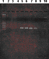
Figure 2 shows the band corresponding to the 111 bp of the PCR amplified product in Salmonella spp.
. PCR amplification results for the Salmonella agona strain. Lane 1 = Molecular size standard (1 Kb DNA Ladder); subsequent lanes represent 50°C, 45°C, and 40°C as annealing temperature, respectively. Lanes 2, 3, and 4 = negative controls; 5, 6, and 7 = positive controls; 8, 9, and 10 = Salmonella agona strain (101).
14 EAST1 toxin in ECOR collection
Results from Lai, Wang, and Uhlin (51) indicate that astA was subjected to some genetic exchanges, such as intragenic recombination, rather than horizontal genetic transfer.
Hemolytic strains resemble a minor category of E. coli called cell detaching E. coli, which are potentially diarrheagenic. Results from this study (51) further prove the relatedness between target cell toxicity and detachment and the expression of highly active hemolysin. In this work (51), the authors found that obvious cell detachment and toxicity correlated well with the expression of highly active a hemolysin, but not with the existence of the astA gene or the silent hlyA gene, under the conditions used.
Since the first astA sequence from EAggEC strain 17-2 was published (86,113,117) and subsequently sequenced, its homologues from other diarrheagenic E. coli show that all their astA genes were nearly identical to each other but differed from the original one (2,17).
Comparison of the astA sequences from EAggEc (117), ETEC (113,115), EPEC (117), EPEC-related E. coli (117), and ECOR collection (51) made it possible to propose a common nucleotide sequence shared by EAggEC, ETEC, EPEC, and ECOR strains as the astA consensus sequence, whilst those which differed in deduced amino acid sequence were variants (including the astA sequence from strains 17-2, N1 and ECOR strains 32, 33 and 35 (51,86,117).
Savarino et al. (86) first designed a synthetic peptide spanning the region of EAST1 from residue 8 to 29 with enterotoxigenic activity. With the functional domain, positions 8-11, 20, 21, 23, and 33 are hot spots for nucleotide changes. It is interesting that sequence analysis of astA from ECOR strains are located in this functional region. At the 8th and 9th (arg) residues, both changes in the third bare do not cause the deduced amino acid substitution, whilst the ala residue at positions 11 and 23 leads to substitutions with theronine and speculate the biological significance of the amino acid change from tiny, hydrophobic, non-polar alanine to the small, less hydrophobic, polar threonine or to the small, aliphatic, less hydrophobic valine. It would also be interesting to check the expression of astA in vivo and in vitro when specific antiserum becomes available, and its activity with the Ussing chamber (86).
Due to the heterogeneous status of the ECOR collection for carrying various virulence factors, it seems appropriate to conclude that some of the ECOR strains of both human and animal origin are potential pathogens with features of all-detaching E. coli (51).
15 Future perspective for the understanding of the role of EAST1 toxin in diarrheal disease
McVeigh et al. (62) worked with culture filtrates from ETEC strains 27D and HB101 (pAM1) for enterotoxic activity by both the suckly mouse bioassay and the in vitro Ussing chamber assay. ETEC 27D culture filtrates induced fluid accumulation in suckling mice and also increased Isc in the Ussing chamber assay. Culture filtrates from HB101 (pAM1) exhibited activity in the Ussing chamber assay, but failed to elicit a response in the suckling mouse assay.
Presence of astA correlated with enterotocic activity in Ussing chamber but not in the suckling mouse assay; this is consistent with earlier findings for the astA allele of EAggEC strain 17-2 (85).
McVeigh et al. (62) have shown that a point mutation was made in astA to look for ablation of toxic activity. Plasmid pAM13 was constructed by introducing a stop codon in place of serine 16 (TGG to TAG); this alternation being silent with respect to ORF1. Culture filtrates from HB101 (pAM13) showed no enterotoxic activity as detected in the Ussing chamber assay. It is interesting to say that this finding indicated that the enterotoxic activity is specifically encoded by astA and not by ORF1.
In this same study, the authors (62) have extended the observations of Yamamoto and Echeverria (113) showing that human ETEC often carry multiple genomic copies of astA. The same observations were found in EHEC serotype O157:H7 (27). They determined the nucleotide sequence of astA from two ETEC strains of human origin, 27D and 83/7, and showed that the plasmid allele from one strain, ETEC 27D, expressed toxic activity. The EAST1 coding sequences of both strains were identical to each other and to that previously reported for prototype ETEC H10407 (113), and were nearly identical to that originally reported for EAggEC 17-2 (86).
McVeigh et al. (62) have isolated and characterized the astA allele from the CFA/I, STIb bearing plasmid containing astA on a 5.5 kb insertion fragment and a 2.4 kb derivative fragment both expressed enterotoxic activity, as shown by electrogenic stimulation of rabbit ileal tissue mounted in Ussing chambers. Mutation of the toxin allele, whereby serine 16 was replaced with an amber codon, resulted in a loss of enterotoxin expression. This mutation was silent with respect to ORF1, an overlapping gene on the same strand but in an alternate reading frame to astA. This finding provides incontrovertible evidence that enterotoxicity is attributable to EAST1 and not to the product of ORF1.
These findings add to the evidence that some ETEC strains have the potential to express EAST1, a factor distinct from other members of its more firmly established arsenal of enterotoxins. With respect to the secretogenic capacity, ETEC strains apparently approach the lost pathogen interface with a considerable degree of redundancy. Whether the ability to produce multiple enterotoxins confers pathogenic advantage to ETEC is not completely understood. Some epidemiological data suggest that strains producing both LT and ST are more likely to cause disease or more severe disease than strains producing only one of these toxins (58). The independent and additive role of EAST1 in ETEC virulence or other diarrheogenic E. coli, if any, may best be addressed by carefully designed epidemiological studies supplemented by studies in the human challenge model with well-defined wild-type strains and EAST1 isogenic derivatives.
The ETEC 27D plasmid allele of astA is part of IS1414, an element with highly characteristic features of an insertion sequence. IS1414 is 1,330 bp long having one ORF (ORF1) that spans its core region with 30-bp imperfect IR sequences at its ends. It is flanked by 8-bp direct repeat sequences that typically arise from duplication of target DNA upon IS insertion. ORF1 encodes a protein that is highly similar to the predicted transposase proteins of IS285 of Y. pestis, IS1356 of B. cepacia, and ISRm3 of R. mililoti. A protein of the same size or that predicted for the product of ORF1 was expressed in vitro from IS1414 DNA template.
In the one region of IS1414, the entirety of the astA gene is embedded in the coding region of ORF1, but in the +1 frame relative to the larger gene. This kind of configuration suggests a remarkably economic arrangement for the storage and dissemination of genetic information of importance to bacterial virulence or survival. It could be compared with DNA fluidity and plasticity in the bacterial chromosome and/or plasmid. It represents the rarest type of overlapping gene and one that, to our knowledge, has not yet been found in a bacterial system. A different overlapping gene arrangement has recently been reported for EAggEC. Two tandem genes identical to those encoding Shigella enterotoxin 1 were found within and on the gene encoding a protein involved in intestinal colonization (Pic) (45).
It is interesting to speculate how IS1414 came to acquire these two overlapping genes. The existence of related IS elements that do not contain the astA sequence imply that acquisition of the toxin gene occurred some time after divergence of the ancestrally related transposase. It is possible that EAST1 then arose by overprinting, using de novo translation of the ORF1 tranposase sequence in a different frame. Origination of a new coding sequence by overprinting predicts unusual codon usage for the new gene. Consistent with this prediction, astA has an atypically low CAI (0.132), indicative of deviation away from preferred E.coli codon usage, while ORF1 shows a greater degree of synonymous codon bias toward E. coli (CAI, 0.362). Bestowal of some advantage to these progeny would favor maintenance and dissemination of the toxin gene, in this case facilitated by IS1414 transposition.
The presence of two overlapping gene pore constraints on evolution impedes the tendency of the individual gene to separate after duplication may be expected. It could be possible that a recombination event in EAggEC strain 17-2 at this transition point led to disruption of ORF1 and loose of integrity of IS1414, without affecting the EAST1 coding sequence.
We can think, at this point, in the particular role of EAST1; if astA expression confers a particular advantage on survival of E. coli expressing additional virulence factors (such as may be the case with EAggEC), degradation of IS1414 function by deletion within the transposase gene might ensure the continued association of astA with after factors encoded by the EAggEC virulence plasmid (20). If, on the after hand, astA expression is neutral in a certain background, where EAST1 activity may be redundant to other toxin, maintenance of IS1414 function might ensure a reservoir for continued dissemination of this gene into new host strains, and to understand this novel mechanism of bacterial virulence evolution.
Yamamoto and Echeverria (113) show that the nucleotide sequences upstream from the EAST1 gene were highly conserved among the CFA/I+ and CFA/II+ ETEC strains. In contrast, the nucleotide sequences downstream from EAST1 gene were somewhat divergent between the downstream sequences on pCS1 and chromosome of sequence of the CFA/I+ ETEC strain different from that of a diffusely adhering E. coli strain, 73-1, by 2 bases.
The authors have also examined the distribution of the EAST1 gene among ETEC strains with various adhesions by PCR. The astA gene was strongly associated with CFD/II+ strains in such a way that 1S CS1+ CS3+, 12CS2+ CS3+, and 10 CS3+ strains were all EAST1-gene positive, irrespective of the toxin type (100%+; n=37). The association of the EAST1 gene with CFA/IV+ (CS6+) strains was also strong (90%+; n=10). For CFD/I+ strains, only 8(53,3%) of 15 strains tested were (+) for the EAST1 gene. The H results (7 strains) were all LTH and STIb+, although one LTH and STIb+ strain was EAST1+.
In contrast, most of CS17+ strains were H for EAST1 (10% positive; n=10). E. coli strains isolated from 50 healthy volunteers were all +1 (0% positive; n=92).
It is interesting to see that in this study, the authors found a strong association of the EAST1 gene with human-colonizing ETEC strains that possessed adhesins such as CFA/II, CFA/III (CS6), and CFA/I.
The association of the EAST1 gene with ETEC strains possessing major adhesins (CFA/I, CFA/II, and CFA/III) (111) together with the fact that multiple copies of the EAST1 gene exist in one of the CFA/I+ ETEC strains, suggests the possibility that the EAST1 gene is an important constituent of ETEC strains that are pathogenic for humans (113). It is also interesting the fact that the EAST1 gene was also present in most animal ETEC strains with K88 adhesin but not in animal ETEC strains with some other adhesins (such as K99). This could indicate the association of the EAST1 gene with the adherence factor type of ETEC. Since the EAST1 gene is widely distributed among different categories of diarrhea-associated E. coli, it may be on a transposon. The EAST1 gene sequences are (or have derived from) a part of the genes involved in transposition events.
16 Conclusions
The essence of the pathogen is in the sequences of the relatively small number of effector molecules. Closely related virulence factors can work within different pathogens because the delivery systems are very similar. Within the specific host species, the kinds of differences that one sees between Salmonella and Shigella, for example, are the expression of distinct differences in effector function. The life of an adapted pathogen, therefore, can be visualized by this scenario as a series of steps of gene activation in response to a complex set of simultaneous environmental conditions (55).
The distinct pathogenic signature, however, is how the microorganism has crafted the effectors of virulence to its own particular utility. Once dismissed as a harmless inhabitant of the intestinal tract, E. coli is now seen as a pathogenic species with remarkable versatility in its ability to cause disease in humans and animals. Pathogen-specific virulence factors have been discovered that adversely affect a wide range of eukaryotic cell processes including protein synthesis, cell division, ion secretion, and transcription (66).
The significance of EAST1 in pathogenesis is unknown. According to the three-stage model of EAggEC pathogenesis proposed by Nataro and Kaper (66), EAST1 might be involved at the third stage where the elaboration of an EAggEC cythotoxin could damage the intestinal cells. Probably, astA may confer an ecological advantage to intestinal E. coli isolates contributing to pathogenesis.
If astA expression confers a particular advantage to the survival of E. coli, and Salmonella expressing additional virulence genes is not yet completely established, and the ability to produce multiple enterotoxins confers pathogenic advantages to bacteria is not entirely clear, but as a prokaryote, they do not expend energy with any advantage to them. So, probably the presence and function of astA sequence might ensure a reservoir for continued dissemination of the gene into new host strains. Comprehension of these phenomena will provide us with hints for the understanding of this mechanism of bacterial virulence evolution.
Received November 11, 2001
Accepted January 10, 2002
- 1 ARBER W. Genetic variation: molecular mechanisms and impact on microbial evolution. FEMS Microbiol. Rev., 2000, 24, 1-7.
- 2 BAUDRY B., SAVARINO SJ., VIAL P., KAPER JB., LEVINE MM. A sensitive and specific DNA probe to identify Enteroaggregative Escherichia coli, a recently discovered diarrhoeal pathogen. J. Infect. Dis, 1990, 161, 1249-51.
- 3 BAUMLER A J., TSOLIS RM., FICHT TA., ADAMS LG. Evolution of host adaptation in Salmonella enterica Infect. Immun., 1998, 66, 4575-87.
- 4 BENJAMIN P., FEDERMAN M., WANKE CA. Characterization of an invasive phenotype associated with enteroaggregative Escherichia coli. Infect. Immun, 1995, 63, 3417-21.
- 5 BERN C., MARTINES J., DE ZOYSA I., GLASS RI. The magnitude of the global problem of diarrhoeal diseases: a ten-year update. Bull. World. Health. Organ., 1992, 70, 705-14.
- 6 BERTIN Y., MARTIN C., GIRARDEAU JP., POHL P., CONTREPOIS M. Association of genes encoding P fimbrial, CS31A antigen and EAST1 toxin among CNF-1 producing Escherichia coli strains from cattle with septicemia and diarrhea. FEMS Microbiol. Lett, 1998, 162, 235-9.
- 7 BOERLIN P., CHEN S., COLBOURNE JK. Evolution of enterohemorrhagic Escherichia coli hemolysin plasmids and the locus for enterocyte effacement in Shiga toxin-producing E. coli. Infect. Immun, 1998, 66, 2553-61.
- 8 BOUDAILLEZ B., PERQUIN P., MARIANI-KURKDJIAN P., BEF D., CUVELIER B., CAPEK I., TRIBOUT B., BINGEN E., PIUSSAN C. Possible person-to-person transmission of Escherichia coli O111- associated hemolytic uremic syndrome. Pediatr. Nephrol, 1996, 1, 36-9.
- 9 BRUNDER W., SCHMIDT H., KARCH H. Katp, a novel catalase-peroxidase encoded by the large plasmid of enterohaemorrhagic Escherichia coli O157:H7. Microbiology, 1996, 142, 3305-15.
- 10 BRY L., FALK PG., MIDTVEDT T., GORDON JI. A model of host microbial interactions in an open mammalian ecosystem. Science, 1996, 273, 1380-3.
- 11 CAMPOS LC., WHITTAM TS., GOMES TAT., ANDRADE JRC., TRABULSI LR. Escherichia coli serogroup O111 includes several clones of diarrheagenic strains with different virulence properties. Infect. Immun, 1994, 62, 3282-8.
- 12 CENSINI S., LANGE C., XIANG Z., CRABTREE JE., GHIARA P., BORODOVSKY M. Cag, a pathogenicity island of Helicobacter pylori, encodes type I specific and disease-associated virulence factors. Proc. Natl. Acad. Sci USA, 1996, 93, 14648-53.
- 13 CHOI C., WAN-SEOB C., HAN-KOOK C., JUNG T., KIM J., CHAE C. Prevalence of the enteroaggregative Escherichia coli heat-stable enterotoxin 1 (EAST1) gene in isolates in weaned pigs with diarrhea and/or edema disease. Vet Microbiol, 2001, 81, 65-71.
- 14 CHRISTENSEN H., NORDENTROFT S., OLSEN JE. Phylogenetic relationships of Salmonella based on RNA sequences. Int. J. Syst. Bacteriol., 1998, 48, 605-10.
- 15 COOKSON ST., NATARO JP. Characterization of HEp-2 cell projection formation induced by diffusely adherent Escherichia coli Microb. Pathog, 1996, 21, 421-34
- 16 COSSART T. Host-pathogen interactions. Subversion of the mammalian cell cytoskeleton by invasive bacteria. J. Clin. Invest, 1997, 99, 2307-11.
- 17 CRAVIOTO A., TELLO A., NAVARRO A., RUIZ J., VILLAFAN H., URIBE F., ESLAVA C. Association of Escherichia coli HEp-2 adherence patterns with type and duration of diarrhea. Lancet, 1991, 337, 262-4.
- 18 CURRIE MG., FOK KF., KATO J., MOORE RJ., HAMRA FK., DUFFIN KL., SMITH CE. Guanylin: an endogenous activator of intestinal guanylate cyclase. Proc. Natl. Acad. Sci. USA, 1992, 89, 947-51.
- 19 CZECZULIN JR., BALELPUR S., HICKS S., PHILLIPS A., HALL R., KOTHARY MH., NAVARRO-GARCIA F., NATARO JP. Aggregative adherence fimbria II, a second fimbrial antigen mediating aggregative adherence in enteroaggregative Escherichia coli. Infect. Immun., 1997, 65, 4135-45.
- 20 CZECZULIN JR., WHITTAM TS., HENDERSON IR., NAVARRO-GARCIA F., NATARO JP. Phylogenetic analysis of enteroaggregative and diffusely adherent Escherichia coli. Infect. Immun., 1999, 67, 2692-9.
- 21 DAOUST JY. Salmonella species. In: DOYLE MP., BEUCHAT LR., MONTVILLE TJ. Eds. Food Microbiology: fundamentals and frontiers. Washington: DC ASM Press, 1997: 129-58.
- 22 DALE JW. Molecular genetics of bacteria. Chichester: John Wiley & Sons, 1994: 287.
- 23 ESLAVA C., NAVARRO-GARCIA F., CZECZULIN JR., HENDERSON IR., CRAVIOTO A., NATARO JP. Pet, an autotransporter enterotoxin from enteroaggregative Escherichia coli. Infect. Immun, 1998, 66, 3155-63.
- 24 ESLAVA C., VILLASECA J., MORALES R., NAVARRO A., CRAVIOTO A. Identification of a protein with toxigenic activity produced by enteroaggregative Escherichia coli In: GENERAL MEETING OF THE ASM, 93, Washington, 1993. Abstracts... Washington: DC, 1993. 44.
- 25 FALKOW S. The evolution of pathogenicity in Escherichia , Shigella and Salmonella In: Neidhradt FC. Ed. Cellular and Molecular Biology 2 ed. Washington: DC ASM Press, 1996: 2723-9.
- 26 FALKOW S. Perspectives series: host-pathogens interaction. Invasion and intracellular sorting of bacteria: searching for bacterial genes expressed during host-pathogen interactions. J. Clin. Invest., 1997, 100, 239-43.
- 27 FASANO A. Cellular microbiology: how enteric pathogens socialize with their intestinal host. ASM News, 1997, 63, 259-65.
- 28 FENG PK., LAMPEL A., KARCH J. Genotypic and phenotypic changes in the emergence of Escherichia coli O157:H7. J. Infect. Dis., 1998, 177, 1750-3.
- 29 FINLAY BB. Molecular and cellular mechanisms of Salmonella pathogenesis. Curr. Top. Microbiol. Immunol, 1994, 192, 163-85.
- 30 FINLAY BB., FALKOW S. Common themes in microbial pathogenicity revisited. Microbiol. Mol. Biol. Rev, 1997, 61, 136-69.
- 31 FRANCIS CL., STARNBACH MN., FALKOW S. Morphological and cytoskeletal changes in epithelial cells occur immediately upon interaction with Salmonellla typhimurium grown under low oxygen conditions. Mol. Microbiol, 1992, 6, 3077-87.
- 32 GÁLAN JE. Molecular genetic bases of Salmonella entry into host cells. Mol. Microbiol, 1996, 20, 263-27.
- 33 GALAN JE. Interaction of Salmonella with host cells through the centrisome 63 type III secretion system. Curr. Opin. Microbiol, 1997, 2, 46-50.
- 34 GARCIA-DEL PORTILLO F., FOSTER W., FINLAY BB. Role of acid tolerance response genes in Salmonella typhimurium virulence. Infect. Immun, 1993, 61, 4489-92.
- 35 GIOPPO NMR., ELIAS JR. WP., VIDOTTO MC., LINHARES RE., SARIDAKIS HO., GOMES TAT., TRABULSI LR., PELAYO JS. Prevalence of HEp-2 cell-adherent Escherichia coli and characterization of Enteroaggregative E. coli and chain-like adherent E. coli isolated from children with and without diarrhoea in Londrina, Brazil. FEMS Microbiol. Lett, 2000, 190, 293-8.
- 36 GOLDBERG MB, SANSONETTI PJ. Shigella subversion of the cellular cytoskeleton: a strategy for epithelial colonization. Infect. Immun, 1993, 61, 4941-6.
- 37 GOMES TAT., VIEIRA MAM., WACHSMUTH IK., BLAKE PA., TRABULSI LR. Serotype-specific prevalence of Escherichia coli strains with EPEC adherence factor genes in infants with and without diarrhea in São Paulo, Brazil. J. Infect. Dis, 1989, 160, 131-5.
- 38 GONÇALVES AG., CAMPOS LC., GOMES TAT., RODRIGUES J., SPERANDIO V., WHITTAM T., TRABULSI LR. Virulence properties and clonal structure of strains of Escherichia coli O119 serotypes. Infect. Immun, 1997, 65, 2034-40.
- 39 GRIFFIN PM., TAUXE RV. The epidemiology of infections caused by Escherichia coli O157:H7, and other enterohemorrhagic E. coli, and the associated hemolytic uremic syndrome. Epidemiol. Rev, 1991, 13, 60-98.
- 40 GROISMAN EA., OCHMAN H. Pathogenicity islands: bacterial evolution in quantum leaps. Cell, 1996, 87, 791-4.
- 41 GULIG PA., DANBARA H., GUINEY DG., LAX AJ., NOREL F., RHEN M. Molecular analysis of spv virulence genes of the Salmonella virulence plasmids. Mol. Microbiol, 1993, 7, 825-30.
- 42 HACKER J., BLUM-OEHLER G., MUHLDORFER I., TSCHAPE H. Pathogenicity islands of virulent bacteria: structure, function and impact on microbial evolution. Mol. Microbiol, 1997, 23,1089-97.
- 43 HEDBERG CW., SAVARINO SJ., BESSER JM. PAULUS CJ., THELEN VM., MYERS LJ., CAMERON DN., BARRET TJ., KAPER JB., OSTERHOLM MT. An outbreak of foodbreak illness caused by Escherichia coli O39:NM, an agent not fitting into the existing scheme for classifying diarrheagenic E. coli. J. Infect. Dis., 1997, 176, 1625-8.
- 44 HENDERSON B., POOLE S., WILSON M. Bacteial modulins: a novel class of virulence factors which cause host tissue pathology by inducing cytokine synthesis. Microbiol. Rev, 1996, 60, 316-41.
- 45 HENDERSON IA., HICKS S., NAVARRO-GARCIA F., ELIAS WP., PHILIPS AD., NATARO JP. Involvement of the enteroaggregative Escherichia coli plasmid-encoded toxin in causing human intestinal damage. Infect. Immun, 1999, 67, 5338-44.
- 46 HICKS S., CANDY DCA., PHILLIPS AD. Adhesion of enteroaggregative Escherichia coli to pedriatic intestinal mucosa in vitro Infect. Immun., 1996, 64, 4751-60.
- 47 ITOH Y., NAGANO I., KUNISHIMA M., EZAKI T. Laboratory investigation of enteroaggregative Escherichia coli associated with a massive outbreak of gastrointestinal illness. J. Clin. Microbiol, 1997, 35, 2546-50.
- 48 JERSE AE., YU J., TALL BD., KAPER JB. A genetic locus of EPEC necessary for the producing of attaching and effacing lesions on tissue culture cells. Proc. Natl. Acad. Sci USA, 1990, 87, 7839-43.
- 49 KAPER JB. Enterohemorrhagic Escherichia coli. Curr. Opin. Microbiol., 1998, 1, 103-8.
- 50 KUBORI T., MATSUSHIMA Y., NAKAMURA D., URABIL J., LARA-TEJERO M., SUCHAN A., GALAN JE., AIZAWA SI. Supramolecular structure of the Salmonella typhimurium type III protein secretion system. Science, 1998, 280, 602-5.
- 51 LAI XH., WANG SY., UHLIN BE. Expression of cytotoxicity by potential pathogens in the standard E. coli collection of reference (ECOR) strains. Microbiology, 1999, 145, 3295-303.
- 52 LAMMEL C., STEPHENS RS. A cell biology approach to microbial pathogenesis. Cell. Microbiol, 1999, 1, 75-7.
- 53 LCCLERC JE., LI B., PAYNE WL. High mutation frequencies among E. coli and Salmonella pathogens. Science, 1996, 274, 1208-11.
- 54 LEE CA. Pathogenicity islands and the evolution of bacterial pathogens. Infect. Agents Dis, 1996, 5, 1-7.
- 55 LERDEBERG J. Paradoxes of the host-parasite relationships. ASM News, 1999, 65, 811-6.
- 56 LEVINE MM., FERRECCIO C., PRADO V., CAYAZZO M., ABREGO P., MARTINEZ J., MAGGI L., BALDINI MM., MARTIN W., MANEVAL D., KAY B., GUERS L., LIOR H., WASSERMAN SS., NATARO JP. Epidemiologic studies of Escherichia coli diarrheal infections in a low socio-economic level peri-urban community in Santiago, Chile. Am. J. Epidemiol., 1993, 138, 849-69.
- 57 LEVINE MM., XU J., KAPER JB., LIOR H., PRADO V., NATARO J., KARCH H., WACHSMUTH K. A DNA probe to identify EHEC O157: H7 and other serotypes that cause hemorrhagic colitis and hemolytic uremic syndrome. J. Infect. Dis, 1987, 156, 175-82.
- 58 LOPEZ-VIDAL Y., CALVA JJ., TRUJILLO A. Enterotoxins and adhesins of enterotoxigenic E. coli: are they risk factors for acute diarrhea in community? J. Infect. Dis., 1990, 162, 442-7.
- 59 MADIGAN MT., MARTINKO JM., PARKER J. Brock: microbiology of microorganisms. 8 ed. London: Prentice-Hall International (UK), 1997: 986.
- 60 MARTINEZ MB., TADDEI CR., FASANO A., TRABULSI JR. Detection of toxins in diffuse adhering E. coli (DAEC) isolated from children with diarrhea. In: Interscience Conference on Antimicrobial Agents and chemoterapy, 39, San Francisco, 1999. Abstracts...San Francisco: ASM, 1999.
- 61 MATHEWSON JJ., JOHNSON PC., DUPONT HL., SATTERWHITE TK., INSOR DK. Pathogenicity of enteroadherent E.coli in adult volunteers. J. Infect. Dis, 1986, 154, 524-7.
- 62 McVEIGH A., FASANO A., DANIEL A., SCOTT S., JEACIC SL., MOSELEY DC., ROBERTSON SJ., SAVARINO SJ. IS1414, and Escherichia coli insertion sequence with a heat-stable enterotoxin gene embedded in a transposase-like gene. Infect. Immun, 2000, 68, 5710-5.
- 63 MONTEIRO-NETO V., CAMPOS LC., FERREIRA AJP., GOMES TAT., TRABUSI LR.. Virulence properties of Escherichia coli O111:H12 strains. FEMS Microbiol. Lett, 1996, 146, 123-8.
- 64 MORABITO S., KARCH H., MARIANI-KURKDJIAN P., SCHMIDT H., MINELLI F., BINGEN E., CAPRIOLI A. Enteroaggregative, Shiga Toxin-producing Escherichia coli O111:H2 associated with an outbreak of hemolytic-uremic syndrome. J. Clin. Microbiol, 1998, 36, 840-2.
- 65 MOXLEY RA., FRANCIS DH. Natural and experimental infection with an attaching and effacing strain of Escherichia coli in calves. Infect. Immun, 1986, 53, 339-46.
- 66 NATARO J., KAPER J. Diarrheagenic Escherichia coli. Clin. Microbiol. Rev., 1998, 11, 142-201.
- 67 NATARO JP., DENG Y., COOKSON S., CRAVIOTO A., SAVARINO SJ., GUERS LD., LEVINE MM., TACKET CO. Heterogeneity of enteroaggregative E. coli virulence demonstrated in volunteers. J. Infect. Dis., 1995, 171, 465-8.
- 68 NATARO JP., DENG Y., MENEVAL DR., GERMAN AL., MARTIN WC., LEVINE MM. Aggregative adherence fimbriae I of enteroaggregative Escherichia coli mediate adherence to HEP-2 cells and hemagglutination of human erythrocytes. Infect. Immun., 1992, 60, 2297-304.
- 69 NAVARRO-GARCIA F., SEARS C., ESLAVA C., CRAVIOTO A., NATARO J. Cytoeskeletal effects induced by Pet, the serine protease enterotoxin of enteroaggregative E. coli. Infect. Immun., 1999, 67, 2184-92.
- 70 NHIEU GT., SANSONETTI PJ. Mechanism of Shigella entry into epithelial cells. Curr. Opin. Microbiol, 1999, 2, 51-5.
- 71 NISHIKAWA Y., OGASAWARA J., NISHIKAWA A., HELANDER K., HARIKI K. An outbreak of gastroenteritis in Japan due to E. coli 0166. Emerg. Infect. Dis,1999, 171, 465-8.
- 72 NORIEGA FR., LIAO FM., FORMAL SB., FASANO A., LEVINE MM. Prevalence of Shigella entetotoxin 1 among Shigella clinical isolates of diverse serotypes. J. Infect. Dis., 1995, 172, 1408-10.
- 73 NUNES SL., GOMES TAT., VIEIRA MM., GUTH B. Occurrence of DNA sequences associated with EAST1 toxin in ETEC isolated in Brazil. Microb. Pathog, 1999, 92 B, 166.
- 74 OBRIEN AD., HOLMES RK. Protein toxins of E. coli and Salmonella In: NEIDHARDT FC. Ed. E .coli and Salmonella Cellular and Molecular Biology. 2 ed. Washington: ASM Press, 1996: 2788-802.
- 75 OBRIEN AD., THESH VL., DONOHUE-HOLFE A, JACKSON MP., OLSNES S., SANDVIG K., LINDBERG AA., KEUSCH GT. Shiga toxin: biochemistry, genetics, mode of action, and role in pathogenesis. Curr. Top. Microbiol. Immunol, 1992, 180, 65-94.
- 76 PAPADOPOULOS D., SCHNEIDER D., MEIH-EISS J., ARBER W., LENSKI RE., BLOT M.. Genomic evolution during a 10,000-generation experiment with bacteria. Proc. Natl. Acad. Sc USA, 1999, 96, 3807-12.
- 77 PATON AW., RATCLIFF RM., DOYLE RM., SEYMOUR-MURRAY J., DAVOS D., LANSER JÁ., PATON JC. Molecular microbiological investigation of an outbreak of hemolytic-uremic syndrome caused by dry fermented sausage contaminated with Shiga-like toxin producing E. coli. J. Clin. Microbiol, 1996, 34, 1622-7.
- 78 PELAYO JS. Propriedades de virulência de amostras de Escherichia coli enteropatogênica atípica São Paulo: Universidade de São Paulo, Instituto de Ciências Biomédicas, 1997. 132 p. [Tese - Doutorado].
- 79 PERNA NT., MAYHEW GF., POSFAI G. Molecular evolution of a pathogenicity island from enterohemorrhagic E. coli O157:H7. Infect. Immun, 1998, 66, 3810-7.
- 80 RODRIGUES J., SCALETSKY ICA., CAMPOS LC., GOMES TAT., WHITTAM TS., TRABULSI LR. Clonal structure and virulence factors in strains of Escherichia coli of the classic serogroup O55. Infect. Immun, 1996, 64, 2680-6.
- 81 SALYERS A., DIXIE D. Bacterial pathogenesis: a molecular approach New York: ASM Press, 1994: 418.
- 82 SANDVIG K., VAN DEURS B. Endocitosis and intracellular sorting of ricin and Shiga toxin. FEBS Lett, 1994, 346, 99-102.
- 83 SARIDAKIS H.O. Non-production of Shiga-like toxins by Escherichia coli serogroup O26. Rev. Microbiol., 1994, 25, 154-5.
- 84 SANGER JM., CHANG R., ASHTON F., KAPER JB., SANGER JW. Novel form of actin-based motility transports lacter on the surface of infected cell. Cell. Motil. Cytoskeleton, 1996, 34, 279-87.
- 85 SAVARINO SJ., FASANO A., ROBERTSON DC., LEVINE MM. Enteroaggregative Escherichia coli elaborate a heat-stable enterotoxin demonstrable in vitro rabbit intestinal model. J. Clin. Invest., 1991, 87, 1450-5.
- 86 SAVARINO SJL, FASANO A., WATSON J., MARTÍN M., LEVINE MM., GUANDALINI S., GUERRY P. Enteroaggregative Escherichia coli heat-stable enterotoxin 1 represents another subfamily of E. coli heat-stable toxin. Proc. Nat. Acad. Sci. USA, 1993, 90, 3093-7.
- 87 SAVARINO SJ., FOX P., DENG Y., NATARO JP. Identification and characterization of a gene cluster mediating enteroaggregative Escherichia coli aggregative adherence fimbria I biogenesis. J. Bacteriol., 1994, 176, 4949- 57.
- 88 SAVARINO SJ., McVEIGH A., WATSON J., MOLINA J., CRAVIOTO A., ECHEVERRIA P., BHAN MK., LEVINE MM., FASANO A. Enteroaggregative Escherichia heat-stable enterotoxin is not restricted to enteroaggregative E. coli. J. Infect. Dis., 1996, 173, 1019-22.
- 89 SAVKOVIC SD., KOUTSOURIS A., HECHT G. Attachment of a noninvasive enteric pathogen enteropathogenic Escherichia coli, to cultured human intestinal epithelial monolayers induces transmigration of neutrophils. Infect. Immun, 1996, 64, 4480-7.
- 90 SCHMIDT H., BEUTIN L, KARCH H. Molecular analysis of the plasmid-encoded hemolysin of Escherichia coli O157:H7 strain EDL 933. Infect. Immun, 1995, 63, 1055-61.
- 91 SCHULZ S., LOPEZ MJ., KULM M., GARBERS DL. Disruption of the guanylyl cyclase-C gene leads to a paradoxical phenotype of viable but heat-stable enterotoxin-resistant mice. J. Clin. Investig, 1997, 100, 1590-5.
- 92 SCOTLAND SM., SMITH HR., ROWE B. Escherichia coli 0128 strains from infants with diarrhea commonly show localized adhesion and positivity in the fluorescent actin staining test but do not hybridize with an enteropathogenic Escherichia coli adherence factor probe. Infect. Immun., 1991, 59, 1569-71.
- 93 SCOTLAND SM., WILLSHAW GA., SMITH HR., ROWE B. Properties of strains of Escherichia coli O126:H11 in relation to their enteropathogenic or enterohemorrhagic classification. J. Infect. Dis, 1991, 162, 1069-74
- 94 SILVA MLM., SCALETSKY ICA., VIOTTO LH. Non-production of cytotoxin among enteropathogenic strains of Escherichia coli isolated in São Paulo, Brazil. Rev. Microbiol, 1983, 14, 161-2
- 95 SMITH HR., CHEASTY T., ROWE B. Enteroaggregative Escherichia coli and outbreaks of gastroenteritis in UK. Lancet, 1997, 350, 814-5.
- 96 SMITH HR., SCOTLAND SM., STOKES N., ROWE B. Examination of strains belonging to enteropathogenic Escherichia coli serogroups for genes encoding EPEC adherence factor and Vero cytotoxins. J. Med. Microbiol, 1990, 31, 235-40.
- 97 SO M., CcCARTHY BJ. Nucleotide sequence of transposon Tn 1681 encoding a heat-stable toxin (ST) and its identification in enterotoxigenic Escherichia coli strains. Proc. Nat. Acad. Sci. USA, 1980, 77, 4011-5.
- 98 SOUSA CP. Distribuição e Expressão do gene astA (toxina EAST1) em enterobactérias São Paulo: Universidade de São Paulo, Instituto de Ciências Biomédicas, 1998. 112p. [Tese Doutorado].
- 99 SOUSA CP. Frequency and expression of the astA gene (EAST1 toxin) in Escherichia coli and Salmonella In: GENERAL MEETING OF AMERICAN SOCIETY FOR MICROBIOLOGY, B/D151, Chicago, 1999. Abstract... Chicago: ASM, 1999, 85.
- 100 SOUSA CP., CAMPOS LC., TRABULSI LR. Frequency of the astA gene (EAST-1 toxin) in strains of different serotypes of diarrheagenic Escherichia coli In: GENERAL MEETING OF ASM, B-252, Miami Beach, 1997. Abstract...Miami Beach: ASM, 1997. 72
- 101 SOUSA CP., DUBREUIL JD. Distribution and expression of the astA gene (EAST1 toxin) in Escherichia coli and Salmonella Int. J. Med. Microbiol, 2001, 291, 15-20.
- 102 TIZIPORI S., MONTANARO J., ROBINS-BROWNE RM., VIAL P., GIBSON R., LEVINE MM. Studies with enteroaggregative E. coli in the gnotobiotic piglet gastroenteritis model. Infect. Immun, 1992, 60, 5302-6.
- 103 TREVORS JT. Evolution of gene transfers in bacteria. World J. Microbiol. Biotechnol, 1999, 15, 1-6.
- 104 VIAL PA., ROBINS-BROWNE R., LIO H., PRADO V., NATARO JP., MANEVAL D., ELSAYED A., LEVINE MM. Characterization of enteroadherent-aggregative Escherichia. coli, a putative agent of diarrheal disease. J. Infect. Dis., 1988, 158, 0-79.
- 105 VILA J., GENE A., VARGAS M. A case-control study of diarrhea in children caused by Escherichia. coli producing heat-stable enterotoxin (EAST 1). J. Med. Microbiol, 1998, 47, 889-91.
- 106 WANKE CA., SCHORLING JB., BARRET LJ., DE SOUZA MA., GUERRANT RL. Potential role of adherence traits of Escherichia coli in persistent diarrhea in an urban Brazilian slum. Pediatr. Infect. Dis. J, 1991, 10, 746-51.
- 107 WELCH RA. Pore-forming cytolysins of Gram-negative bacteria. Mol. Microbiol., 1991, 5, 521-8.
- 108 WHITTAM TS. Genetic variation and evolutionary processing in natural populations of Escherichia coli In: NEIDHARDT FC. Ed. Escherichia coli and Salmonella: cellular and molecular biology. Washington: ASM Press, 1996: 2708-20.
- 109 WHITTAM TS., WOLFE ML., WACHSMUTH IK., ORSKOV I., WILSON RA. Clonal relationship among Escherichia coli strains that cause hemorrhagic colitis and infantile diarrhea. Infect. Immun, 1993, 61, 1619-29.
- 110 WILLIAMS PH., CLARKE SC. Why do microbes have toxins? J. Appl. Microbiol. Symp, 1998, 84, 1S-6S.
- 111 WOLF MK., TAYLOR DN., BOEDEKER EC., HYANS KC., MANEVAL DR., LEVINE MM., TAMURA K., WILSON RA., ECHEVERRIA P. Characterization of Enterotoxigenic Escherichia coli isolated from U.S troops deployed to the Middle East. J. Clin. Microbiol, 1993, 31, 851-6.
Publication Dates
-
Publication in this collection
09 Dec 2003 -
Date of issue
2003
History
-
Accepted
10 Jan 2002 -
Received
11 Nov 2001