 Endovascular management of severe renal trauma: a therapeutic challenge
Abstract
Severe renal trauma is a rare and potentially life-threatening condition that requires rapid and individualized management. Selective embolization has proven to be an effective and minimally invasive option for hemorrhage control and renal function preservation. We present two cases of severe renal trauma, one blunt and one penetrating, successfully treated with superselective embolization. In the first case, microcoils were used for segmental branch occlusion; in the second, cyanoacrylate glue (Hystoacryl + Lipiodol, 33%) was applied. Both patients achieved hemodynamic stabilization, laboratory recovery, and preserved renal function without complications. Endovascular management proved to be a safe and decisive treatment, avoiding nephrectomy and reinforcing the essential role of interventional radiology in the management of high-complexity renal trauma.
Keywords:
renal trauma; therapeutic embolization; interventional radiology; endovascular procedures
Resumo
O trauma renal grave é uma condição rara e potencialmente fatal que requer abordagem rápida e individualizada. A embolização seletiva tem se mostrado uma alternativa eficaz e minimamente invasiva para o controle do sangramento e preservação da função renal. Apresentam-se dois casos de trauma renal grave, um contuso e outro penetrante, tratados com sucesso por embolização superseletiva. No primeiro, utilizaram-se micromolas para oclusão de ramos segmentares; no segundo, aplicou-se cola (Hystoacryl + Lipiodol) a 33%. Ambos os pacientes evoluíram com estabilização hemodinâmica, recuperação laboratorial e preservação funcional do rim afetado, sem complicações. O tratamento endovascular mostrou-se seguro e determinante para evitar a nefrectomia, destacando o papel da radiologia intervencionista como ferramenta essencial no manejo dos traumas renais de alta complexidade.
Palavras-chave:
trauma renal; embolização terapêutica; radiologia intervencionista; procedimentos endovasculares
INTRODUCTION
Renal trauma accounts for approximately 1%-3% of all trauma cases and is most commonly caused by blunt injuries, such as motor vehicle accidents or falls.1 Although most renal injuries can be managed conservatively, severe cases require immediate intervention to control bleeding and preserve renal function. According to the World Society of Emergency Surgery–American Association for the Surgery of Trauma (WSES-AAST) guidelines, management should consider the patient’s hemodynamic status and the anatomical classification of the injury, with selective embolization representing an effective alternative for high-grade injuries (grades III–V).1,2
Selective arterial embolization has emerged asa minimally invasive alternative to open surgery, with technical success rates exceeding 90% and clinical success rates of up to 94.5%, as demonstrated in the multicenter iRESCUE study.3-5 These outcomes reflect advances in microcatheters and embolic agents, which allow greater precision and a lower risk of segmental ischemia.
When selective embolization is not feasible, particularly in extensive or multiple injuries, total renal artery embolization may be performed using temporary agents, such as Gelfoam, or permanent agents, such as PVA particles, microspheres, coils, n-butyl cyanoacrylate (NBCA) glue, and Onyx.6,7
Despite these advances, endovascular management of severe renal trauma remains challenging, requiring individualized decision-making regarding both the technique and embolic agent, as well as close collaboration between surgical and radiological teams.
This article presents two cases of severe renal trauma (one blunt and one penetrating) successfully treated with selective embolization, highlighting critical aspects of decision-making and multidisciplinary management.
PART I – CLINICAL STATUS
Case 1
A 52-year-old previously healthy man was admitted to the emergency department after a high-energy motor vehicle accident (pedestrian-motorcycle collision). On admission, he presented with multiple fractures and was evaluated by a multidisciplinary team, including general surgery and orthopedics, with no immediate indication for surgical intervention. He subsequently developed severe abdominal pain and signs of hemodynamic instability (blood pressure: 90/60 mmHg; heart rate: 125 bpm), in addition to a decline in hematimetric indices (hemoglobin: 8.8 g/dL; hematocrit: 27%).
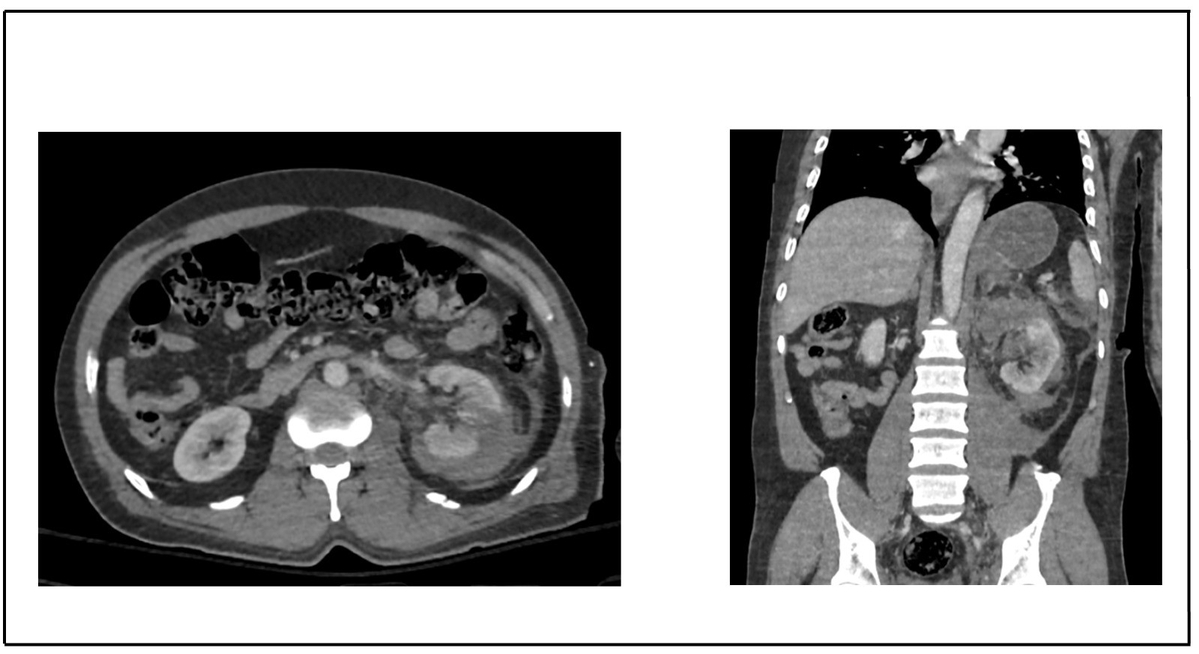
Contrast-enhanced abdominal computed tomography (CT) demonstrated a normally positioned left kidney with poorly defined contours, and heterogeneous density due to hypoenhanced areas involving the entire upper pole and part of the middle third, associated with an extensive perirenal hematoma. A grade IV left renal laceration was diagnosed, without active contrast extravasation (Figure 1).
Contrast-enhanced abdominal computed tomography demonstrating a grade IV renal injury according to the WSES-AAST classification.
Given the radiological findings and hemodynamic instability, arteriography followed by selective arterial embolization was indicated as the initial therapeutic approach.
Case 2
A 22-year-old previously healthy man was admitted to the emergency department after a gunshot wound to the right thoracoabdominal region and right upper limb. On admission, he presented with tachypnea, hemodynamic stability, and hematuria. CT revealed a right-sided pneumothorax and liver injury, and a right-sided closed thoracic drainage procedure was performed.
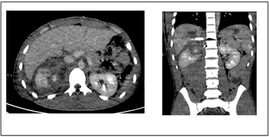
An exploratory laparotomy was subsequently performed, revealing a nonpulsatile, nonexpanding zone 2 retroperitoneal hematoma on the right side, compatible with renal trauma, in addition to diaphragmatic and hepatic injuries, which were treated with diaphragmatic repair and hepatic hemostasis. The retroperitoneal hematoma was considered stable, and therefore was not explored during surgery in order to avoid additional risks, such as increased bleeding or hemodynamic instability. The patient evolved with a decline in hematimetric indices (hemoglobin: 10.7 g/dL → 5.3 g/dL), requiring transfusion of two units of packed red blood cells. A repeat CT scan demonstrated a grade IV right renal injury (WSES-AAST), with a hypoenhanced upper pole and contrast extravasation (Figure 2). Arteriography with the possibility of selective embolization was indicated, considering the patient’s relative stability and active bleeding.
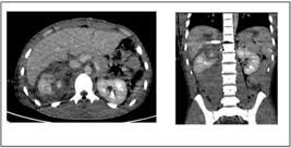
Contrast-enhanced abdominal computed tomography demonstrating a grade IV renal injury according to the WSES-AAST classification.
PART II – WHAT WAS DONE
Case 1
Selective arteriography of the left renal artery, performed using a Mikaelson catheter, revealed vascular amputation and vasospasm in branches of the superior segmental arteries (Figure 3A). Selective microcatheterization was performed using an Echelon microcatheter and an Avigo 0.014” microguidewire, followed by embolization with microcoils, resulting in occlusion of the affected branches (Figure 3BE).
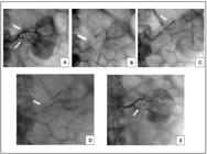
(A) Diagnostic renal arteriography identifying vascular truncation (white arrows); (B) selective microcatheterization of the first injured vessel (white arrow); C) embolization with microcoils (white arrow); (D) selective microcatheterization of the second injured vessel (white arrow); (E) final control arteriography (white arrow).
Following the procedure, the patient showed immediate clinical improvement, with hemodynamic stabilization and relief of abdominal pain, without complications. Laboratory recovery was observed, with maintenance of hematimetric indices and renal function, thereby avoiding laparotomy for nephrectomy. Subsequently, the patient remained under orthopedic follow-up for fracture management and was discharged from the hospital without the need for further endovascular interventions. The patient remains under outpatient follow-up for 1 year, with favorable clinical evolution, without recurrent bleeding or the need for additional interventions.
Case 2
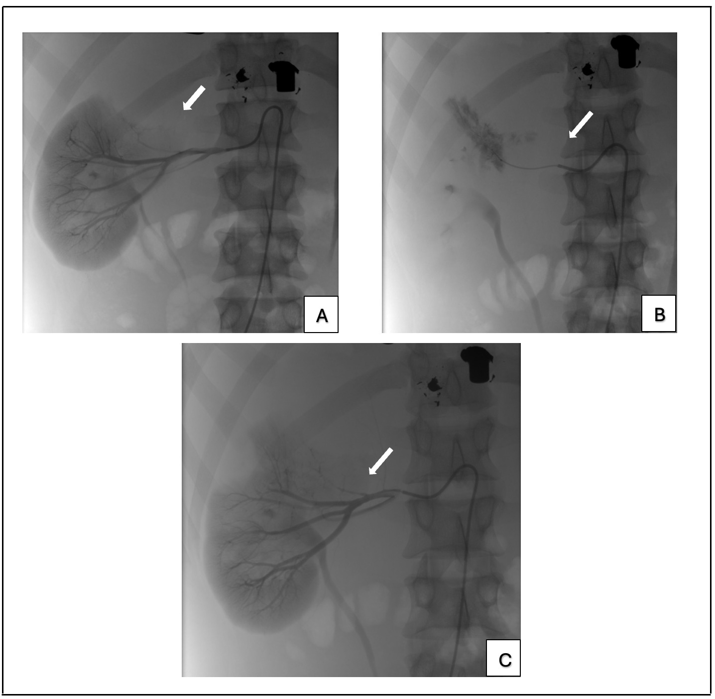
Selective arteriography of the right renal artery, performed using a Simmons 1 catheter, identified vascular irregularity in the superior renal segment and a small contrast extravasation in the right kidney (Figure 4A). Selective microcatheterization of the superior segmental artery was performed using a Rebar 27 microcatheter and a Command 0.014” microguidewire, followed by embolization with 33% glue (Hystoacryl + Lipiodol) (Figure 4BC). Control arteriography demonstrated complete occlusion of the injured artery, with preservation of most of the right renal parenchyma (Figure 4D).
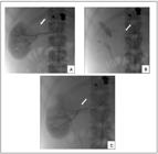
(A) Diagnostic renal arteriography demonstrating vascular injury in the upper segment of the right kidney, with contrast extravasation (white arrow); (B) selective microcatheterization of the affected arterial branch, confirming the site of active bleeding (white arrow); (C) embolization with glue (Hystoacryl + Lipiodol, 33%), resulting in occlusion of the injured vessel (white arrow).
The patient showed immediate clinical improvement, with hemodynamic stabilization and no need for further blood transfusion. He evolved without complications and was discharged from the hospital 72 hours after the procedure, without the need for additional interventions. The patient remains under outpatient follow-up for 2 years, with favorable clinical evolution and no need for further interventions.
DISCUSSION
The cases presented in this study reinforce the fundamental role of selective arterial embolization as a safe and effective strategy in the management of severe renal trauma, providing rapid hemorrhage control, preservation of renal function, and reduction in the need for open surgery.1-4
This approach is particularly valuable in critically ill patients, in whom laparotomy may worsen the clinical condition because of the risk of aggravating hemodynamic instability or precipitating abdominal compartment syndrome, although it does not preclude it from being performed subsequently if indicated.1,8 Endovascular treatment allows total or partial renal preservation in most cases; however, in situations of persistent hemodynamic instability, complete embolization or even nephrectomy may be required as damage-control measures to preserve life.
The WSES-AAST and Brazilian Society of Angiology and Vascular Surgery (SBACV) guidelines recommend endovascular interventions in hemodynamically stable patients with isolated injuries (grades I–IV), prioritizing hemorrhage control with renal preservation whenever feasible.1,8 In more complex scenarios, such as the cases presented in this study, multidisciplinary integration is essential to balance risks and benefits and to avoid complications such as unnecessary ischemia or delays in damage control.8
The choice of endovascular approach should be based on careful assessment of clinical stability, injury grade, and the anatomical conditions of the renal artery.2 In both reported cases, selective embolization was indicated because the injuries were classified as grade IV according to the WSES-AAST classification, with active contrast extravasation in one patient and initial hemodynamic instability in the other. Early decision-making regarding intervention was crucial to therapeutic success and avoided the need for open surgery.
Recent literature reinforces the high efficacy of renal embolization, with consistent results in terms of hemorrhage control and functional preservation.5-7,9 The multicenter iRESCUE study reported high success rates attributed to the use of modern microcatheters and embolic agents, which enable superselective occlusion and reduce complications such as segmental ischemia and postprocedural hypertension.5-7
Embolization outcomes are comparable to conservative management with respect to renal function, including urea, creatinine, and glomerular filtration rate, while presenting a lower risk of surgical complications and chronic kidney disease. However, immediate laparotomy remains indicated in hemodynamically unstable patients.2,10 In the cases described in this study, the early decision to proceed with embolization was possible because of the involvement of multidisciplinary teams, allowing rapid and safe intervention.
The review by Lopez-Gonzalez and Zurkiya11 further supports the role of interventional radiology, highlighting the use of microcatheters and superselective techniques as strategies to precisely treat vascular injuries while minimizing ischemia in unaffected areas. The integration of endovascular and surgical approaches, particularly in patients with contraindications to open surgery or transient hemodynamic instability, expands therapeutic possibilities and improves clinical outcomes.
In both cases, the choice of microcoils and glue (Hystoacryl + Lipiodol) was appropriate for the injury pattern, ensuring complete occlusion of the injured branches while preserving remaining renal perfusion, with rapid clinical recovery and no complications. This approach is consistent with current recommendations, which prioritize superselective embolization in stable patients with high-grade injuries.2,9,10
Despite its benefits, there are risks to embolization. The primary reported complications include segmental ischemia, arterial hypertension, and acute kidney injury.2,4,11 The risk increases with larger contrast volumes and more extensive embolized areas.12,13 In the cases reported in this study, the contrast volume was less than 100 mL, and no renal dysfunction occurred, reinforcing the safety of the procedure when carefully indicated.
Endovascular management of renal trauma requires adequate infrastructure and a trained team, as the interval between diagnosis and intervention is a determining factor for outcomes. The integrated collaboration between the surgical and radiological teams was essential to the favorable outcomes observed in both patients, demonstrating that individualized therapeutic decision-making and the availability of interventional radiology are critical factors for successful treatment.
CONCLUSION
In the cases presented in this study, selective embolization proved to be safe, effective, and decisive for hemorrhage control and preservation of renal function, avoiding the need for nephrectomy and reducing complications. Endovascular treatment proved to be a minimally invasive alternative with excellent outcomes, enabling rapid clinical recovery and early hospital discharge.
These findings reinforce the role of interventional radiology as an essential strategy in the management of severe renal trauma, provided it is performed by a trained team in an adequately equipped setting.
Ethics statement
This study was approved by the Research Ethics Committee of Hospital da Restauração (Opinion Number: 7.848.435; CAAE: 90963625.2.0000.5198). The requirement for a free and informed consent form was waived by the Committee because this was a retrospective study based on medical record review.
DATA AVAILABILITY
All data generated or analyzed during this study are included in this published article and/or its supplementary material.
-
How to cite:
Leal IZ, Galvão RS, Buril GO, Rolim DD, Albuquerque Neto CC, Guedes AJS, et al. Endovascular management of severe renal trauma: a ther-apeutic challenge. J Vasc Bras. 2026;25:e20250201. https://doi.org/10.1590/1677-5449.202502012
-
Financial support:
None.
-
The study was conducted at the Department of Hemodynamics of Hospital da Restauração, Recife, PE, Brazil.
-
Ethics committee approval:
The study protocol was approved by the Hospital da Restauração Research Ethics Committee under registration number CAAE 90963625.2.0000.5198, opinion number 7.848.435.
References
-
1 Coccolini F, Moore EE, Kluger Y, et al. Kidney and uro-trauma: WSES-AAST guidelines. World J Emerg Surg. 2019;14(1):54. https://doi.org/10.1186/s13017-019-0274-x PMid:31827593.
» https://doi.org/10.1186/s13017-019-0274-x -
2 Smith TA, Eastaway A, Hartt D, Quencer KB. Endovascular embolization in renal trauma: a narrative review. Ann Transl Med. 2021;9(14):1198. https://doi.org/10.21037/atm-20-4310 PMid:34430639.
» https://doi.org/10.21037/atm-20-4310 -
3 Rossi UG, Cariati M. Blunt renal artery trauma: a therapeutic dilemma. Turk J Urol. 2018;45(3):233-6. https://doi.org/10.5152/tud.2018.91145 PMid:31846422.
» https://doi.org/10.5152/tud.2018.91145 -
4 Contegiacomo A, Amodeo EM, Cina A, et al. Renal artery embolization for iatrogenic renal vascular injuries management: 5 years’ experience. Br J Radiol. 2020;93(1106):20190256. https://doi.org/10.1259/bjr.20190256 PMid:31794248.
» https://doi.org/10.1259/bjr.20190256 -
5 Öcal O, Puhr-Westerheide D, Mühlmann M, et al. iRESCUE - Interventional embolization of Renal arteries after SurgiCal or traUmatic injury with hemorrhage. Eur J Radiol. 2021;136:109540. https://doi.org/10.1016/j.ejrad.2021.109540 PMid:33476964.
» https://doi.org/10.1016/j.ejrad.2021.109540 -
6 Kopačin V, Turk T, Pavlović O, Perković J, Krajina V, Pavoković D. The role of interventional radiologists in the treatment of complications in urologic patients. Acta Clin Croat. 2023;62(Suppl 2):153-7. https://doi.org/10.20471/acc.2023.62.s2.23 PMid:38966013.
» https://doi.org/10.20471/acc.2023.62.s2.23 -
7 Jardinet T, Bonne L, Oyen R, Maleux G. Initial experience with the microvascular plug in selective renal artery embolization. Vasc Endovascular Surg. 2020;54(3):240-6. https://doi.org/10.1177/1538574419897500 PMid:31928203.
» https://doi.org/10.1177/1538574419897500 -
8 Góes AMO, Parreira JG, Kleinsorge GHD, et al. Brazilian guidelines on diagnosis and management of traumatic vascular injuries. J Vasc Bras. 2023;22:e20230042. https://doi.org/10.1590/1677-5449.202300422 PMid:38021277.
» https://doi.org/10.1590/1677-5449.202300422 -
9 Wang B, Wen C, Song S, et al. Safety and efficacy of Endovascular Management of high-grade blunt renal injury. J Interv Med. 2022;5(1):23-7. https://doi.org/10.1016/j.jimed.2021.12.003 PMid:35586283.
» https://doi.org/10.1016/j.jimed.2021.12.003 -
10 Xu H, Min X, Li Y, Yang L, Ren Y. A comparative study of conservation, endovascular embolization therapy, and surgery for blunt renal trauma. Med Sci Monit. 2020;26:e922802. https://doi.org/10.12659/MSM.922802 PMid:32372763.
» https://doi.org/10.12659/MSM.922802 -
11 Lopez-Gonzalez DB, Zurkiya O. Interventional radiology in renal trauma. Semin Intervent Radiol. 2021;38(1):113-22. https://doi.org/10.1055/s-0041-1726006 PMid:33883808.
» https://doi.org/10.1055/s-0041-1726006 -
12 Piasecki P, Ząbkowski T, Brzozowski K, et al. The assessment of the risk of acute kidney injury in patients undergoing an urgent endovascular treatment due to severe renal bleeding. Cardiovasc Intervent Radiol. 2018;41(3):398-405. https://doi.org/10.1007/s00270-017-1800-4 PMid:29038877.
» https://doi.org/10.1007/s00270-017-1800-4 - 13 Sgalambro F, Giordano AV, Carducci S, et al. The role of interventional radiology in hepatic and renal hemorrhage embolization: single center experience and literature review. Acta Biomed. 2021;92(S5):e2021405. PMid:34505844.
Edited by
-
Editor-in-Chief responsible
Dr. Winston Bonetti Yoshida