 Free-living amoebae: a journey into historical aspects and to current discoveries
Abstract
Free-living amoebas (FLA) are ubiquitous protists found in the environment. They have shown exceptional resistance to environmental challenges and play significant roles in controlling microbial populations through their predatory behaviour and microbicidal activity in both soil and aquatic ecosystems environments. However, although rare, a limited group of FLA can cause serious infections in the central nervous system and other diseases, particularly in immunocompromised individuals with high mortality rates. They can also cause keratitis in otherwise healthy individuals. This review offers a comprehensive overview of freshwater naked amoebae but does not cover all aspects in detail. Its goal is to provide a historical context for our current understanding while addressing the most critical elements of FLA biology, their pathogenic potential, and their interactions with important human pathogens.
Key words:
freshwater amoebae; Acanthamoeba sp.; host-parasite interaction; taxonomy
Amoebae are a group of protists that exhibit irregular shapes by extending pseudopods (cytoplasm expansion), supported by their cytoskeleton, composed of actin-myosin or tubulin elements. These pseudopods are used for movement or capture food.1,2 Amoebae can be found across all branches of the eukaryotic trees,3,4,5 as some amoeboid organisms have genetic relationships closer to plants or metazoans.6 Free-living amoebae (FLAs) are non-monophyletic, single-celled eukaryotic commonly found in natural and artificial aquatic environments, as well as in moist soil and within the microfauna of vertebrates and invertebrates.7,8,9,10,11 Notably, some FLAs can enter a temporary multicellular phase when food is scarce.12 They are extremely diverse and widely distributed across all continents, including the harsh conditions of Antarctica.9,13,14,15 Despite being studied since the advent of microscopy, our understanding of these organisms remains limited.
Revisiting the history of free-living amoeba and reflecting on Aragão’s research and hierarchical classification
The identification and classification of FLAs play a crucial role in understanding their diversity, evolutionary relationships, and ecological significance.9 Research on FLAs has a significant historical legacy that dates to the mid-eighteenth century. In 1755, the German naturalist August Johann Rosel von Rosenhof (1705-1759) provided detailed illustrations and descriptions of a freshwater amoeba through a microscope, naming it “small proteus”, which resembled Amoeba proteus according to his illustrations.16 Later, the Swedish naturalist Carl Linnaeus (1707-1778) classified it as Chaos proteus.17 In 1838, the term “amoeba” was first attributed to this group of organisms by Christian Gottfried Ehrenberg (1785-1876), who established the genus Amoeba in Germany.18 Two years later, Felix Dujardin (1801-1860) described “Limax ameba”, in France.19 In 1838, Christian Gottfried Ehrenberg (1795-1876), initiated the first attempt to classify amoeboid protists, creating sections based on pseudopod structure (lobopoda, filopodia, reticulopodia and axopoda) and classifying families such as Amoebae and Arcellina.20,21,22 Between 1841 and 1858, a new classification was developed that used the term rhizopods to distinguish amoebae based on the presence or absence of shells, separating freshwater from marine representatives. Subsequently, amoeboid protists were primarily classified based on differences in pseudopod structure. The Rhizopoda class was shared into three orders: Lobosa, Radiolaria, and Reticulosa.22,23 Over the subsequent decades, the description of new species, classes, orders, and families, often accompanied by multiple synonyms, led to confusion in distinguishing historical amoeboid species. In 1879, Joseph Leidy (1823-1891) proposed grouping all “common” giant freshwater amoebae into a single species, suggesting the name A. proteus, marking a period of extensive synonymy.24 In 1899, Franz Schardinger (1853-1920) described an amoeba that could transform into a flagellate form, naming it Amoeba gruberi.25 Later, in 1900, French scientist Pierre Augustin Dangeard (1862-1947) described the mitotic process in amoebae, highlighting the complexity of their classification: “nothing is more difficult and challenging than the classification of amoeba”.26 This sentiment was echoed by other researchers,27,28 including Henrique de Beaurepaire Rohan Aragão (1879-1956), who conducted one of Brazil’s first studies on freshwater amoebae. His research, published in the inaugural edition of the Memórias do Instituto Oswaldo Cruz in 1909, meticulously detailed the biology and a unique form of nuclear division in an amoeba isolate found in freshwater near the Oswaldo Cruz Foundation campus laboratory. Aragão observed two distinct nuclear division processes: the first involved small rod-shaped chromosomes scattered irregularly along the spindle, fusing to form a solid mass of chromatin, representing the daughter plates, while the second involved chromosomes arranged in a definitive equatorial plate. He also described the amoeba’s morphological details, including its trophozoite form, size, pseudopodia, nucleus and vacuoles. This amoeba was named Amoeba Diplomitica following microscopic analyses of fresh and stained samples using ferric hematoxylin.29 However, it was not recognised as a valid species according to the rules of that time, as the identified characteristics were insufficient to define a species, especially due to the inappropriate cytological staining technique used.30 Although not officially recognised, Aragão’s research represents one of many similar studies in protozoology, particularly regarding freshwater amoebae (amphizoic and non-parasitic), paving the way for the work of many scientists in understanding the FLAs. Over the years, research has focused on establishing practical identification rules based on comprehensive analyses of morphological data, ultrastructure, and isoenzyme patterns, particularly, regarding amoeba movement, cytoplasmic flow, nuclear structure, division, and life cycle, which significantly contributed to our current understanding of the morphology of FLAs.31-37
In the 1930s, there was a decline in research related to in the FLAs. However, some researchers described the phagocytic capacity of freshwater amoebae in bacterial cultures, as well as their selective phagocytosis of red blood cells in cell cultures.38,39 Cytopathic effects were observed also observed in cell culture, although these were initially considered environmental contaminants.40,41 Subsequent studies revealed the presence of amoebae in human nose, throat, and lung fluids.42,43 When these amoebae were introduced into the noses of mice, they infiltrated the mucous membrane, migrated to the brain, and caused fatal meningitis, resulting in the death of all affected animals.44 This discovery prompted further research focusing on the detection of potentially pathogenic FLAs, such as Acanthamoeba sp. and Naegleria sp., which are responsible for primary amoebic meningoencephalitis and granulomatous amoebic meningoencephalitis. The occurrence of human infections by free-living amoebae was demonstrated a few years later.45,46,47 In the 1960s, the first fatal meningitis due to Naegleria fowleri was recorded.46 Subsequently, brain abscesses were reported,48,49,50 and in 1974, a case of corneal infection caused by Acanthamoeba sp. was documented.51 Notably, a retrospective study revealed that the first cases of primary amoebic meningoencephalitis occurred in 1937.52Balamuthia mandrillaris was identified in the brain of a baboon with meningitis in the United States in 198653 and was officially characterised in 1993.54 In addition, there are other species of free-living amoebae associated with human infectious diseases, including Sappinea pedata, Vermamoeba vermiformes (formerly Hartmannella vermiformis), Vanellas sp., Vahlkampfia sp., and, more recently, Paravahlkampfia francinae.55,56,57,58 These reports underscore the critical need for ongoing research and surveillance in the field of infectious diseases caused by FLAs.
Amoebic organisms represent a general form of life rather than a specific taxonomic group. Although these organisms may exhibit morphological similarities, such similarities can result from phenotypic convergence or be influenced by the conditions of the culture medium and the source of isolation. Freshwater amoebae were traditionally categorised within the subphylum Sarcodina and superclass Rhizopodea.59 However, analyses of small ribosomal subunit RNA (SSU-rRNA) have shown that Sarcodina does not form a monophyletic group.60 This finding has prompted a new era in the classification of these protozoans, particularly at the end of the 20th century. The nomenclature of amoebae has been repeatedly, often radically, based on advancements in microscopic techniques, ultrastructure, and DNA analysis.4,5 Recently, the International Society of Protozoologists proposed a new classification system based on morphological, biochemical, DNA sequencing, and phylogenetic approaches.5 The integration of these data has provided new perspectives on the identification of freshwater amoebae and allowed for a more validated classification, leading to revisions and improvements in the classification of FLAs and their relationships among higher-level groups. As a result, FLAs have been classified into four clades (Amoebozoa, Excavata, Rhizaria and Opisthokonta) in the eukaryotic tree of life.4,5 Many low-level taxa have been established, and the analysis of SSU-rDNA sequences has significantly enhanced our understanding of freshwater amoebae distributions.60,61 Furthermore, results based on multigene analyses indicate that Naked Lobose Amoeba (Amoebozoa: Lobosa), the most common group in moist soils and freshwater habitats, are robust, particularly regarding the genera and species responsible for opportunist infections in animals.62,63,64 Among these, Acanthamoeba spp. Balamuthia mandrilaris and Vannella sp. are classified within Supergroup Amoebozoa, Class Lobosea (sub-division: Acanthamoebidae and Discosea) and Sappinia (sub-division:Thecamoebidae), while N. fowleri and P. francinae are classified under Excavata (Heterolobosia:Vahlkampfiidae)5 (Table).
In contrast, non-parasitic amoebae species, particularly those found in wet soils and aquatic environments, are seldom studied despite their widespread environmental presence and morphological diversity. Research in this area remains slow and challenging due to a lack of DNA sequences in databases, a shortage of trained taxonomists, limited culture types, and insufficient background information.63 FLAs, such as filose and reticulose cercozoans, are represented in the Rhizaria class, with a scattered distribution across different phylogenetic groups. The estimated diversity of FLAs related to Rhizaria and their reliable affiliations remain unresolved.5 Some authors have postulated that their diversity may be attributed to prokaryotic lateral gene transfer or that the sampling taxa has been extremely limited.65,66
It is noteworthy that the diversity of these amoebas is crucial for understanding their biological and ecological roles in the environment. Additionally, in the last decade, new opportunistic free-living amoebae species have been identified as causing diseases in humans and other animals through molecular diagnostics, such as real-time polymerase chain reaction (PCR) and DNA sequencing.55,56,58,67
Opportunistic free-living amoebae
Pathogenic and opportunistic free-living amoebae belonging to the genera Naegleria, Balamuthia, Acanthamoeba, Sappinia, and Vahlkampfia are mitochondria-bearing, aerobic, eukaryotic protists.9,11,53,57,67 These amoebae can exist as free-living organisms in nature and sometimes parasitise host tissue and are therefore called amphizoic. Infections in humans and other animals are generally not well known or clinically recognised, with the majority of diagnoses made post-mortem, leading to many cases going unrecognised.
Acanthamoeba spp. and B. mandrillaris can cause an insidious chronic granulomatous disease known as granulomatous amebic encephalitis (GAE) in both immunocompetent and immunosuppressed individuals and animals.
Balamuthia mandrillaris leads to a chronic and subacute central nervous system (CNS) disease that develops over a period ranging from two weeks to two years. Many patients with GAE have developed skin lesions on the face, limbs, and hydrocephalus, ulcers in the sinus cavities and rhinitis. Otitis media can occur, and the infection may disseminate to the lungs, kidneys, adrenal glands and uterus.68-72 However, CNS involvement does not always occur. Since B. mandrillaris was identified from the brain tissue of a mandrill baboon that died of meningoencephalitis at the San Diego Zoological, cases have been reported worldwide.9,11,54 In contrast, N. fowleri can cause a severe and often fatal disease called primary amoebic meningoencephalitis (PAM), which results in necrotising, hemorrhagic inflammation of the brain. This disease typically occurs suddenly and progresses rapidly, with a survival rate of less than 5%. PAM usually affects children and young adults who were previously healthy and have a history of swimming in heated pools, man-made lakes, or contact with water or mud. However, there have been cases where the disease occurred without any contact with water.9,53,58N. fowleri, a thermophilic FLA, demonstrates a preference for warm environments and is ubiquitously present in soil, water, and air. It has been found on every continent except Antarctica. This amoeba is commonly found in lakes, hot springs, heated pools, as well as in industrial waste and areas rich in organic matter. Naegleria spp. have also been found in moist areas in hospital physiotherapy departments, dust samples in hospitals in Brazil, and dental treatment units. The amoeba can tolerate temperatures of up to 46ºC and exists in three distinct forms: trophozoite, cyst, and a flagellated form. The flagellated form, which is elliptical and has two flagella, is a transient phase responsible for the spread of the microorganism due to its high mobility. When trophozoites come into contact with water, they undergo a process called exflagellation, transforming into the flagellated form.9,11,54
Despite a few reports, other species are associated with the disease.9Sappinia pedata and Vahlkampfiid amoeba have been confirmed as newly recognised human pathogens. S. pedata can cause GAE, but only one case has been reported, with the patient surviving without long-term consequences.57 The Vahlkampfiid amoeba, classified as Tetramitus ovis by DNA sequencing, has been isolated from contact lens cases.73Paravahlkampfia sp. was isolated from intestinal tracts of lizards and corneal keratitis samples of patients without contact lenses.66 Additionally, P. francinae was isolated from the cerebrospinal fluid (CSF) of patients suspected of having viral meningitis. Cell culture revealed an amoeba with morphology similar to that of the Vahlkampfid group, and P. francinae was identified by DNA sequencing.55
FLAs, the genus Acanthamoeba has been the subject of extensive research, providing consistent data on its distribution, biology, pathogenesis, and ecological impact on the environment.9,53,73,74,75
Acanthamoeba spp. are ubiquitous protists widely distributed in diverse environments, including fresh water, ocean sediment, dust, moist soil, air, invertebrates and invertebrate microfauna.8,9,11,54 Their life cycle consists of two stages, a vegetative trophozoite stage, in which the organism feeds and multiplies, and a metabolically inactive cyst stage, which enables the amoeba not only to survive without nutrients and withstand desiccation and heat, but also to resist disinfection and treatment.76 Generally, Acanthamoeba spp. feed on bacteria, algae and fungi under aerobic and anaerobic conditions and under extreme conditions concerning pH, salinity and temperature. The trophozoites have a structure similar to professional phagocyte cells. They contain granuloplasm with cell organelles and hyaloplasm that produce pseudopods. The nucleus has a prominent, dense central nucleolus, with finely granular chromatin and two Golgi complexes on opposite sides of the nucleus in one plane. The mitochondria are oval or round-shaped with typically tubular cristae and intra-crystal inclusions. Additionally, there are smooth and rough endoplasmic reticula, free ribosomes, digestive vacuoles, lysosomes, and microtubules. The cytoplasm contains lipid droplets, polysaccharide reserves, and a prominent contractile vacuole that functions in the osmoregulation of the cell.77,78,79 A characteristic feature of this genus is the presence of spiny surface projections called acanthopodia, which are built up by actin fibres and networks.80 They play a crucial role in the infection process when interacting with the host cell surfaces. However, when environmental conditions become unfavourable, the trophozoite transforms into highly resistant cysts. These cysts have a double-layered wall and an outer ectocyst, allowing them to maintain viability in the natural environment for at least 25 years.81
The genus Acanthamoeba currently consists of around 30 species and genotypes (T1 to T23), which are identified based on 18S rDNA sequencing.82 Some of these species and genotypes have the potential to cause diseases, leading to severe and often fatal conditions such as GAE and pneumonia, renal infections, skin injuries, and sinusitis in immunosuppressed individuals. The T4 genotype appears to be the most prevalent in various environments and is the most common genotype found in human infections.82Acanthamoeba keratitis amoebic occurs in immunocompromised individuals. The greatest populations at risk are soft contact lens wearers, who have been mainly described in industrialised countries. At the onset of the infection, the clinical diagnosis is difficult to assess due to the lack of distinct pathological signs, mainly due to the similarity with herpetic keratitis and with bacterial or fungal infections, leading to insufficient treatment and worsening of clinical conditions. The prognosis is usually unfavourable when clinical suspicion is late and may result in vision loss.67,69,83Acanthamoeba infections outside of the cornea have been rarely described. GAE occurs in patients with immune suppression and chronic diseases such as HIV infection, haematological neoplasms, organ transplantation, use of steroids or other immunosuppressive therapy, systemic lupus erythematosus, diabetes mellitus, prolonged and excessive use of antibiotics, chronic alcoholism, liver cirrhosis, malnutrition, pregnancy, surgical trauma, burns, wounds, or radiotherapy.9,11,58,70,76
Freshwater amoebae as host for other microorganisms
Freshwater amoebae typically inhabit biofilms, where they consume a variety of microorganisms, including bacteria, algae, nematodes, fungi, cyanobacteria, rotifers ciliates, and other protozoa.74,75 They capture their prey through phagocytosis, ingesting them and transferring them to lysosomal compartments for digestion. However, some of these microorganisms have evolved mechanisms to evade the microbicidal activity of amoebae, similar to those used by pathogens for survival within macrophages. Recent studies have described the implications of these interactions between FLA and their endosymbiontss on public and environmental health.75,84 A variety of FLAs are known to act as vehicles for endosymbionts. For instance, B. mandrillaris can serve as a host for the pathogenic Legionella pneumophila and support the intracellular growth of a Chlamydia-like bacterium, Simkania negevensis.85,86 Similarly, N. fowleri can provide an intracellular environment conducive to the multiplication of L. pneumophila. Notably, the association of N. fowleri with L. pneumophila has shown no increase in pathogenic potential in mice following intranasal inoculation nor an enhancement of bacterial virulence.87 Additionally, Protochlamydia naegleriophila sp. nov., recognised as an etiological agent for pneumonia, has been isolated from Naegleria lovaniensis,88 while V. cholerae can survive in Naegleria sp. amoeba, after encystation.89 Moreover, V. vermiformis interacts with fungi such as Aspergillus fumigatus, Candida spp., and Fusarium oxysporum, as well as with bacteria such as Campylobacter jejuni and Mycobacterium leprae, which can remain viable and virulent within V. vermiformis cysts.56,90
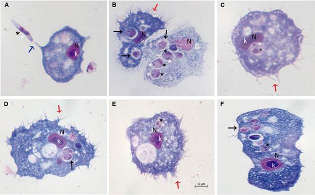
In recent decades, Acanthamoeba spp have garnered increasing attention due to their ability to serve as hosts or reservoirs for a variety of microbes, including viruses (e.g., mimivirus, coxsackieviruses, adenoviruses, poliovirus, echovirus, enterovirus, vesicular stomatitis virus, etc.), bacteria (e.g., Aeromonas, Bacillus, Bartonella, Burkholderia, Coxiella, Campylobacter, Chlamydophila, E. coli, Flavobacterium, Helicobacter, Legionella, Listeria, Staphylococcus, Mycobacterium, Pasteurella, Prevotella, Porphyromonas, Pseudomonas, Rickettsia, Salmonella, Shigella, Vibrio, etc.), protists (e.g., Cryptosporidium and Toxoplasma gondii), and yeast (e.g., Cryptococcus, Blastomyces, Sporothrix, Histoplasma, Streptomyces, etc.).73,74,90-91,92,93,94,95,96 Several pathogens can survive within A. castellanii using mechanisms similar to those employed by macrophages. For example, L. pneumophila relies on the Dot/Icm type IV secretion system for intracellular proliferation in both human macrophages and Acanthamoeba spp.96 Additionally, L. pneumophila is phagocytosed by Acanthamoeba via coiling phagocytosis, but it can hinder phagosome-lysosome fusion.97 Similarly, Vibrio cholerae can evade amoeboid phagosome killing by neutralising proteolytic enzymes, altering local pH, and counteracting reactive nitrogen and oxygen species that might otherwise eradicate internalised bacteria.98 Moreover, the invasion of A. castellanii or macrophages by Mycobacterium spp. shows significant similarities at the transcriptional and post-translational levels.99A. castellanii is also capable of engulfing pathogenic fungi, such as Cryptococcus neoformans, Histoplasma capsulatum, Blastomyces dermatitides, Fusarium solani, and Sporothrix schenckii.74 Notably, from the host’s perspective, macrophages and amoebae exhibit comparable interaction patterns, which may have significant implications for understanding infections in mammals.74 Recently, it has been reported that Leishmania spp. can evade the biochemical/lytic activity of A. castellanii and replicate within the amoeba100 (Figure).

Photomicrographs of Acanthamoeba castellanii infected with Leishmania spp., parasite ratio of 1:10, stained with Panoptic fast staining. (A) L. major; (B, C and F) L. braziliensis; (D) L. amazonensis; (E) L. infantum. V: vacuole; N: amoebae nucleus; K: amoeba karyosome; Black star: Leishmania nucleus; Write star: Leishmania kinetoplast; Black arrowheads: intracellular the rounded/oval body of Leishmania inside vacuoles or in the cytosol; Red arrowheads: pine-like pseudopods (acanthopodia); Blue arrowheads: interactions via flagella; Scale bar 10 µm.
These interactions suggest that the innate immune pathway is well conserved and that Leishmania spp. may interact within hosts through universal receptors found in professional phagocytes, including Acanthamoeba spp. Both A. castellanni and A. polyphaga can engulfed L. braziliensis, L. amazonensis, L. infantum, and L. major promastigotes, facilitating their intracellular conversion to an amastigote-like state and supporting their intracellular multiplication via primitive metabolic pathways.100 Studies have shown that interactions between pathogens and Acanthamoeba can preserve virulence factors and enhance microbial pathogenicity. Consequently, Acanthamoeba species may act as universal hosts for certain pathogenic microorganisms, connecting environmental factors to host virulence.101 Interestingly, another example of amoebae is Neoparamoeba spp. that harbours a kinetoplastid is Perkinsiella amoebae-like endosymbionts. These endosymbionts are believed to be related to the kinetoplastid Ichthyobodo, a free-living flagellate. These endosymbionts seem to be mutually beneficial for both organisms, as suggests a metabolic and cellular interdependence between the amoeba and the kinetoplastid.102
Conclusion remarks and perspectives
This review explored historical aspects of current discoveries on FLA. Despite the relatively low number of infections caused by FLA, it is important to recognise that the reported frequency may be significantly underestimated due to the lack of a diagnostic test with adequate specificity and sensibility. Understanding the pathogenesis and pathophysiology of these infections at the molecular, cellular, and clinical levels is essential. Enhancing our knowledge of these pathogens will help in developing more effective diagnostic, preventive, and therapeutic interventions. From the host’s perspective, macrophages and amoebas exhibit similar interaction patterns. This similarity may have important implications for understanding infections in mammals. By investigating the molecular basis of the interactions between amoebas and various microorganisms - both pathogenic and non-pathogenic - we can gain insights into the receptors, gene expression, and underlying molecular mechanisms involved. These insights could reveal functions that are also present in mammalian cells. Additionally, studying amoebas may provide a complementary perspective on the evolutionary pathways that pathogens have taken to adapt to different hosts.
REFERENCES
-
1 Schilde C, Schaap P. The Amoebozoa. Methods Mol Biol. 2013; 983: 1-15. https://doi: 10.1007/978-1-62703-302-2_1.
» https://doi.org/10.1007/978-1-62703-302-2_1 -
2 Hess S. The amoebae of Idionectes vortex (Cutosea, Amoebozoa): motility, cytoskeleton architecture and extracellular scales. J Eukaryot Microbiol. 2021; 68(6): e12869. https://doi: 10.1111/jeu.12869.
» https://doi.org/10.1111/jeu.12869 -
3 Pawlowski J, Burki F. Untangling the phylogeny of amoeboid protists. J Eukaryot Microbiol. 2009; 56(1): 16-25. https://doi: 10.1111/j.1550-7408.2008.00379.x.
» https://doi.org/10.1111/j.1550-7408.2008.00379.x -
4 Adl SM, Simpson AG, Lane CE, Lukes J, Bass D, Bowser SS, et al. The revised classification of eukaryotes. J Eukaryot Microbiol. 2012; 59(5): 429-93. https://doi: 10.1111/j.1550-7408.2012.00644.x. Erratum: J Eukaryot Microbiol. 2013; 60(3): 321.
» https://doi.org/10.1111/j.1550-7408.2012.00644.x -
5 Adl SM, Bass D, Lane CE, Lukes J, Schoch CL, Smirnov A, et al. Revisions to the classification, nomenclature, and diversity of eukaryotes. J Eukaryot Microbiol. 2019; 66(1): 4-119. https://doi: 10.1111/jeu.12691.
» https://doi.org/10.1111/jeu.12691 -
6 Bradley SG, Marciano-Cabral F. Diversity of free-living 'naked' amoeboid organisms. J Ind Microbiol Biotechnol. 1996; 17(3-4): 314-21. https://doi.org/10.1007/BF01574706
» https://doi.org/10.1007/BF01574706 - 7 Frank W. Limax-amoeba from cold-blooded vertebrates. Ann Soc Belg Med Trop. 1974; 54(4-5): 343-9.
- 8 Franke E, Mackiewicz J. Isolation of Acanthamoeba and Naegleria from the intestinal contents of freshwater fishes and their potential pathogenicity. J Parasitol. 1982; 68: 164.
-
9 Schuster FL, Visvesvara GS. Free-living amoebae as opportunistic and non-opportunistic pathogens of humans and animals. Int J Parasitol. 2004; 34: 1001-27. https://doi.org/10.1016/ j.ijpara.2004.06.004
» https://doi.org/10.1016/ j.ijpara.2004.06.004 -
10 Visvesvara GS, Stehr-Green JK. Epidemiology of free-living ameba infections. J Protozool. 1990; 37(4): 25-33S. https://doi: 10.1111/j.1550-7408.1990.tb01142.x.
» https://doi.org/10.1111/j.1550-7408.1990.tb01142.x -
11 Visvesvara GS, Moura H, Schuster FL. Pathogenic and opportunistic free-living amoebae: Acanthamoeba spp., Balamuthia mandrillaris, Naegleria fowleri, and Sappinia diploidea. FEMS Immunol Med Microbiol. 2007; 50(1): 1-26. http://doi: 10.1111/j.1574-695X.2007.0023 2.x.
» https://doi.org/10.1111/j.1574-695X.2007.0023 -
12 Fan S, Shen Y, Qian L. Social life of free-living amoebae in aquatic environment-comprehensive insights into interactions of free-living amoebae with neighboring microorganisms. Front Microbiol. 2024; 19: 15: 1382075. https://doi:10.3389/fmicb .2024.138 2075.
» https://doi.org/10.3389/fmicb -
13 Thompson AR. Phagotrophic protists (protozoa) in Antarctic terrestrial ecosystems: diversity, distribution, ecology, and best research practices. Polar Biol. 2021; 44: 1467-84. https://doi.org/10.1007/s00300-021-02896-3
» https://doi.org/10.1007/s00300-021-02896-3 -
14 Salazar-Ardiles C, Asserella-Rebollo L, Andrade DC. Free-living Amoebas in extreme environments: the true survival in our planet. Biomed Res Int. 2022; 18: 2359883. https://doi: 10.1155/2022/2359883.
» https://doi.org/10.1155/2022/2359883 -
15 Chaúque BJM, da Silva TCB, Dos Santos DL, Benitez GB, Chaúque LGH, Benetti AD, et al.Global prevalence of free-living amoebae in solid matrices - A systematic review with meta-analysis. Acta Trop. 2023; 247: 107006. https://doi: 10.1016/j. actatropica.2023.107006.
» https://doi.org/10.1016/j - 16 Rosel von Rosenhof AJ. Der kleine proteus. Der monat.-herausgeg, Insekten- Belustigungen. 1755; 3: 622.
- 17 Linnaeus C. Systema Naturae. 12th edn. London: Brit Mus Nat History; 1767.
- 18 Ehrenberg CG. Die infusionsthierchen als vollkommene organismen. Leipzig; 1838.
- 19 Dujardin F. Histoire naturelle des zoophytes. Infusoires. Paris: Libraire Encyclopedique de Roret; 1841.
- 20 Carpenter WB. On the systematic arrangement of the Rhizopoda. Natural History Review. 1861; 456-472.
- 21 Siebold CTE von. Bericht uber die Leistungen in der Naturgeschichte der Wurmer, Zoophyten und Protozoen wahrend des Jahres 1843 und 1844. Arch Naturgesch. 1845; 11: 256-96.
- 22 Schulze FE. Rhizopodien studien. Arch Mikrosk Anat. 1875; 11: 583-596.
- 23 Leidy J. Notice of a remarkable Amoeba. Proc Acad Nat Sci Philadelphia. 1874; 24: 142-3.
- 24 Leidy J. Fresh-water rhizopodes of North America. Rep US Geol Surv Territories. 1879; 12.
- 25 Schardinger F. Entwicklungskreis eine Amoeba lobosa (Gymnamoeba): Amoeba gruberi. Sitzungsberichte. 1899; 108: 713-34.
- 26 Dangeard PA. Fetude de la Karyokine'se chez 1* Amoeba hyalina sp. nova. Botaniste. 1900; 7: 49.
- 27 Bovee EC. Morphological identification of free living Amoebida. Proc Iowa Acad Sci. 1953; 60: 599-615.
-
28 Page FC. A revised classification of the Gymnamoebia (Protozoa: Sarcodina). Zool J Linn Soc. 1976; 58(1): 61-77. https://doi.org/10.1111/j.1096-3642.1976.tb00820.x
» https://doi.org/10.1111/j.1096-3642.1976.tb00820.x - 29 Aragäo HB. Sobre a Amoeba diplomitotica n. sp. Contribuição para o estudo da divizão nuclear nas amebas. Mem Inst Oswaldo Cruz. 1909; 1(1): 33-43.
-
30 Singh BN, Das SR. Studies on pathogenic and non-pathogenic small free-living amoebae and the bearing of nuclear division on the classification of the order amoebida. Philos Trans R Soc Lond B Biol Sci. 1970; 22; 259(832): 435-76. https://doi: 10.1098/rstb.1970.0063
» https://doi: 10.1098/rstb.1970.0063 - 31 Zulueta A. Promitosis y sindieresis. Dos modos de división nuclear coexistentes en amebas del grupo "Limax". Trabajos del Museo Nacional de Ciencias Naturales. 1917; 33: 66 pp.
- 32 Schaeffer A. Taxonomy of the Amoebas: with descriptions of thirty-nine new marine and freshwater species. Washington, DC: Carnegie Institute of Washington; 1926; 116 pp.
- 33 Mast SO, Doyle WL. Structure, origin and function of cytoplasmic constituents in Amoeba proteus. I. Structure Arch Protistenkd. 1935; 86: 156-80.
- 34 Obetz SW, Daniels EW. Electron microscopic studies of mitosis in Amoebae. I. Amoeba proteus. J. Biophys Biochem Cytol. 1960; 8(1): 207-20.
-
35 Andersen N, Holder H. The genera Amoebae. Science. 1949; 110(2848): 114-5. doi: 10.1126/science.110.2848.114.
» https://doi.org/10.1126/science.110.2848.114 - 36 Danielli JF, Lorch IJ, Ord MJ, Wilson EG. Nucleus and cytoplasm in cellular inheritance. Nature. 1955; 76: 1114-15.
-
37 Pappas GD. Electron microscope studies on amoebae. Ann NY Acad Sci. 2007; 78(2): 448-73. https://doiI:10.1111/j.1749-6632.1959.tb56117.x
» https://doiI:10.1111/j.1749-6632.1959.tb56117.x - 38 Shinn LE, Hadley PB. Note on the spontaneous contamination of a bacterial culture by an organism resembling Hartmannella castellani. J Infect Dis. 1936; 58: 23.
-
39 Chi L, Voguel JE, Shelokov A. Selective phagocytosis of nucleated erythrocytes by cytotoxic amebae in cell culture. Science. 1959; 25; 130(3391): 1763. https://doi: 10.1126/science.130. 3391.1763
» https://doi: 10.1126/science.130. 3391.1763 - 40 Moore AE, Hlinka J. Hartmannella sp. (Acanthamoeba) as a tissue culture contaminant. J Natl Cancer Inst. 1968; 40(3): 569-81.
-
41 Casemore DP. Contamination of virological tissue culture with a species of free-living soil amoeba. Clin Pathol. 1969; 22(3): 254-7. https://doi: 10.1136/jcp.22.3.254.
» https://doi.org/10.1136/jcp.22.3.254 - 42 Wang SS, Feldman HA. Occurrence of Acanthamoeba in tissue cultures inoculated with human pharyngeal swabs. Antimicrob Agents Chemother. 1961; 1: 50.
-
43 Wang SS, Feldman HA. Isolation of Hartmannella species from human throats. N Engl J Med. 1967; 30; 277(22): 1174-9. https://doi: 10.1056/NEJM196711302772204
» https://doi: 10.1056/NEJM196711302772204 -
44 Culbertson CG, Smith JW, Minner JR. Acanthamoeba: observations on animal pathogenicity. Science. 1958; 27; 127(3313): 1506. http://doi: 10.1126/science.127.3313.1506
» http://doi: 10.1126/science.127.3313.1506 - 45 Butt CG. Amebic meningoencephalitis. Am J Clin Pathol. 1964; 42: 516.
-
46 Fowler M, Carter RF. Acute pyogenic meningitis probably due to Acanthamoeba spp.: a preliminary report. Br Med J. 1965; 2(5464): 740-2. https://doi: 10.1136/bmj.2.5464.734-a.
» https://doi.org/10.1136/bmj.2.5464.734-a -
47 Shookhoff HB. Meningoencephalitis due to free-living amoebas normally found in soil. Ann Intern Med. 1969; 70(6): 1276-7. http://doi: 10.7326/0003-4819-70-6-1276.
» https://doi.org/10.7326/0003-4819-70-6-1276 -
48 Shumaker JB. Primary amebic meningoencephalitis. J Infect Dis. 1970; 121(1): 89-90. http://doi: 10.1093/infdis/121.1.89.
» https://doi.org/10.1093/infdis/121.1.89 -
49 Carter RF. Description of Naegleria sp. isolated from two cases of primary amebic meningoencephalitis and of the experimental pathological change induced by it. J Pathol. 1970; 100(4): 217-44. http://doi: 10.1002/path.1711000402.
» https://doi.org/10.1002/path.1711000402 -
50 Chang SL. Small, free-living amebas: cultivation, quantitation, identification, classification, pathogenesis and resistance. Curr Top Comp Pathobiol. 1971; 1: 201-54. doi: 10.1016/b978-0-12-153401-1.50010-7.
» https://doi.org/10.1016/b978-0-12-153401-1.50010-7 -
51 dos Santos JG. Fatal primary amebic meningoencephalitis: a retrospective study in Richmond, Virginia. Am J Clin Pathol. 1970; 54(5): 737-42. http://doi:10.1093/ajcp/54.5.737.
» https://doi.org/10.1093/ajcp/54.5.737 -
52 Visvesvara GS, Martinez AJ, Schuster FL, Leitch GJ, Wallace SV, Sawyer TK, et al. Leptomyxid ameba, a new agent of amebic meningoencephalitis in humans and animals. J Clin Microbiol. 1990; 228(12): 2750-6. http://doi: 10.1128/jcm.28.12.2750-2756.1990.
» https://doi.org/10.1128/jcm.28.12.2750-2756.1990 -
53 Visvesvara GS, Schuster FL, Martinez AJ. Balamuthia mandrillaris, N. G., N. Sp., agent of amoebic meningoencephalitis in humans and other animals. J Eukaryot Microbiol. 1993; 40(4): 504-14. http://doi: 10.1111/j.1550-7408.1993.tb04943.x.
» https://doi.org/10.1111/j.1550-7408.1993.tb04943.x -
54 Visvesvara GS, Sriram R, Qvarnstrom Y, Bandyopadhyay K, Da Silva AJ, Pieniazek NJ, et al. Paravahlkampfia francinae n. sp. masquerading as an agent of primary amoebic meningoencephalitis. J Eukaryot Microbiol. 2009; 56(4): 357-66. https://doi: 10.1111/j.1550-7408.2009.00410.x.
» https://doi.org/10.1111/j.1550-7408.2009.00410.x -
55 Siddiqui R, Makhlouf Z, Khan NA. The increasing importance of Vermamoeba vermiformis. J Eukaryotic Microbiol. 2021; 68: e12857. https://doi: 10.1111/jeu. 12857.
» https://doi.org/10.1111/jeu -
56 Gelman BB, Popov V, Chaljub G, Nader R, Rauf SJ, Nauta HW, et al. Neuropathological and ultrastructural features of amebic encephalitis caused by Sappinia diploidea. J Neuropathol Exp Neurol. 2003; 62(10): 990-8. https://doi: 10.1093/jnen/62.10.990.
» https://doi.org/10.1093/jnen/62.10.990 -
57 Diaz JH. Increasing intracerebral infections caused by free-living amebae in the United States and worldwide. J Neuroparasitol. 2010; 1: 23-32. https:/doi:10.4303/jnp/N10080.
» https://doi.org/10.4303/jnp/N10080 -
58 Levine ND, Corliss JO, Cox FE, Deroux G, Grain J, Honigberg BM, et al. A newly revised classification of the protozoa. J Protozool. 1980; 27(1): 37-58. https://doi: 10.1111/j.1550-7408.1980.tb04228.x.
» https://doi.org/10.1111/j.1550-7408.1980.tb04228.x -
59 Vinogradov SN, Bailly X, Smith DR, Tinajero-Trejo M, Poole RK, Hoogewijs D. Microbial eukaryote globins. Adv Microb Physiol. 2013,; 63: 391-446. https://doi.org/10.1016/B978-0-12-407693-8.00009-1
» https://doi.org/10.1016/B978-0-12-407693-8.00009-1 -
60 Cavalier-Smith T, Fiore-Donno AM, Chao E, Kudryavtsev A, Berney C, Snell EA, et al. Multigene phylogeny resolves deep branching of Amoebozoa. Mol Phylogenet Evol. 2015; 83: 293-304. https://doi.org/10.1016/j.ympev.2014.08.011
» https://doi.org/10.1016/j.ympev.2014.08.011 -
61 Smirnov A, Nassonova E, Berney C, Fahrni J, Bolivar I, Pawlowski J. Molecular phylogeny and classification of the lobose Amoebae. Protist. 2005; 156(2): 129-42. https://doi.org/10.1016/j.protis.2005.06.002
» https://doi.org/10.1016/j.protis.2005.06.002 -
62 Fahrni JF, Bolivar I, Berney C, Nassonova E, Smirnov A, Pawlowski J. Phylogeny of lobose Amoebae based on actin and small-subunit ribosomal RNA genes. Mol Biol Evol. 2003; 20(11): 1881-6. https://doi.org/10.1093/molbev/msg201
» https://doi.org/10.1093/molbev/msg201 -
63 Tice AK, Shadwick LL, Fiore-Donno AM, Brown MW, Spiegel FW, Cavalier-Smith T, et al. Expansion of the molecular and morphological diversity of Acanthamoebidae (Centramoebida, Amoebozoa) and identification of a novel life cycle type within the group. Biol Direct. 2016; 11: 69. https://doi.org/10.1186/s13062-016-0171-0
» https://doi.org/10.1186/s13062-016-0171-0 -
64 Burki F, Roger AJ, Brown MW, Simpson AGB. The new tree of eukaryotes. Trends Ecol Evol. 2020; 35(1): 43-55. https://doi.org/10.1016/j.tree.2019.08.008
» https://doi.org/10.1016/j.tree.2019.08.008 -
65 van Hooff JJE, Eme L. Lateral gene transfer leaves lasting traces in Rhizaria. bioRxiv [Preprint]. 2023. Available from: https://www.biorxiv.org/content/10.1101/2023.01.27.525846v1
» https://www.biorxiv.org/content/10.1101/2023.01.27.525846v1 -
66 Tolba ME, Huseein EA, Farrag HM, Mohamed Hel D, Kobayashi S, Suzuki J, et al. Allovahlkampfia spelaea causing keratitis in humans. PLoS Negl Trop Dis. 2016; 14: 10(7): e0004841. https://doi:10.1371/journal.pntd.0004841.
» https://doi.org/10.1371/journal.pntd.0004841 -
67 Khurana S, Sharma C, Radotra BD, Mewara A, Tanwar P, Datta P, et al. Molecular diagnosis of encephalitis/meningoencephalitis caused by free-living Amoebae from a tertiary center in India. Pathogens. 2022; 9: 11(12): 1509. https://doi: 10.3390/pathogens11121509.
» https://doi.org/10.3390/pathogens11121509 -
68 Moran S, Mooney R, Henriquez FL. Diagnostic considerations for non- Acanthamoeba amoebic keratitis and clinical outcomes. Pathogens. 2022; 8: 11(2): 219. http://doi: 10.3390/pathogens11020219.
» https://doi.org/10.3390/pathogens11020219 -
69 Siddiqui R, Khan NA. Balamuthia amoebic encephalitis: an emerging disease with fatal consequences. Microb Pathog. 2008; 44(2): 89-97. http://doi: 10.1016/j.micpath.2007.06.008.
» https://doi.org/10.1016/j.micpath.2007.06.008 -
70 Yamanouchi K, Arima H, Sakamoto Y, Kanto K, Kasai K, Ito K, et al. First report of the isolation of Balamuthia mandrillaris in the northern region of Japan. Parasitol Res. 2018; 117(9): 2895-900. https://doi:10.1007/s00436-018-5980-x
» https://doi.org/10.1007/s00436-018-5980-x -
71 Itoh K, Yagita K, Nozaki T, Katano H, Hasegawa H, Matsuo K, et al. An autopsy case of Balamuthia mandrillaris amoebic encephalitis, a rare emerging infectious disease, with a brief review of the cases reported in Japan. Neuropathol. 2015; 35(1): 64-9. https://doi.org/10.1111/neup.121 51
» https://doi.org/10.1111/neup.121 51 -
72 De Jonckheere JF, Brown S. Isolation of a vahlkampfiid amoeba from a contact lens: Tetramitus ovis (Schmidt, 1913) n. comb. Eur J Protistol. 2005; 41(2): 93-7. https:// doi.org/10.1016/j.ejop.2004.11.006
» https:// doi.org/10.1016/j.ejop.2004.11.006 -
73 Guimaraes AJ, Gomes KX, Cortines JR, Peralta JM, Peralta RH. Acanthamoeba spp. as a universal host for pathogenic microorganisms: one bridge from environment to host virulence. Microbiol Res. 2016; 193: 30-8. https://doi: 10.1016/j.micres.2016.08.001.
» https://doi.org/10.1016/j.micres.2016.08.001 -
74 Mungroo MR, Siddiqui R, Khan NA. War of the microbial world: Acanthamoeba spp. interactions with microorganisms. Folia Microbiol (Praha). 2021; 66(5): 689-99. https://doi: 10.1007/s12223-021-00889-7.
» https://doi.org/10.1007/s12223-021-00889-7 -
75 Khan NA. Acanthamoeba: biology and increasing importance in human health. FEMS Microbiol Rev. 2006; 30(4): 564-95. https://doi: 10.1111/j.1574-6976.2006.00023.x.
» https://doi.org/10.1111/j.1574-6976.2006.00023.x -
76 Bowers B, Korn ED. The fine structure of Acanthamoeba castellanii. I. The trophozoite. J Cell Biol. 1968; 39(1): 95-111. https:// doi: 10.1083/jcb.39.1.95.
» https://doi.org/10.1083/jcb.39.1.95 -
77 Bowers B, Korn ED. The fine structure of Acanthamoeba castellanii (Neff strain). II. Encystment. J Cell Biol. 1969; 41(3): 786-805. https://doi: 10.1083/jcb.41.3.786.
» https://doi.org/10.1083/jcb.41.3.786 -
78 Bowers B, Korn ED. Cytochemical identification of phosphatase activity in the contractile vacuole of Acanthamoeba castellanii. J Cell Biol. 1973; 59(3): 784-91. https://doi: 10.1083/jcb.59.3.784.
» https://doi.org/10.1083/jcb.59.3.784 -
79 González-Robles A, Castañón G, Hernández-Ramírez VI, Salazar-Villatoro L, González-Lázaro M, Omaña-Molina M, et al. Acanthamoeba castellanii identification and distribution of actin cytoskeleton. Exp Parasitol. 2008; 119(3): 411-7. https://doi: 10.1016/j.exppara.2008.04.004.
» https://doi.org/10.1016/j.exppara.2008.04.004 - 80 Mazur T, Hadas E, Iwanicka I. The duration of the cyst stage and the viability and virulence of Acanthamoeba isolates. Trop Med Parasitol. 1995; 46(2): 106-8.
-
81 Otero-Ruiz A, Gonzalez-Zuñiga LD, Rodriguez-Anaya LZ, Lares-Jiménez LF, Gonzalez-Galaviz JR, Lares-Villa F. Distribution and current state of molecular genetic characterization in pathogenic free-living Amoebae. Pathogens. 2022; 11(10): 1199. https://doi: 10.3390/pathogens11101199.
» https://doi.org/10.3390/pathogens11101199 -
82 Visvesvara GS. Amebic meningoencephalitides and keratitis: challenges in diagnosis and treatment. Curr Opin Infect Dis. 2010; 23(6): 590-4. https://doi: 10.1097/QCO.0b013e3283 3ed78b.
» https://doi.org/10.1097/QCO.0b013e3283 -
83 Balczun C, Scheid PL. Free-living Amoebae as hosts for and vectors of intracellular microorganisms with public health significance. Viruses. 2017; 1: 9(4): 65. https://doi: 10.3390/v9040065.
» https://doi.org/10.3390/v9040065 -
84 Shadrach WS, Rydzewski K, Laube U, Holland G, Ozel M, Kiderlen AF, et al. Balamuthia mandrillaris, a free-living ameba and opportunistic agent of encephalitis, is a potential host for Legionella pneumophila bacteria. Appl Environ Microbiol. 2005; 71(5): 2244-9. http://doi: 10.1128/AEM.71.5.2244-2249.2005.
» https://doi.org/10.1128/AEM.71.5.2244-2249.2005 - 85 Michel R, Muller KD, Zoller L, Walochnick J, Hartmann M, Schmid EN. Free-living amoebae serve as a host for the Chlamydia-like bacterium Simkania negevensis. Acta Protozool. 2005; 44: 113-21.
-
86 Newsome AL, Baker RL, Miller RD, Arnold RR. Interactions between Naegleria fowleri and Legionella pneumophila. Infect Immun. 1985; 50(2): 449-52. http://doi: 10.1128/iai.50.2.449-452.1985.
» https://doi.org/10.1128/iai.50.2.449-452.1985 - 87 Michel R, Muller KD, Hauröder B, Zöller L. A coccoid bacterial parasite of Naegleria sp. (Schizopyrenida: Vahlkampfiidae) inhibits cyst formation of its host but not transformation to the flagellate stage. Acta Protozool. 2000; 39: 199-207.
-
88 Thom S, Warhurst D, Drasar BS. Association of Vibrio cholerae with fresh water amoebae. J Med Microbiol. 1992; 36(5): 303-6. http://doi: 10.1099/00222615-36-5-303.
» https://doi.org/10.1099/00222615-36-5-303 - 89 Cabello-Vílchez AM, Mena R, Zuñiga J, Cermeño P, Martín-Navarro CM, González AC, et al. Endosymbiotic Mycobacterium chelonae in a Vermamoeba vermiformis strain isolated from the nasal mucosa of an HIV patient in Lima, Peru. Exp Parasitol. 2014; 145(Suppl.): S127-30. doi: 10.1016/j.exppara.2014.02.014.
- 90 Wright SJL, Redhead K, Maudsley H. Acanthamoeba castellanii, a predator of cyanobacteria. J Gen Microbiol. 1981; 125: 293-300.
-
91 Siddiqui R, Khan N. Acanthamoeba is an evolutionary ancestor of macrophages: a myth or reality? Exp Parasitol. 2012; 130(2): 95-7. https://doi.org/10.1016/j.exppara.2011.11.005
» https://doi.org/10.1016/j.exppara.2011.11.005 -
92 Gómez-Couso H, Paniagua-Crespo E, Ares-Mazás E. Acanthamoeba as a temporal vehicle of Cryptosporidium. Parasitol Res. 2007; 100(5): 1151-4. http://doi: 10.1007/s00436-006-0377-7.
» https://doi.org/10.1007/s00436-006-0377-7 -
93 Casadevall A, Fu MS, Guimaraes AJ, Albuquerque P. The 'amoeboid predator-fungal animal virulence' hypothesis. J Fungi (Basel). 2019; 5(1): 10. http://doi: 10.3390/jof5 0100 10.
» https://doi.org/10.3390/jof5 -
94 Winiecka-Krusnell J, Dellacasa-Lindberg I, Dubey JP, Barragan A. Toxoplasma gondii: uptake and survival of oocysts in free-living Amoebae. Exp Parasitol. 2009; 121(2): 124-31. https://doi.org/10.1016/j.exppara.2008.09.022
» https://doi.org/10.1016/j.exppara.2008.09.022 -
95 Derengowski LDS, Paes HC, Albuquerque P, Tavares AHFP, Fernandes L, Silva-Pereira I, et al. The transcriptional response of Cryptococcus neoformans to ingestion by Acanthamoeba castellanii and macrophages provides insights into the evolutionary adaptation to the mammalian host. Eukaryot Cell. 2013; 12: 761-74. https://doi.org/10.1128/EC.00073-13
» https://doi.org/10.1128/EC.00073-13 -
96 Segal G, Howard AS. Legionella pneumophila utilizes the same genes to multiply within Acanthamoeba castellanii and human macrophages. Infect Immun. 1999; 67(5): 2117-24. http:// doi: 10.1128/iai.67.5.2117-2124.1999.
» https://doi.org/10.1128/iai.67.5.2117-2124.1999 -
97 Al-Khodor S, Price CT, Habyarimana F, Kalia A, Abu Kwaik Y. A Dot/Icm-translocated ankyrin protein of Legionella pneumophila is required for intracellular proliferation within human macrophages and protozoa. Mol Microbiol. 2008; 70(4): 908-23. https://doi.org/10.1111/j.1365-2958.2008.06453.x
» https://doi.org/10.1111/j.1365-2958.2008.06453.x -
98 Espinoza-Vergara G, Hoque MM, McDougald D, Noorian P. The impact of protozoan predation on the pathogenicity of Vibrio cholerae. Front Microbiol. 2020; 21(11): 17 https://doi.org/10.3389/fmicb.2020.00017
» https://doi.org/10.3389/fmicb.2020.00017 -
99 Danelishvili L, Wu M, Stang B, Harriff M, Cirillo SL, Cirillo JD, et al. Identification of Mycobacterium avium pathogenicity island important for macrophage and amoeba infection. Proc Natl Acad Sci USA. 2007; 104(26): 11038-43. http://doi: 10.1073/pnas.0610746104.
» https://doi.org/10.1073/pnas.0610746104 -
100 Santos HLC, Pereira GL, do Reis RB, Rodrigues IC, Masini d'Avila C, Vidal VE. Using Acanthamoeba spp. as a cell model to evaluate Leishmania infections. PLoS Negl Trop Dis. 2024; 18(10): e0012517. http://doi: 10.1371/journal.pntd.0012517.
» https://doi.org/10.1371/journal.pntd.0012517 -
101 Rayamajhee B, Willcox MDP, Henriquez FL, Petsoglou C, Subedi D, Carnt N. Acanthamoeba, an environmental phagocyte enhancing survival and transmission of human pathogens. Trends Parasitol. 2022; 38(11): 975-90. http://doi: 10.1016/j.pt.2022.08.007.
» https://doi.org/10.1016/j.pt.2022.08.007 - 102 Dyková I, Fiala I, Lom J, Lukes J. Perkinsiella amoebae-like endosymbionts of Neoparamoeba spp., relatives of the kinetoplastid Ichthyobodo. Europ J Protistol. 2003; 39: 37-52.