Evaluation of the Antioxidant and Osteoinductive Potential of the Leaf Extract from Dysphania ambrosioides L.
ABSTRACT
Objective: To analyze the antioxidant activity of an alcoholic extract from the leaves of Dysphania ambrosioides L. and investigate its influence on in vitro differentiation of MC3T3-E1 pre-osteoblastic cells.
Material and Methods: The content of the extract’s flavonoids, tannins, and polyphenols was determined by spectrophotometry. The antioxidant potential was evaluated through tests of total antioxidant capacity (TAC), reductive activity, sequestration of superoxide radicals, and copper chelation. Cell viability and proliferation were evaluated by reduction of tetrazolium (MTT). Osteoblastic differentiation was assessed by alkaline phosphatase activity, quantification, and matrix mineralization using von Kossa stain.
Results: The phytochemical screening revealed that the extract has a high tannin content (43.27% catechin equivalents), with polyphenols and flavonoids accounting for 6.57% and 6.24%, respectively. The extract demonstrated antioxidant potential, particularly for the tests that evaluated TAC (I20mg ascorbic acid equivalents) and copper chelation (79.2% of chelation relative to EDTA). The extract (ISO µg/mL) increased the proliferation of MC3T3 cells by 251% in the first 24h. None of the osteoblastic differentiation tests demonstrated an influence on the treatments with the extract.
Conclusion: The alcoholic extract of D. ambrosioides proved to be a potent antioxidant but did not exhibit osteoinductive potential.
Keywords:
Antioxidants; Cell Differentiation; Plant Extracts
Introduction
The regeneration capacity of the bone is a long process[1]. However, some compounds, such as the ones with antioxidant properties, have the potential to exert a positive influence on bone metabolism since bone physiopathologies are associated with oxidative stress[2]. The capacity to generate an osteoinductive potential is important in several areas of dentistry, such as in surgical or periodontitis cases. This chronic inflammatory disease is highly associated with oxidative stress, which causes progressive bone loss[3, 4].
The osteoinductive potential of compounds can be tested using pre-osteoblastic cell cultures. In this context, pre-osteoblasts cells - MC3T3 offer methodological advantages, as the differentiation of these cells into mature osteoblasts is easily identified by markers of osteoblastic metabolism, such as alkaline phosphatase (ALP) and the degree of mineralization of the extracellular matrix (ECM)[5, 6].
The search for compounds derived from natural plants has gained attention over the years due to the potential therapeutic action for treating various disorders. However, a myriad of molecules has yet to be explored. In this aspect, traditional folk wisdom has guided scientific investigations that seek to ensure the population’s safe, effective use of medicinal plants and stimulate their use as therapeutic alternatives and isolate compounds for the development of plant-based drugs[7]. Dysphania ambrosioides L., or Chenopodium ambrosioides, is known as wormseed, Jesuit’s tea, Mexican tea, Payqu, Epazote, or Mastruz. Based on widespread knowledge, this plant is widely used by the population as an anti-parasitic, expectorant, oral infection treatment and wound-healing agent, and for the repair of fractures. For that, booth aerial parts and roots of the plant have been used in different preparations such as infusions, decoctions, and maceration[8, 9, 10].
Works published in the last decade using animal models have shown that the extract obtained from this plant has prevented bone loss in collagen-induced arthritis, induced bone repair in a rodent model, and exhibited anti-resorptive properties in a ligature-induced periodontitis model in rats[10, 11, 12, 13]. However, little is known about the effects of these phytochemical molecules on osteoblastic cell activity, as no studies are currently published in the literature. Considering the potential of D. ambrosioides L. in the bone repair process, this in vitro study investigated its antioxidant activity, its influence on the differentiation of MC3T3 pre-osteoblasts, and its effect on bone matrix mineralization. Additionally, a phytochemical analysis of the leaf extract was performed.
Material and Methods
Vegetal Material
The material was collected in the municipality of Lagoa Seca, state of Paraiba, Brazil (7°10′15″ S, 35°51′14″ W). A voucher specimen was deposited in the Manuel de Arruda Câmara Herbarium (nº 042/ACAM) at the State University of Paraíba, Campina Grande, Paraíba. The leaves were dehydrated in a hothouse, where air circulated and ground.
Acquisition of the Extract
The ground leaves were first submitted to a maceration process and remained at rest for five days in a percolator containing 98% (v/v) ethyl alcohol, followed by filtration. The extract was then vacuum-evaporated and freeze-dried.
Phytochemical Analysis
The content of fiavonoids, tannins, and polyphenols of the alcoholic extract from the leaves of Dysphania ambrosioides L. was determined using a spectrophotometer (UV mini – 1240, Shimadzu Corp., Kyoto, Japan). Determination of total polyphenol content[14], total flavonoid content[15], and condensed tannin content were performed[16].
In Vitro Antioxidant Activity
Total antioxidant capacity (TAC), reduction activity, sequestration of superoxide radicals, and copper chelation were performed to evaluate the antioxidant activity. The TAC test enables the evaluation of the ability of a sample to donate electrons to oxidized compounds with reactive potential. The assay is based on reducing molybdenium+6 from an ammonium molybdate solution to molybdenium+5, which forms a complex with phosphate under acid pH and a temperature of 95 °C. This phosphomolybdenum complex has a greenish color that can be monitored spectrophotometrically at 695 nm[17]. Antioxidant potential was determined concerning the ascorbic acid standard based on the slope equation applied to the calibration curve (r2 = 0.98): y = 35.89 × −0.0355. Masses of 0.1 mg and 1 mg were used for this test. It was calculated that 1 g of sample has TAC equal to 120 mg of ascorbic acid. The plotting of the calibration curve enabled the determination of the mass of ascorbic acid at which the most excellent absorbance was obtained, which was then defined as the reference.
The reduction activity evaluates the potassium ferricyanide reduction to potassium ferrocyanide, which generates a greenish blue color (Prussian blue) monitored at 700 nm in the presence of iron chloride. This assay used masses of 0.05 mg, 0.01 mg, 0.025 mg, 0.5 mg, and 1 mg. The reduction power of the samples was quantified using the methods described elsewhere[18, 19].
The copper chelation method is based on the sample’s chelation capacity of copper ions. This test is performed with a violet reagent, which binds to free copper ions in the solution, generating a violet color monitored at 632 nm[20]. Different sample masses (0.05 mg, 0.01 mg, 0.025 mg, 0.5 mg, and 1 mg). The determination of the percentage of chelation of copper ions by the sample was established by comparison to the blank, which represents 0% chelation (maximum absorbance for violet monitored at 632 nm). For such, the following formula was used: (Ab-Aa / Ab) × 100, in which Ab is the absorbance of the blank, and Aa is the absorbance of the sample.
The superoxide radical sequestration assay is based on the capacity of a sample to inhibit the photochemical reduction of nitroblue tetrazolium (NBT) to formazan by the superoxide radical in the riboflavin-light-NBT system at pH 7.4 and room temperature. Formazan causes the occurrence of a change from a pale-yellow color to purple, which is accompanied by spectrophotometry at 560 nm. For such, the method described by Dasgupta and De[21] was followed, and different sample masses were evaluated (0.05 mg, 0.1 mg, 0.25 mg, and 0.5 mg). The percentage of superoxide radical sequestration was established based on the data obtained for the sample, control (which represents 100% reduction due to the absence of antioxidant) and the blank (which represents 100% sequestration), using the following formula: % sequestration = (Ac – Aa) / (Ac – Ab), in which Ac is absorbance of the control, Aa is absorbance of the sample and Ab is absorbance of the blank.
Cell Viability and Proliferation Test by Reduction of Tetrazolium Dye (MTT)
Cell viability and proliferation were evaluated using the MTT (3-[4,5-dimethylthiazol-2-yl]-2,5 diphenyl tetrazolium bromide) assay based on the spectrophotometric determination of formazan formation produced by viable MC3T3 cells[22]. Three MTT assays were performed at 24, 48, and 72 hours. Approximately 5 × 10³ MC3T3 cells were placed in each sterile 96 wells in α-MEM medium supplemented with 10% fetal bovine serum (FBS – Gibco, Thermo Fisher Scientific Inc., Waltham, MA, USA). Cells were placed in a medium without serum for 24 hours. Subsequently, the medium was aspirated, and the cells were added to the medium with 10% FBS in the absence (control) and presence of the extract at concentrations of 50 µg/mL, 100 µg/mL, 150 µg/mL, 200 µg/mL, and 250 µg/mL. Next, the MTT salt (1 mg/mL) was added to the cells, and the plates were incubated for another four hours. Ethyl alcohol A. G. (100 µL) was added to dissolve the precipitated formazan crystals, and absorbance was quantified in a 96-well plate reader at 570 nm. The assay was conducted in triplicate, and the results were obtained by comparing the treated and control cells. The relative percentage for the treatments was determined using the following formula: (Mt/Mc) × 100, in which Mt is the mean absorbance value for the treatments, and Mc is the mean absorbance value for the controls. Thus, values above 100% represent a more significant number of viable cells, and values below 100% indicate that the treatment affected cell viability.
Evaluation of Alkaline Phosphatase (ALP) Activity and Quantification
Initially, 5×10⁴ MC3T3/mL cells were added to each well of the culture plates. After cell adhesion, the α-MEM medium was aspirated and replaced with the differentiation medium in the absence (control) or presence of the extract at concentrations of 50 µg/mL, 100 µg/mL, and 150 µg/mL. The supernatants were collected from the wells 7, 14, and 21 days after the onset of treatment and stored at −20 ºC until the assays. ALP activity was evaluated using a commercial liquiform ALP kit (Labtest Diagnostica S.A., Lagoa Santa, MG, Brazil). In alkaline pH, the ALP present in the sample de-phosphorilizes the p-nitrophenyl phosphate reagent, releasing inorganic phosphate and p-nitrophenol. This is monitored at 405 nm, and the absorbance generated is directly proportional to the enzymatic activity of ALP. The initial absorbance reading was performed after 1 minute of reaction (A1), and another reading was done after the second minute (A2). ALP was quantified using the following formula: (A2-A1/2) × CF, in which CF is the correction factor calculated for the conditions described for Test 2764, and the result is expressed in units of ALP per liter (U/L).
Analysis of the Degree of Mineralization of the Matrix Using von Kossa Staining
The assay described by McGee-Russell[23] enables the viewing of mineralized nodules through the reaction between calcium phosphate and silver nitrate, forming silver phosphate, which is subsequently reduced to metallic silver by UV radiation. Thus, the nodules become easily identified under a microscope as black deposits on the cell layer. Each well of the three plates – one for each interval (7, 14, and 21 days) – was submitted to fixation with paraformaldehyde 4% at pH 7.4 for 20 minutes. Next, the cultures were covered with a 5% silver nitrate solution and exposed to UV radiation for 30 minutes. After rinsing in distilled water, the cultures were covered with a 5% sodium thiosulfate solution for 2 minutes to remove excess iron in the non-precipitated form and rinsed again with distilled water. The cells were counterstained with a solution of hematoxylin (1 mg/mL) for 5 minutes, rinsed with distilled water, and covered with phosphate-buffered saline. An inverted light microscope was used to photograph each well for the comparative analyses.
Statistical Analysis
The results were expressed as mean and standard deviation. Data was analyzed with GraphPad Prism software (version 8.02) using analysis of variance (AN OVA), followed by the student’s t-test for the MTT assays and the quantification of ALP. Ap-value < 0.05 was considered indicative of a statistically significant difference.
Results
Phytochemical Analysis
The phytochemical characterization revealed a high tannin content with 43.27% catechin equivalents. Total polyphenols corresponded to 6.57% gallic acid equivalents, and fiavonoids corresponded to 6.24% quercetin equivalents.
In Vitro Antioxidant Activity
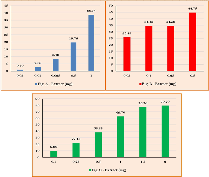
The antioxidant activity of D. ambrosioides leaf extract was demonstrated by the TAC equivalent to 120 mg of ascorbic acid (Figure 1A) and the chelation of copper ions, relative to 79.2% of EDTA chelation (Figure lB). Meanwhile, the percentage of superoxide radical sequestration showed a dose-dependent result (Figure 1C).

Antioxidant potential of the alcoholic extract from the leaves of Dysphania ambrosioides L. (A) Relative percentage for reduction activity of extract; (B) Relative percentage for sequestration of superoxide radicals by extract; (C) Relative percentage of copper chelation by sample
Cell Viability and Proliferation Tests by Reduction of Tetrazolium Dye (MTT)
All assays differed significantly from the control group (p<0.05) on the three tests (24, 48, and 72h), except for the 250 µg/mL - treatment (Figure 2). Based on the results, concentrations of 50, 100, and 150 µg/mL of the sample were chosen for the cell differentiation tests.

Percentage of cell proliferation in each treatment (concentration of extract in µg/mL) in relation to the control for each period.
ALP Activity and Quantification
No significant differences between the controls and treatments, between treatments on the same day or among treatments on different days were found (p>0.05), as stated in Figure 3.

Quantification of alkaline phosphatase for each treatment in 7, 14, and 21 days of differentiation.
Degree of Mineralization of the Matrix Using von Kossa Staining
The images obtained for the 7 days served as the control since the MC3T3 cells are in the maturation phase of the ECM in this period, and it is, therefore, possible only to observe cells in semi-confluence. Likewise, no mineralized nodules were found in any treatments or controls at 14 days. At 21 days, some regions were stained, indicating the presence of calcium phosphate in the medium, but no typical mineralized nodules were identified (Figures 4, 5, and 6).

Photomicrographs of MC3T3 cultures at 7 days of treatment with extract stained using the von Kossa method (magnification: 40x). Control (A); 50 µg/mL (B); 100 µg/mL (C); 150 µg/mL (D).

Photomicrographs of MC3T3 cultures at 14 days of treatment with extract stained using the von Kossa method (magnification: 40x). Control (A); 50 µg/mL (B); 100 µg/mL (C); 150 µg/mL (D).
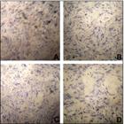
Photomicrographs of MC3T3 cultures at 21 days of treatment with extract stained using the von Kossa method (magnification: 40x). Control (A); 50 µg/mL (B); 100 µg/mL (C); 150 µg/mL (D).
Discussion
In the present study, the tests performed on the extract from the leaves of D. ambrosioides indicate pronounced antioxidant activity, as demonstrated by the TAC and chelation of copper ions results. Antioxidant compounds have either direct or indirect action on bone metabolism. Such compounds affect osteoprogenitor cell recruitment and favor osteoblastic differentiation and function[24-26]. Thus, D. ambrosioides could have antiresorptive potential, which is an aspect that needs to be investigated.
The initial screening for determining polyphenols, fiavonoids, and tannins content demonstrated that all these classes of compounds are well represented. The results underscored a high tannin content and the less expressive content of total polyphenols and flavonoids. Flavonoids and polyphenols are vegetables’ main antioxidant compounds and have been appointed as preventers of osteoporosis[26,27], and tannins are often related to healing processes[24, 28]. Altogether, the phytocomplex might be responsible for the extract activities.
Besides the antioxidant potential, the anabolic potential of the D. ambrosioides extract was also investigated using cell culture methods. For such, it is crucial to determine the degree of toxicity and solubility of the compounds and their effect on the proliferation and morphology of the cell line. Regarding cell viability and proliferation, the 250 µg/mL treatment in the 72h period was the only group to return a value below 100%, indicating influence on cell viability. Nonetheless, other researchers have studied the toxicity potential of the D. ambrosioides extract. For instance, Carneiro et al.[13] have demonstrated safety parameters of its use in in vivo models, in which the extract did not cause toxicity in rats’ liver, kidney, or stomach.
Accentuated cell proliferation was found in all other tested concentrations, reaching nearly triple the cell population at 150 µg/mL in the first 24 h (251% growth). Additionally, all treatments differed significantly from the control group regardless of the established time. These findings suggest that the extract can accelerate and stimulate pre-osteoblast proliferation, which is essential to the initial bone healing and remodeling phase. The concentrations of 50, 100, and 150 µg/mL were chosen for the cell differentiation assay based on MTT assay results.
The ALP enzyme is associated with the mineralization of the extracellular matrix. Still, its action is most remarkable in the second phase of osteoblastic differentiation since it is responsible for the maturation of the matrix that will subsequently be mineralized[29]. ALP promotes the hydrolysis of organic phosphates (β-glycerophosphate in the case of cell cultures), thereby increasing the concentration of phosphate ions at mineralization sites, which, together with the calcium ions in the surrounding medium, promotes the formation of calcium phosphate deposits[30]. Due to this central role and the ease of its biochemical and histological determinations, ALP has become the marker of choice in evaluating the phenotype or maturation of the development of mineralized tissue cells[29]. In the present study, the statistical analysis revealed no significant differences among treatments regardless of treatment time (p>0.05). Controversially, Pinheiro Neto et al.[31] noted a more excellent ALP activity during early fracture healing under the effect of a D. ambrosioides graft, enhancing bone regeneration.
The results indicate that the extract’s action mechanism must not be directly related to osteoblastic differentiation, as no significant differences were found in the quantification of ALP between the treated cells and controls. Moreover, no mineralized nodules were found 21 days after the onset of treatment. These findings do not confirm the hypothesis of the osteoconductive potential of D. ambrosioides, which is based on folk medicine in communities where this plant is used topically on bone injuries to accelerate the regeneration process[7, 24].
The degree of mineralization of the ECM formed by the culture cells was reflected by the mineralized area identified with an inverted microscope after treatment with silver nitrate using the von Kossa staining method. At 21 days after the onset of treatment for cell differentiation, the control group exhibited discrete grayish spots, which may represent the beginning of nodular formation. MC3T3 cells take approximately 21 days to differentiate and can exhibit mineralized nodules beginning in the second week of differentiation. These results are in accordance with Penha et al.[11] findings, in which the authors observed histological bone mineralization and repair after 30 days of treatment with D. ambrosioides extract.
Even though the findings do not confirm the osteoconductive action of the extract, one must not discard the possibility that the extract is beneficial to bone health during the repair of injuries or the maintenance/recovery of normal bone mass since the extract of D. ambrosioides has already shown positives results regarding the increase bone density[11, 31, 32]. In addition, studies have also demonstrated the protective potential of the extract. Carneiro et al.[13], for instance, showed that D. ambrosioides was able to reduce bone loss by 58%, marked by a reduction in the number of osteoclasts in rats with periodontitis.
Even though antioxidant activity is correlated to combat oxidative stress, which is beneficial to bone health, reactive oxygen species (ROS) in low concentrations also act as modulators of osteoclasts and osteoblasts. Morphogenetic proteins (BMBs) in osteoblasts generate ROS, vital to the activation of ALP. Moreover, ROS induces mitogen-activate kinase proteins, which are important to the osteoclastic differentiation process[33]. Thus, previous studies have associated D. ambrosioides antioxidant potential through the 1,1-Diphenyl-2-picrylhydrazyl (DPPH) radical method with the enhancement of bone regeneration[31].
As the treated cells in the present study were not under oxidative stress, the presence of a potent antioxidant may have negatively affected osteoblastic differentiation. Arakaki et al.[33] found that MC3T3-E1 cells incubated in the presence of N-acetylcysteine antioxidants, in which the production of ROS was significantly suppressed, mineralization was also suppressed, demonstrating that ROS play an essential role in the regulation of osteoblastic differentiation[25, 34, 35]. Thus, further investigations are encouraged.
Moreover, compounds can positively influence bone health in different ways. Some can promote an anabolic effect on osteoblasts, stimulating proliferation, differentiation, and mineralization, whereas others have an anti-catabolic effect, acting on the differentiation and function of osteoclasts. For instance, pro-angiogenic compounds are also helpful to the regeneration process of bone injuries[11, 13, 24, 31]. This is explained by the fact that the regeneration of a bone injury is a complex process involving several steps. Thus, besides promoting pronounced cell proliferation, which is important to the healing of bone tissue, D. ambrosioides may also influence other processes that were not analyzed herein. Other analyses need to be performed focusing on antiresorptive and osteoinductive activities to demonstrate its effect on bone health, the fracture healing process, and angiogenic activity.
Conclusion
The alcoholic extract from the leaves of D. ambrosioides proved to be a potent antioxidant but did not exhibit osteoinductive potential.
Financial Support
Data Availability
The data used to support the findings of this study can be made available upon request to the corresponding author.
References
-
1 Saul D, Menger MM, Ehnert S, Nüssler AK, Histing T, Laschke MW. Bone healing gone wrong: Pathological fracture healing and non-unions — Overview of basic and clinical aspects and systematic review of risk factors. Bioeng 2023; 10(1):85. https://doi.org/10.3390/bioengineering10010085
» https://doi.org/10.3390/bioengineering10010085 -
2 Bacevic M, Brkovic B, Albert A, Rompen E, Radermecker RP, Lambert F. Does oxidative stress play a role in altered characteristics of diabetic bone? A systematic review. Calcif Tissue Int 2017; 101(6):553-563. https://doi.org/10.1007/s00223-017-0327-7
» https://doi.org/10.1007/s00223-017-0327-7 -
3 Ko KI, Sculean A, Graves DT. Diabetic wound healing in soft and hard oral tissues. Transl Res 2021; 236:72-86. https://doi.org/10.1016/j.trsl.2021.05.001
» https://doi.org/10.1016/j.trsl.2021.05.001 -
4 Chen M, Cai W, Zhao S, Shi L, Chen Y, Li X, et al. Oxidative stress-related biomarkers in saliva and gingival crevicular fluid associated with chronic periodontitis: A systematic review and meta-analysis. J Clin Periodontol 2019; 46(6):608-622. https://doi.org/10.1111/jcpe.13112
» https://doi.org/10.1111/jcpe.13112 -
5 Cai W, Sun B, Song C, Liu F, Wu Z, Liu Z. Resveratrol induces proliferation and differentiation of mouse pre-osteoblast MC3T3-E1 by promoting autophagy. BMC Complement Med Ther 2023; 23(1):121. https://doi.org/10.1186/s12906-023-03943-8
» https://doi.org/10.1186/s12906-023-03943-8 -
6 Sekaran S, Vimalraj S, Thangavelu L. The physiological and pathological role of tissue nonspecific alkaline phosphatase beyond mineralization. Biomolecules 2021; 11(11):1564. https://doi.org/10.3390/biom11111564
» https://doi.org/10.3390/biom11111564 -
7 Newman DJ, Cragg GM. Natural products as sources of new drugs over the nearly four decades from 01/1981 to 09/2019. J Nat Prod 2020; 83(3):770-803. https://doi.org/10.1021/acs.jnatprod.9b01285
» https://doi.org/10.1021/acs.jnatprod.9b01285 -
8 Kandsi F, Lafdil FZ, Hachlafi N El, Jeddi M, Bouslamti M, Fadili M El, et al. Dysphania ambrosioides (L.) Mosyakin and Clemants: Bridging traditional knowledge, photochemistry, preclinical investigations, and toxicological validation for health benefits. Naunyn Schmiedebergs Arch Pharmacol 2024; 397(2):969-1001. https://doi.org/10.1007/s00210-023-02658-4
» https://doi.org/10.1007/s00210-023-02658-4 -
9 Lemhadri A, Achtak H, Lamraouhi A, Louidani N, Benali T, Dahbi A, et al. Diversity of medicinal plants used by the local communities of the Coastal Plateau of Safi Province (Morocco). Front Biosci 2023; 15(1):1. https://doi.org/10.31083/j.fbs1501001
» https://doi.org/10.31083/j.fbs1501001 -
10 Ribeiro RV, Bieski IGC, Balogun SO, Martins DTO. Ethnobotanical study of medicinal plants used by Ribeirinhos in the North Araguaia microregion, Mato Grosso, Brazil. J Ethnopharmacol. 2017; 205:69-102. https://doi.org/10.1016/j.jep.2017.04.023
» https://doi.org/10.1016/j.jep.2017.04.023 -
11 Penha ESD, Lacerda-Santos R, Carvalho MGF, Oliveira PT. Effect of Chenopodium ambrosioides on the healing process of the in vivo bone tissue. Microsc Res Tech 2017; 80(11):1167-1173. https://doi.org/10.1002/jemt.22913
» https://doi.org/10.1002/jemt.22913 -
12 Pereira WS, da Silva GP, Vigliano MV, Leal NRF, Pinto FA, Fernandes DC, et al. Anti-arthritic properties of crude extract from Chenopodium ambrosioides L. leaves. J Pharm Pharmacol 2018; 70(8):1078-1091. https://doi.org/10.1111/jphp.12926
» https://doi.org/10.1111/jphp.12926 -
13 Carneiro DTO, da Silva MD, Vasconcelos KVP, Dias R, Costa V, Vasconcelos RF, et al. Chenopodium Ambrosioides Linn mitigates bone loss in rats with periodontitis. J Dent 2024; 25(1):59-67. https://doi.org/10.30476/dentjods.2023.95767.1891
» https://doi.org/10.30476/dentjods.2023.95767.1891 -
14 Chandra S, De Mejia EG. Polyphenolic compounds, antioxidant capacity, and quinone reductase activity of an aqueous extract of Ardisia compressa in comparison to mate (llex paraguariensis) and green (Camellia sinensis) teas. J Agric Food Chem 2004; 52(11):3583-3589. https://doi.org/10.1021/jf0352632
» https://doi.org/10.1021/jf0352632 -
15 Meda A, Lamien CE, Romito M, Millogo J, Nacoulma OG. Determination of the total phenolic, flavonoid and proline contents in Burkina Fasan honey, as well as their radical scavenging activity. Food Chem 2005; 91(3):571-577. https://doi.org/10.1016/j.foodchem.2004.10.006
» https://doi.org/10.1016/j.foodchem.2004.10.006 -
16 Makkar HPS, Becker K. Vanillin-Hcl method for condensed tannins: Effect of organic solvents used for extraction of tannins. J Chem Ecol 1993; 19(4):613-621. https://doi.org/10.1007/BF00984996
» https://doi.org/10.1007/BF00984996 -
17 Prieto P, Pineda M, Aguilar M. Spectrophotometric quantitation of antioxidant capacity through the formation of a phosphomolybdenum complex: Specific application to the determination of vitamin E 1. Anal Biochem 1999; 269(2):337-341. https://doi.org/10.1006/abio.1999.4019
» https://doi.org/10.1006/abio.1999.4019 -
18 Oyaizu M. Antioxidative activities of browning products of glucosamine fractionated by organic solvent and thin-layer chromatography. J Jpn Soc Food Sci 1988; 35(11):771-775. https://doi.org/10.3136/nskkk1962.35.11_771
» https://doi.org/10.3136/nskkk1962.35.11_771 -
19 Athukorala Y, Kim KN, Jeon YJ. Antiproliferative and antioxidant properties of an enzymatic hydrolysate from brown alga, Ecklonia cava. Food Chem Toxicol 2006; 44(7):1065-1074. https://doi.org/10.1016/jfct.2006.01.011
» https://doi.org/10.1016/jfct.2006.01.011 -
20 Saiga A, Soichi T, Nishimura T. Antioxidant activity of peptides obtained from porcine myofibrillar proteins by protease treatment. J Agric Food Chem 2003; 51(12):3661-3667. https://doi.org/10.1021/jf021156g
» https://doi.org/10.1021/jf021156g -
21 Dasgupta N, De B. Antioxidant activity of Piper betle L. leaf extract in vitro. Food Chem 2004; 88(2):219-224. https://doi.org/10.1016/j.foodchem.2004.01.036
» https://doi.org/10.1016/j.foodchem.2004.01.036 -
22 Mosmann T. Rapid colorimetric assay for cellular growth and survival: Application to proliferation and cytotoxicity assays. J Immunol Methods 1983; 65(1-2):55-63. https://doi.org/10.1016/0022-1759(83)90303-4
» https://doi.org/10.1016/0022-1759(83)90303-4 -
23 Mcgee-Russell SM. Histochemical methods for calcium. J Histochem Cytochem 1958; 6(1):22-42. https://doi.org/10.1177/6.1.22
» https://doi.org/10.1177/6.1.22 -
24 Hanga-Farcas A, Miere Groza F, Filip GA, Clichici S, Fritea L, Vicas LG, et al. Phytochemical compounds involved in the bone regeneration process and their innovative administration: A systematic review. Plants 2023; 12(10):2055. https://doi.org/10.3390/plants12102055
» https://doi.org/10.3390/plants12102055 -
25 Tao H, Li X, Wang Q, Yu L, Yang P, Chen W, et al. Redox signaling and antioxidant defense in osteoclasts. Free Radic Biol Med 2024; 212:403-414. https://doi.org/10.1016/j.freeradbiomed.2023.12.043
» https://doi.org/10.1016/j.freeradbiomed.2023.12.043 -
26 Su Z, Yao B, Liu G, Fang J. Polyphenols as potential preventers of osteoporosis: A comprehensive review on antioxidant and anti-inflammatory effects, molecular mechanisms, and signal pathways in bone metabolism. J Nutr Biochem 2024; 123:109488. https://doi.org/10.1016/j.jnutbio.2023.109488
» https://doi.org/10.1016/j.jnutbio.2023.109488 -
27 Dias MC, Pinto DCGA, Silva AMS. Plant flavonoids: Chemical characteristics and biological activity. Molecules 2021; 26(17):5377. https://doi.org/10.3390/molecules26175377
» https://doi.org/10.3390/molecules26175377 -
28 Zhao Y, Li J, Liu L, Wang Y, Ju Y, Zeng C, et al. Zinc-based tannin-modified composite microparticulate scaffolds with balanced antimicrobial activity and osteogenesis for infected bone defect repair. Adv Healthc Mater 2023; 12(20):e2300303. https://doi.org/10.1002/adhm.202300303
» https://doi.org/10.1002/adhm.202300303 -
29 Nakamura T, Nakamura-Takahashi A, Kasahara M, Yamaguchi A, Azuma T. Tissue-nonspecific alkaline phosphatase promotes the osteogenic differentiation of osteoprogenitor cells. Biochem Biophys Res Commun 2020; 524(3):702-709. https://doi.org/10.1016/j.bbrc.2020.01.136
» https://doi.org/10.1016/j.bbrc.2020.01.136 -
30 Yevlashevskaya OS, Scheven BA, Walmsley AD, Shelton RM. Differing responses of osteogenic cell lines to β-glycerophosphate. Sci Rep 2023; 13(1):14472. https://doi.org/10.1038/s41598-023-40835-w
» https://doi.org/10.1038/s41598-023-40835-w -
31 Pinheiro Neto VF, Ribeiro RM, Morais CS, Campos MB, Vieira DA, Guerra PC, et al. Chenopodium ambrosioides as a bone graft substitute in rabbits radius fracture. BMC Complement Altern Med 2017; 17(1):350. https://doi.org/10.1186/s12906-017-1862-5
» https://doi.org/10.1186/s12906-017-1862-5 -
32 Pereira WS, da Silva GP, Vigliano MV, Leal NRF, Pinto FA, Fernandes DC. Anti-arthritic properties of crude extract from Chenopodium ambrosioides L. leaves. J Pharm Pharmacol 2018; 70(8):1078-1109. https://doi.org/10.1111/jphp.12926
» https://doi.org/10.1111/jphp.12926 -
33 Arakaki N, Yamashita A, Niimi S, Yamazaki T. Involvement of reactive oxygen species in osteoblastic differentiation of MC3T3-E1 cells accompanied by mitochondrial morphological dynamics. Biomed Res 2013; 34(3):161-166. https://doi.org/10.2220/biomedres.34.161
» https://doi.org/10.2220/biomedres.34.161 -
34 Agidigbi TS, Kim C. Reactive oxygen species in osteoclast differentiation and possible pharmaceutical targets of ROS-mediated osteoclast diseases. Int J Mol Sci 2019; 20(14):3576. https://doi.org/10.3390/ijms20143576
» https://doi.org/10.3390/ijms20143576 -
35 Zhu C, Shen S, Zhang S, Huang M, Zhang L, Chen X. Autophagy in bone remodeling: A regulator of oxidative stress. Front Endocrinol 2022; 13:898634. https://doi.org/10.3389/fendo.2022.898634
» https://doi.org/10.3389/fendo.2022.898634