Salivary Biochemical and Microbial Factors Associated with Dental Caries Risk in Primary School Children in Aceh, Indonesia
ABSTRACT
Objective: To evaluate the association of salivary biochemical parameters, Streptococcus mutans load, and gtfB gene expression as a microbial virulence indicator with dental caries among primary school children in Banda Aceh, Indonesia.
Material and Methods: A cross-sectional study included 150 children aged 6-12 years. Dental caries was assessed using the dmft/DMFT index. Unstimulated saliva was analyzed for calcium, phosphate, pH, flow rate, and buffering capacity. S. mutans counts were quantified, and gtfB gene expression was quantified using SYBR Green-based qPCR. Behavioral factors (toothbrushing frequency and sweet intake) and socioeconomic status were recorded. Data were analyzed using chi-square tests, independent t-tests, and multivariate logistic regression (95% CI).
Results: Dental caries was significantly associated with frequent consumption of sweets, inadequate toothbrushing, and low socioeconomic status (p < 0.05). Caries-positive children exhibited lower salivary calcium and pH, reduced flow rate, and higher S. mutans counts and gtfB expression. Logistic regression identified low calcium, low pH, high S. mutans load, and frequent sweet intake as significant risk factors.
Conclusion: Salivary biochemical parameters and microbial virulence indicators are significantly associated with caries risk, supporting the integration of salivary, microbial, behavioral, and socioeconomic indicators for early caries risk assessment.
Keywords:
Dental Caries; Saliva; Biomarkers; Streptococcus mutans; Dental Caries Susceptibility.
Introduction
Dental caries remains a significant public health concern worldwide, particularly among school-aged children undergoing rapid dietary, behavioral, and oral microbiome transitions [1]. This multifactorial disease develops through the interaction of host susceptibility, oral hygiene behaviors, dietary patterns, and microbial activity, rather than a single diagnostic determinant. Among cariogenic microorganisms, Streptococcus mutans plays a central role through its capacity to produce acids that initiate enamel demineralization and form structured biofilms that support bacterial adhesion and persistence [2].
Saliva is an essential component of oral homeostasis. It contributes to mechanical cleansing, acid buffering, antimicrobial defense, and remineralization through minerals such as calcium and phosphate [3]. Several studies have reported that reduced salivary calcium, phosphate, and pH are associated with a higher risk of dental caries in children [4]. In addition to biochemical factors, the expression of microbial genes, such as gtfB, in Streptococcus mutans has been shown to influence glucan synthesis and biofilm formation, thereby indicating enhanced cariogenic potential [5].
The virulence of S. mutans is primarily mediated by glucosyltransferase enzymes encoded by genes including gtfB and gtfC, which are essential for synthesizing extracellular polysaccharides that contribute to biofilm maturation [6]. Experimental evidence demonstrating that deletion of these genes attenuates biofilm formation underscores their importance in cariogenicity [5]. However, while many studies have examined isolated biochemical or microbial parameters, relatively few have evaluated their combined role as integrated risk indicators for dental caries in pediatric populations [7].
This knowledge gap is particularly relevant in lowand middle-income countries, including Indonesia, where the prevalence of untreated childhood caries remains high, and access to preventive dental services is often limited. Socioeconomic disparities, dietary habits, and oral health behaviors may further modify biological risk, underscoring the need for context-specific evidence to inform early prevention strategies.
In response to these needs, the present study investigates the association between salivary biochemical parameters and microbial indicators, including calcium, phosphate, pH, salivary flow rate, buffering capacity, Streptococcus mutans load, and gtfB gene expression, with the presence of dental caries among primary school children in Banda Aceh, Indonesia. By integrating clinical, biochemical, behavioral, socioeconomic, and microbial data, this study aims to identify key factors associated with caries occurrence within a multifactorial risk framework, rather than proposing a saliva-based diagnosis. Early identification of at-risk children through saliva-based screening could support targeted preventive strategies and reduce the long-term burden of oral disease [8].
The null hypothesis posits that there is no significant association between salivary or microbial indicators and dental caries status in children. This study provides a novel, region-specific contribution by simultaneously evaluating salivary biochemical composition, microbial gene expression, and behavioral and socioeconomic characteristics in a pediatric population from Aceh, Indonesia, an area underrepresented in the current literature. Unlike previous studies that assessed biological factors in isolation, this research provides a comprehensive, context-sensitive evaluation of salivary and microbial risk indicators in a Southeast Asian population.
Material and Methods
Study Design and Population
This laboratory-based, cross-sectional observational study was conducted among primary school children aged 6-12 years in Banda Aceh, Indonesia. A total of 150 participants were recruited using purposive sampling to ensure representation across age groups, sex, and socioeconomic background. Schools were selected based on administrative approval, accessibility, and willingness to participate, to ensure feasibility and heterogeneity of participants. Because non-random sampling was used, the findings are context-specific and not intended for population-wide generalization; they should be interpreted within the selected sample framework. The sample size was determined based on feasibility and consistency with previous pediatric salivary studies. It was considered sufficient to achieve statistical power ≥0.80 for group comparisons and logistic regression analyses at α = 0.05 (R2). Children were classified as caries-positive (dmft/DMFT ≥1) or caries-free (dmft/DMFT = 0) [9].
Inclusion and Exclusion Criteria
Eligible participants were healthy children aged 6-12 years whose parents or guardians provided written informed consent. Children with systemic diseases affecting salivary secretion, xerostomia, or recent systemic antibiotic use within the preceding 2 weeks were excluded. Although antibiotic effects on the oral microbiota may persist beyond 2 weeks, this exclusion window aligns with commonly used pediatric microbiome protocols; information on antibiotic use in the previous 3 months was also recorded. The potential influence of residual antibiotic exposure is acknowledged as a limitation of the study.
Caries Examination (dmft/DMFT Index)
Dental caries status was assessed using the WHO Oral Health Survey guidelines and the dmft/DMFT index [10]. Clinical examinations were performed under adequate lighting with sterile dental instruments. Lesions were evaluated using standardized visual-tactile criteria. Two calibrated examiners conducted the assessments, and interand intra-examiner reliability was confirmed using Cohen's Kappa coefficient ≥ 0.80, indicating diagnostic consistency.
Saliva Collection and Processing
Unstimulated whole saliva was collected between 09:00 and 11:00 AM to reduce circadian variation. Participants refrained from eating or drinking for at least one hour before collection. Saliva was collected over five minutes by passive drooling into sterile tubes. A minimum volume of 1.5-2.0 mL was required to complete all biochemical, microbiological, and molecular analyses. Samples were transported on ice, homogenized, and systematically divided into predefined sub-aliquots for bacterial culture, biochemical assays, pH/buffer analysis, and RNA extraction, with remaining samples stored at -80°C [11].
Salivary pH, Flow Rate, and Buffering Capacity
Salivary pH was measured with a calibrated digital pH meter, and three consecutive readings were obtained and averaged for analysis. Unstimulated salivary flow rate was calculated as milliliters per minute. Buffering capacity was measured using a commercial CRT® buffer strip, with 15-20 µL of saliva applied and color changes interpreted after 5 minutes according to the manufacturer's scale (low, medium, and high) [12].
Biochemical Assays for Calcium and Phosphate
Salivary calcium and phosphate concentrations were quantified using standardized colorimetric assays [13]. Calcium was measured using the Arsenazo III method, while phosphate was assessed using a molybdate-based reaction. Absorbance was read spectrophotometrically, and concentrations were derived from calibration curves. All measurements were performed in duplicate to ensure analytical reliability.
Bacterial Growth Assay
Streptococcus mutans counts were determined using TYS20B agar, which is selective for S. mutans. A 100-µL saliva aliquot was serially diluted, plated using the spread-plate method, and incubated anaerobically at 37°C for 48 hours. Characteristic colonies were enumerated and expressed as CFU/mL of saliva [14].
Quantitative PCR for gtfB Gene Expression
RNA was extracted from a standardized saliva volume of ≥200 µL using a commercial kit [15]. RNA purity was verified spectrophotometrically, and only samples meeting quality thresholds were analyzed. cDNA synthesis and qPCR were performed using SYBR Green chemistry, with primer specificity confirmed by a single melting-curve peak and amplification efficiency ranging from 95% to 102%. Relative expression of gtfB was calculated using the ∆∆Ct method with 16S rRNA as the reference gene.
Questionnaire Assessment of Behavioral, Dietary, and Socioeconomic Factors
Behavioral, dietary, and socioeconomic information was collected using a structured interviewer-administered questionnaire completed by parents or caregivers. Questionnaire items were adapted from WHO Child Oral Health Survey tools and Indonesian pediatric instruments and were pilot-tested for clarity and content validity. The frequency of sugar intake, oral hygiene practices, and socioeconomic indicators was recorded. Nutritional status was classified using WHO BMI-for-age Z-scores [16].
Data Analysis
Statistical analyses were performed using SPSS and R software. Data normality was assessed before analysis. Group comparisons between caries-positive and caries-free children were conducted using appropriate parametric or non-parametric tests. Correlation analyses (Pearson or Spearman) were used to examine relationships between salivary parameters and dmft/DMFT scores. Multivariate logistic regression identified independent caries-associated factors, and all inferential analyses were reported with 95% confidence intervals. Random Forest and Decision Tree models were developed using 10-fold cross-validation and 1,000 bootstrap replications to evaluate predictive performance.
Ethical Considerations
Ethical approval for this study was obtained from the Health Research Ethics Committee of the Faculty of Dentistry, Universitas Syiah Kuala (Approval No. 482/KE/FKG/2024). Written informed consent was obtained from all parents or legal guardians, and verbal assent was obtained from all children before participation.
Results
A total of 150 primary school children aged 6-12 years were included in the analysis and categorized into caries-positive (n=97) and caries-free (n=53) groups based on dmft/DMFT scores.
The characteristics of the participating children are summarized in Table 1. The mean age of the sample was 9.1 ± 1.8 years, with a nearly equal distribution of males and females. Most children (72.0%) had normal nutritional status, while 16.7% were underweight and 11.3% were overweight or obese. Caries prevalence differed significantly by parental education (p = 0.009) and socioeconomic status (p = 0.004), as determined by chi-square testing. No statistically significant differences were observed for age, sex, or nutritional status between caries-positive and caries-free groups (p > 0.05).
Dietary and oral hygiene behaviors are presented in Table 2. Fifty-three children were caries-free. Daily sugar consumption was significantly more frequent among caries-positive children (78.4%) compared with caries-free children (42.6%) (p < 0.001). Sweet food intake frequency was also higher in the caries-positive group (median 3 times/day) than in the caries-free group (median 1 time/day) (p < 0.001). Toothbrushing frequency differed significantly, with children brushing ≤1 time/day showing higher caries prevalence (p = 0.013). In contrast, the use of fluoridated toothpaste did not differ significantly between groups (p > 0.05).
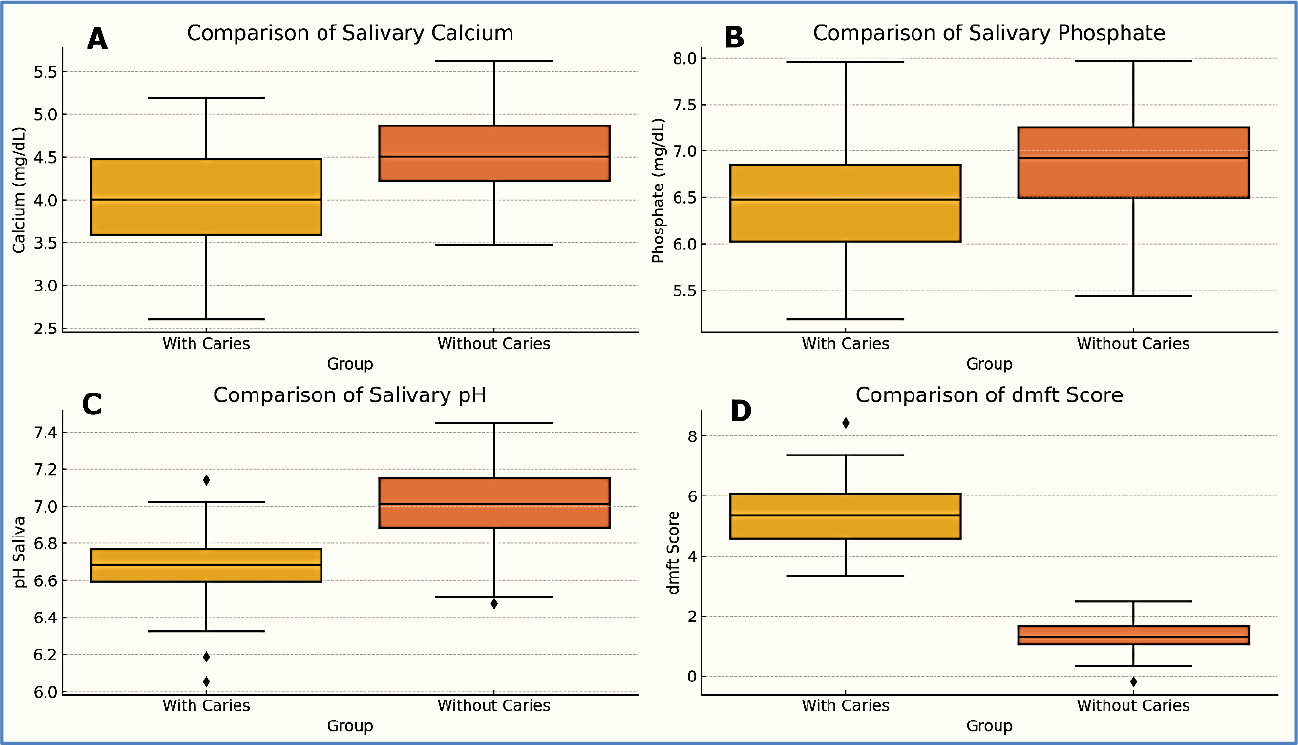
Salivary biochemical parameters are shown in Table 3 and Figure 1. Caries-positive children had significantly lower salivary calcium (2.84 ± 0.71 mg/dL) compared with caries-free children (3.29 ± 0.83 mg/dL) (independent t-test, p = 0.001). Phosphate levels were also lower in the caries-positive group (4.51 ± 0.92 mg/dL vs. 5.02 ± 1.03 mg/dL; p = 0.006). Salivary pH was significantly reduced in caries-positive children (6.74 ± 0.23) compared with caries-free children (6.89 ± 0.21; p < 0.001). Salivary flow rate was likewise lower in the caries-positive group (0.39 ± 0.12 mL/min vs. 0.45 ± 0.10 mL/min; p = 0.002). Buffering capacity differed significantly between groups (p = 0.003).

Comparisons of salivary calcium, phosphate, pH, and dmft scores between caries-positive and caries-free children. Children without caries exhibited higher salivary calcium and phosphate concentrations and higher pH values. Conversely, caries-positive children presented significantly higher dmft scores (p < 0.05), indicating a consistent relationship between reduced salivary mineral content and caries severity.
Microbiological findings are presented in Table 4, with gene expression illustrated in Figure 2. Median Streptococcus mutans counts were significantly higher in caries-positive children (1.3 × 10⁵ CFU/mL) than in caries-free children (3.9 × 10⁴ CFU/mL) (p < 0.001). The proportion of children exceeding the clinical threshold of 105 CFU/mL differed significantly between groups (p < 0.001). gtfB gene expression was significantly higher in caries-positive children (∆∆Ct 1.92 ± 0.65) than in caries-free children (p < 0.001).

Relative expression levels of the gtfB gene in Streptococcus mutans among caries-positive and caries-free children. Expression was quantified using SYBR Green-based quantitative PCR and normalized to 16S rRNA using the ∆∆Ct method. Caries-positive children demonstrated significantly higher gtfB expression (p < 0.001), indicating increased virulence and biofilm-forming potential.
Results of the multivariate logistic regression analysis are shown in Table 5. Low salivary calcium (<3.0 mg/dL), low salivary pH (<6.8), high S. mutans load (>105 CFU/mL), frequent sweet food intake (>2 times/day), and low socioeconomic status were independently associated with caries status (p < 0.01; 95% CI). A high S. mutans load was associated with the largest adjusted odds ratio (OR = 3.41; 95% CI: 1.82-6.41).
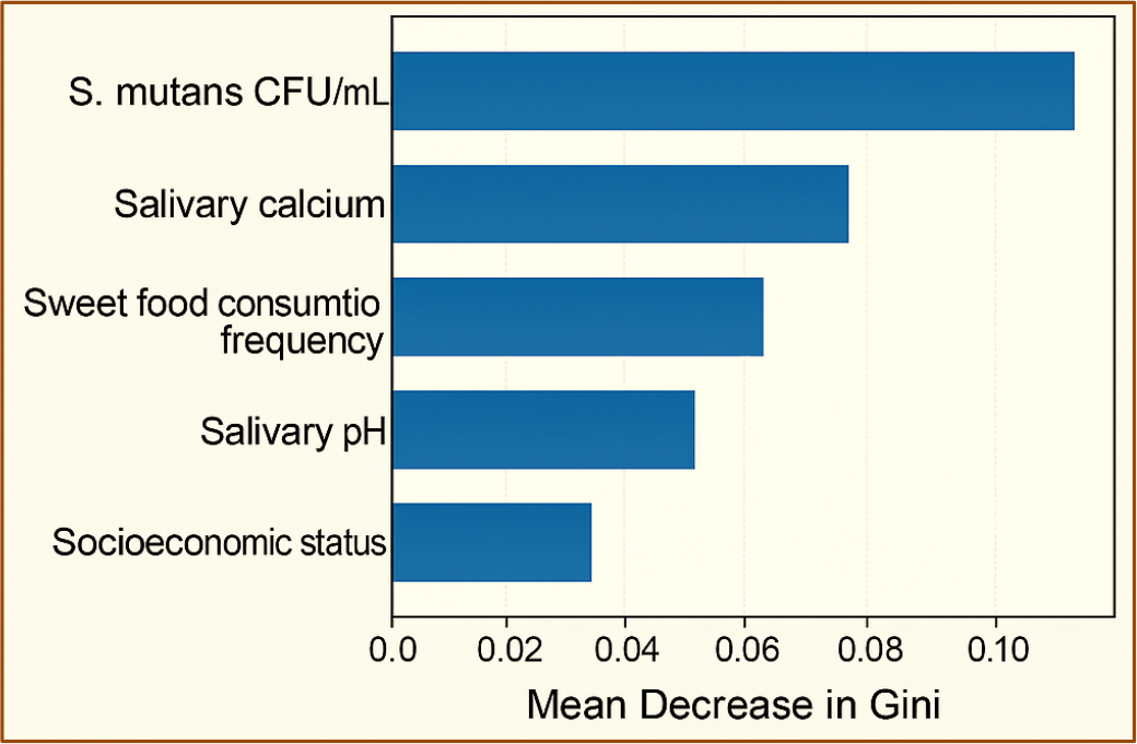
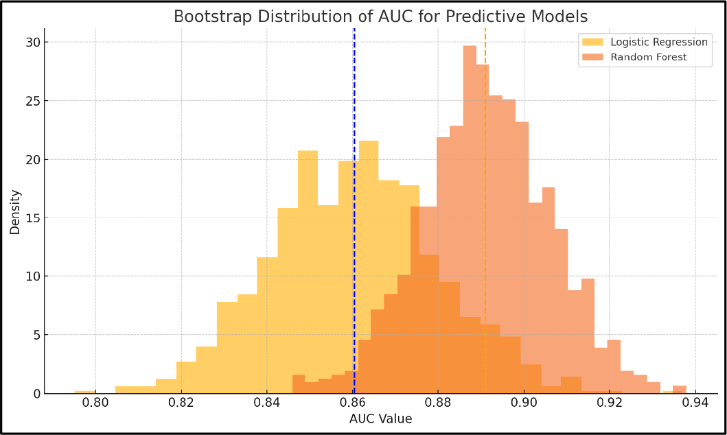
Model performance metrics are summarized in Table 6, and feature importance and ROC distributions are shown in Figures 3 and 4, respectively. The Random Forest model achieved an AUC of 0.905, an accuracy of 0.86, a sensitivity of 0.88, and a specificity of 0.81, compared with an AUC of 0.872 and an accuracy of 0.79 for logistic regression. Bootstrapped AUC distributions demonstrated narrower dispersion for the Random Forest model (Figure 4).

Feature importance ranking of predictors included in the Random Forest classification model for dental caries risk. Importance values represent mean decreases in Gini impurity across 1,000 bootstrap samples. S. mutans CFU/mL, salivary calcium concentration, sweet food consumption frequency, salivary pH, and socioeconomic status were identified as the top five contributors to caries prediction.

Kernel density estimation plots showing the bootstrapped distribution of Area Under the Curve (AUC) values for Logistic Regression (yellow) and Random Forest (orange) models across 1,000 iterations. Dashed vertical lines represent mean AUC values. The Random Forest model exhibited a higher, more concentrated AUC distribution (mean 0.905) than logistic regression (mean 0.872), indicating superior predictive performance and greater model stability.
Feature importance ranking (Figure 3) identified S. mutans CFU/mL, salivary calcium, frequency of sweet food intake, salivary pH, and socioeconomic status as the top-ranked variables.
Discussion
This study examined the combined roles of salivary biochemistry, microbial burden, virulence gene expression, oral hygiene behaviors, dietary habits, and socioeconomic factors in determining dental caries risk among primary school children in Banda Aceh. The findings reinforce the multifactorial nature of dental caries and illustrate how biological, behavioral, and social determinants interact to influence oral health. Rather than framing saliva as a diagnostic tool, this discussion emphasizes its role in a multidimensional caries risk assessment framework, in which salivary and microbial parameters serve as risk indicators rather than diagnostic endpoints.
The demographic and socioeconomic profile of participants (Table 1) showed that caries was significantly more prevalent among children from low-income households and those whose parents had only primary education. These findings are consistent with global evidence indicating that socioeconomic disadvantage is associated with reduced access to preventive dental care, lower oral health literacy, and less favorable dietary patterns [17]. The absence of significant differences in age, sex, and nutritional status between caries groups suggests that contextual and behavioral factors outweighed biological demographics in this population. Dietary and oral hygiene behaviors (Table 2) further support this interpretation, as frequent sugar consumption and infrequent toothbrushing were strongly associated with caries prevalence. These results align with extensive literature identifying sugar intake as a major driver of acidogenic bacterial activity and enamel demineralization and inadequate plaque control as a key contributor to caries development [18,19].
Salivary biochemical profiles (Table 3; Figure 2) demonstrated that children with caries exhibited significantly lower salivary calcium, phosphate, pH, and flow rate, indicating a less protective oral environment. The strong inverse association between salivary calcium and dmft scores (Figure 3) highlights calcium as an exceptionally robust indicator of caries susceptibility. This finding is consistent with previous studies showing that adequate salivary calcium and phosphate levels support enamel remineralization and counteract acid challenges [3]. The weaker association observed for phosphate compared with calcium may reflect modulation by pH and salivary flow rate, as suggested in prior research. Lower salivary pH and reduced buffering capacity further support earlier evidence that acidic, poorly buffered saliva promotes demineralization and cariogenic microbial growth [20,21].
Microbiological analysis (Table 4) revealed that caries-positive children had significantly higher Streptococcus mutans counts, frequently exceeding the clinical threshold of 105 CFU/mL. High bacterial load emerged as the strongest independent predictor of caries in multivariate analysis (Table 5), reaffirming the central role of S. mutans in caries initiation and progression. Elevated gtfB gene expression (Figure 1) further indicates enhanced biofilm-forming potential, consistent with previous studies demonstrating the importance of glucosyltransferase-mediated glucan synthesis in cariogenicity [22]. Detailed technical validation of the qPCR assay has been moved to the Appendix and is referenced here only briefly to maintain focus on biological interpretation. Molecular virulence indicators improve understanding of disease mechanisms but should be interpreted alongside salivary conditions and behavioral factors [23].
Although sex and age did not significantly influence salivary or microbial parameters (Tables 1-4), higher dmft scores were observed among younger children (6-8 years) and males (Table 3). These patterns may reflect developmental and behavioral factors, such as lower manual dexterity, higher sugar exposure, and less consistent oral hygiene practices among younger children and boys [24]. However, as sexand age-related differences vary across populations, these findings should be interpreted cautiously and contextualized within local behavioral norms rather than generalized assumptions.
Multivariate analysis (Table 5) identified low salivary calcium, low salivary pH, high S. mutans load, frequent consumption of sweet foods, and low socioeconomic status as independent predictors of caries. These findings underscore the cumulative and interactive nature of caries risk, supporting the view that no single factor alone can explain disease occurrence [25]. Predictive modeling (Table 6; Figures 3-4) showed that the Random Forest classifier outperformed logistic regression, demonstrating superior accuracy, sensitivity, specificity, and AUC. The narrow bootstrapped AUC distribution highlights the robustness of this approach for handling complex, multifactorial datasets, consistent with previous studies applying machine learning to early childhood caries prediction [26].
Notably, the most influential predictors identified by the Random Forest model closely mirrored those observed in logistic regression analysis, namely S. mutans load, salivary calcium, sweet food frequency, salivary pH, and socioeconomic status (Figure 3). This convergence strengthens the internal validity of the findings and highlights the value of integrating biological, behavioral, and social indicators into risk assessment algorithms. From a practical perspective, these results support the potential application of saliva-based risk indicators in school-based screening programs, primary healthcare settings, and community oral health initiatives to facilitate early identification of high-risk children and targeted preventive interventions. Machine-learning approaches such as Random Forests may further enhance early detection by synthesizing diverse data inputs into individualized risk profiles.
This study is limited by its cross-sectional design, which precludes causal inference. Purposive sampling restricts generalizability beyond the Banda Aceh context. Self-reported dietary and oral hygiene data may introduce recall bias. Key confounders, including fluoride exposure and parental oral health literacy, were not assessed, and no formal sample size calculation was performed. Future longitudinal studies with probability-based sampling are needed to validate these findings.
Conclusion
Dental caries in primary school children is associated with a combination of salivary biochemical factors, microbial burden, behavioral habits, and socioeconomic conditions. Lower salivary calcium and pH, higher Streptococcus mutans load, increased gtfB gene expression, frequent intake of sweet foods, and inadequate toothbrushing were significantly associated with the presence of caries. The Random Forest model demonstrated superior predictive performance, highlighting the value of integrating biological, behavioral, and social indicators for caries risk assessment. These findings support a multifactorial, risk-based approach rather than a diagnostic interpretation of saliva, and suggest that saliva-based indicators, combined with analytical models, may aid in the early identification of children at higher caries risk.
-
Financial Support
None.
Acknowledgments
The authors thank the participating schools, students, and parents in Banda Aceh for their cooperation. Special appreciation is extended to the laboratory staff and data collectors for their technical assistance and support during the study.
Data Availability
The data used to support the findings of this study can be made available upon request to the corresponding author.
ν References
-
[1] Syafitri FU. Knowledge assessment of bad habits in children's oral cavity related to malocclusion. J Syiah Kuala Dent Soc 2023; 8(1):16-23. https://doi.org/10.24815/jds.v8i1.33021
» https://doi.org/10.24815/jds.v8i1.33021 -
[2] Mallya PS, Mallya S. Microbiology and clinical implications of dental caries-A review. J Evol Med Dent Sci 2020; 9(48):3670-3675. https://doi.org/10.14260/jemds/2020/805
» https://doi.org/10.14260/jemds/2020/805 -
[3] Enax J, Fandrich P, Schulze zur Wiesche E, Epple M. The remineralization of enamel from saliva: A chemical perspective. Dent J 2024; 12(11):339. https://doi.org/10.3390/dj12110339
» https://doi.org/10.3390/dj12110339 -
[4] Rusu L-C, Roi A, Roi C-I, Tigmeanu CV, Ardelean LC. The influence of salivary pH on the prevalence of dental caries. In: Rusu LC, Ardelean LC. Dental Caries - The Selection of Restoration Methods and Restorative Materials. London: IntechOpen; 2022. https://doi.org/10.5772/intechopen.106154
» https://doi.org/10.5772/intechopen.106154 -
[5] Rezaei T, Mehramouz B, Gholizadeh P, Yousefi L, Ganbarov K, Ghotaslou R, et al. Factors associated with Streptococcus mutans pathogenicity in the oral cavity. Biointerface Res Appl Chem 2023; 13(4):368. https://doi.org/10.33263/BRIAC134.368
» https://doi.org/10.33263/BRIAC134.368 -
[6] Zhang Q, Ma Q, Wang Y, Wu H, Zou J. Molecular mechanisms of inhibiting glucosyltransferases for biofilm formation in Streptococcus mutans. Int J Oral Sci 2021; 13(1):30. https://doi.org/10.1038/s41368-021-00137-1
» https://doi.org/10.1038/s41368-021-00137-1 -
[7] Min H, Zhu S, Safi L, Alkourdi M, Nguyen BH, Upadhyay A, et al. Salivary diagnostics in pediatrics and the status of saliva-based biosensors. Biosensors 2023; 13(2):206. https://doi.org/10.3390/bios13020206
» https://doi.org/10.3390/bios13020206 -
[8] Bruins MJ, Bird JK, Aebischer CP, Eggersdorfer M. Considerations for secondary prevention of nutritional deficiencies in high-risk groups in high-income countries. Nutrients 2018; 10(1):47. https://doi.org/10.3390/nu10010047
» https://doi.org/10.3390/nu10010047 -
[9] Alanzi A, Husain F, Husain H, Hanif A, Baskaradoss J. Does the severity of untreated dental caries of preschool children influence the oral health-related quality of life? BMC Oral Health 2023; 23(1):552. https://doi.org/10.1186/s12903-023-03274-7
» https://doi.org/10.1186/s12903-023-03274-7 -
[10] Lamloum D, Dettori M, La Corte P, Agnoli MR, Cappai A, Viarchi A, et al. Oral health survey in Burundi: Evaluation of the caries experience in schoolchildren using the dmft index. Medicina 2023; 59(9):1538. https://doi.org/10.3390/medicina59091538
» https://doi.org/10.3390/medicina59091538 -
[11] Hossain MS, Alam S, Nibir YM, Tusty TA, Bulbul SM, Islam M, et al. Genotypic and phenotypic characterization of Streptococcus mutans strains isolated from patients with dental caries. Iran J Microbiol 2021; 13(4):449-457. https://doi.org/10.18502/ijm.v13i4.6968
» https://doi.org/10.18502/ijm.v13i4.6968 - [12] Maldupa I, Brinkmane A, Mihailova A. Comparative analysis of CRT Buffer, GC saliva check buffer tests and laboratory titration to evaluate saliva buffering capacity. Stomatologija 2011; 13(2):55-61.
-
[13] Razzaque MS. Salivary phosphate as a biomarker for human diseases. FASEB BioAdv 2022; 4(2):102-108. https://doi.org/10.1096/fba.2021-00104
» https://doi.org/10.1096/fba.2021-00104 -
[14] Fang Y, Ma Q, Zhao W, Liu N, Cheng L, Chen J, et al. A novel selective medium Sucrose-Bacitracin agar 10 for accurate isolation and identification of Streptococcus mutans. BMC Oral Health 2025; 25(1):1332. https://doi.org/10.1186/s12903-025-06675-y
» https://doi.org/10.1186/s12903-025-06675-y -
[15] Gandhi V, O'Brien MH, Yadav S. High-quality and high-yield RNA extraction method from whole human saliva. Biomark Insights 2020; 15:1177271920929705. https://doi.org/10.1177/1177271920929705
» https://doi.org/10.1177/1177271920929705 -
[16] Jaleel A, Chilumula M, Satya SGC, Singnale P, Telikicherla UR, Pandurangi R, et al. The assessment of nutritional status of adolescents aged 15-18 years using BMI cut-offs and BMI Z scores: A secondary analysis of national family health survey-5 (2019-21) data. Cureus 2024; 16(5):e59800. https://doi.org/10.7759/cureus.59800
» https://doi.org/10.7759/cureus.59800 -
[17] Chamut S, Alhassan M, Hameedaldeen A, Kaplish S, Yang AH, Wade CG, et al. Every bite counts to achieve oral health: A scoping review on diet and oral health preventive practices. Int J Equity Health 2024; 23(1):261. https://doi.org/10.1186/s12939-024-02279-0
» https://doi.org/10.1186/s12939-024-02279-0 -
[18] Chen X, Daliri EB-M, Tyagi A, Oh D-H. Cariogenic biofilm: Pathology-related phenotypes and targeted therapy. Microorganisms 2021; 9(6):1311. https://doi.org/10.3390/microorganisms9061311
» https://doi.org/10.3390/microorganisms9061311 -
[19] Church L, Franks K, Medara N, Curkovic K, Singh B, Mehta J, et al. Impact of oral hygiene practices in reducing cardiometabolic risk, incidence, and mortality: A systematic review. Int J Environ Res Public Health 2024; 21(10):1319. https://doi.org/10.3390/ijerph21101319
» https://doi.org/10.3390/ijerph21101319 -
[20] Inchingolo AD, Malcangi G, Semjonova A, Inchingolo AM, Patano A, Coloccia G, et al. Oralbiotica/oralbiotics: The impact of oral microbiota on dental health and demineralization: A systematic review of the literature. Children 2022; 9(7):1014. https://doi.org/10.3390/children9071014
» https://doi.org/10.3390/children9071014 -
[21] Poza-Pascual A, Serna-Muñoz C, Pérez-Silva A, Martínez-Beneyto Y, Cabello I, Ortiz-Ruiz AJ. Effects of fluoride and calcium phosphate-based varnishes in children at high risk of tooth decay: A randomized clinical trial. Int J Environ Res Public Health 2021; 18(19):10049. https://doi.org/10.3390/ijerph181910049
» https://doi.org/10.3390/ijerph181910049 -
[22] Atta L, Mushtaq M, Siddiqui AR, Nur-e-Alam M, Ahmed A, Ul-Haq Z. Functional characterization of residues affecting the catalytic activity of glucosyltransferase from Streptococcus mutans. J Phys Chem B 2025; 129(21):5091-5103. https://doi.org/10.1021/acs.jpcb.4c07136
» https://doi.org/10.1021/acs.jpcb.4c07136 -
[23] Souchet B, Michaïl A, Billoir B, Braudeau J. Biological diagnosis of alzheimer's disease based on amyloid status: An illustration of confirmation bias in medical research? Int J Mol Sci 2023; 24(24):17544. https://doi.org/10.3390/ijms242417544
» https://doi.org/10.3390/ijms242417544 -
[24] Spatafora G, Li Y, He X, Cowan A, Tanner AC. The evolving microbiome of dental caries. Microorganisms 2024; 12(1):121. https://doi.org/10.3390/microorganisms12010121
» https://doi.org/10.3390/microorganisms12010121 -
[25] Eusufzai S, Jamayet N, Ahmed S, Islam M, Ahmad W, Alam M. Development and evaluation of an early childhood caries prediction model: A deep learning-based hybrid statistical modelling approach. Eur Arch Paediatr Dent 2025; 26(5):953-964. https://doi.org/10.1007/s40368-025-01046-1
» https://doi.org/10.1007/s40368-025-01046-1 -
[26] Huang Y. Exploring the principle of multidimensional risk analysis and a case study in two-dimensional risk. Risks 2025; 13(4):79. https://doi.org/10.3390/risks13040079
» https://doi.org/10.3390/risks13040079
Edited by
-
Academic Editor:
Carina Silva-Boghossian