Evaluation of Antimicrobial Action, Cytotoxicity, pH, Flow, and Radiopacity of a New Filling Paste for Primary Teeth
ABSTRACT
Objective: To evaluate the antimicrobial action, cytotoxicity, pH, flow, and radiopacity of an experimental paste for primary teeth.
Material and Methods: Root canals of 50 extracted human teeth were inoculated with Enterococcus faecalis and divided into five groups (n=10): G1 - distilled water (negative control); G2 - zinc oxide-eugenol (ZOE) paste; G3 - calcium hydroxide (Ca(OH)2) paste; G4 - iodoform paste; and G5 - experimental paste containing chlorhexidine, (Ca(OH)2), neomycin sulfate, and barium sulfate. Colony-forming unit counts assessed antimicrobial activity. Cytotoxicity was analyzed in fibroblast cells through cell viability assays. pH and flow were measured with colorimetric strips and dispersion diameter, respectively. Radiopacity was compared to an aluminum scale using digital radiography. Statistical analysis was performed at α=5%.
Results: High antimicrobial activity and radiopacity were observed in groups 2 (ZOE paste), 3 (Ca(OH)2 paste), 4 (iodoform paste), and 5 (experimental paste) (p<0.05). The highest cell viability was found in groups 1 (distilled water), 4 (iodoform paste), and 5 (experimental paste) (p<0.05). Groups 3 and 5 showed the highest pH values, while flow was highest for groups 3 (Ca(OH)2 paste), 4 (iodoform paste), and 5 (experimental paste) (p<0.05).
Conclusion: The experimental paste demonstrated a satisfactory antimicrobial effect, cytocompatibility, an alkaline pH, adequate flow, and radiopacity, comparable to conventional pastes, and has potential for clinical application.
Keywords:
Chlorhexidine; Root Canal Filling Materials; Tooth; Deciduous.
Introduction
The endodontic filling material must possess several properties to be considered suitable for use in primary teeth. Among these properties, antimicrobial activity is essential to neutralize microorganisms in the root canal system, and the absence of cytotoxicity is required to prevent inflammatory reactions in the periapical tissues. It should also have a resorption rate similar to that of the primary root. In addition, the endodontic filling material must be able to raise the pH to inhibit bacterial growth, flow easily into the complex anatomy of primary root canals, and provide sufficient radiopacity for visualization on imaging [1].
According to the literature, there is no consensus on the filling protocol for primary teeth, and the available materials do not meet all requirements for satisfactory fillings. Zinc oxide and eugenol (ZOE)-based paste is associated with severe inflammatory reactions when extravasated into the periapical tissues due to its cytotoxicity, and with deviations from the natural position during the eruption of permanent teeth due to its hardness [2]. In your turn, calcium hydroxide-based pastes present severe reabsorption and low radiopacity [3]. Finally, iodoformed pastes have a strong, characteristic odor, the potential to change the color of dental structures, and cytotoxic effects [3,4]. Given these limitations, there is a need for new alternatives to filling materials for primary teeth.
Recently, an experimental paste for filling of primary teeth was developed, composed of chlorhexidine (CHX), neomycin sulfate, calcium hydroxide (Ca(OH)2), and barium sulfate (BaSO4) (the authors hold the patent for this filling material). Chlorhexidine exhibits recognized broad-spectrum antimicrobial activity and biocompatibility [5]. Similarly, neomycin sulfate is associated with the attenuation of inflammatory reactions and effective antimicrobial action [6]. Calcium hydroxide has antimicrobial and anti-inflammatory actions, biocompatibility, induces pH elevation and periapical repair by promoting mineral tissue deposition, and can inactivate endotoxins [7]. Finally, barium sulfate is the most widely used agent to confer radiopacity on some commercially available pastes containing calcium hydroxide [8]. Considering the properties of each component, this new filling paste was developed for testing as an alternative filling material for primary teeth.
This study aimed to evaluate the antimicrobial action, cytotoxicity, pH, flow, and radiopacity of a new filling paste for primary teeth. The hypotheses were that this new filling paste (i) presents effective antimicrobial activity, (ii) induces low levels of cytotoxicity, (iii) presents alkaline pH, (iv) satisfactory flow, and (v) radiopacity.
Material and Methods
Ethical Clearance and Sample
The present study was approved by the Institutional Review Board (Protocol 5.022.728).
The BioEstat 5.0 statistical package (Fundação Mamirauá, Belém, PA, Brazil) was used to calculate sample sizes for all evaluation tests. A minimum of 10 samples per group was required for the antimicrobial evaluation. At the same time, a minimum of 5 samples per group was required for pH, flow, and radiopacity measurements. It was considered that the test power was 80% and the alpha error rate was 5% to estimate the sample size for the analyzed outcomes. The cytotoxicity evaluation was performed according to previous studies evaluating the antimicrobial activity of intracanal decontamination protocols [9,10].
Antimicrobial Action Evaluation
Fifty single-rooted human extracted permanent teeth were selected, presenting straight roots and complete root apices. The dental crowns were sectioned so that all roots retained a length of 15 mm. The root canals were enlarged in all extensions using the ProTaper system (Dentsply-Maillefer, Ballaigues, Switzerland) up to the F3 file and NaOCl (Farmácia Natupharma Manipulação & Medicamentos, Passo Fundo, RS, Brazil) as the irrigant, to standardize the final root canal diameter. Final irrigation with 3 mL of 17% EDTA (Biodinâmica Química e Farmacêutica, Ibiporã, PR, Brazil) and 5 mL of distilled water (DW) was performed. Finally, the root canals were dried with absorbent paper points.
The 50 roots were fixed with Putty-C Silicone for impression (Silon 2 APS, Dentsply Indústria e Comércio, Petrópolis, RJ, Brazil) in plastic micro-tubes (Axygen Inc, Union City, CA, USA), so that they remained upright with the cervical portion facing upwards. After this, the teeth were divided into five groups (n=10), and each group was placed in a polypropylene box (Heathrow Scientific, Vernon Hills, IL, United States). The samples were sterilized in the box at 120 °C in an autoclave (Kavo Dental, Joinville, SC, Brazil) for 30 min.
The reference strain was Enterococcus faecalis (E. faecalis) (ATCC 19433), which was cultivated in brain-heart infusion (BHI) broth (Acumedia-Neogen Corp., Lansing, MI, USA) for 24 h at 37 °C in a bacteriological incubator (Kavo Dental, Joinville, SC, Brazil). The turbidity degree was adjusted to the McFarland’s 1.0 scale, corresponding to 3.0 ×10⁸ CFU/mL. The optical density of the tube was 0.25, measured at 550 nm. A 100-μL culture aliquot was inoculated into the root canal of each sample until extravasation to the root canal entrance. The culture was maintained for 14 days for biofilm formation, and the remaining volume was replaced every 48 h with sterile BHI. Once a week, one BHI aliquot was collected from a randomly selected specimen from each group and submitted for Gram staining and culture on blood agar, followed by catalase and esculin tests, to verify the absence of other microorganisms.
After contamination, the 50 samples were irrigated with 5 mL of DW and randomly distributed into five groups (n=10), according to the tested protocol:
Group 1 DW (negative control): DW was inserted using a 3 mL disposable syringe (Descarpack Descartáveis do Brasil, São Paulo, SP, Brazil) and a 30-gauge needle (Navi-Tip, Ultradent Products, Inc., South Jordan, UT, USA) until the root canals were filled.
Group 2 ZOE paste: the filling material was prepared by mixing 0.1g of zinc oxide powder with 100 μL of liquid eugenol until a paste-like consistency was reached. Then, the paste was inserted with a Lentulo drill (Dentsply-Maillefer), placed 3 mm short of the working length, and activated until the root canals were filled.
Group 3 Ca(OH)2 paste: the commercially available calcium hydroxide paste (Calen - SSWhite Dufllex, Rio de Janeiro, RJ, Brazil) was dispensed over the plate. After this, the paste was inserted with a Lentulo drill (Dentsply-Maillefer, Ballaigues, Switzerland), placed 3 mm short of the working length, and activated until the root canals were filled.
Group 4 Iodoform paste: the commercially available iodoform paste (Feapex - Fórmula e Ação Farmácia, São Paulo, SP, Brazil) was dispensed over the plate. After this, the paste was inserted into the root canals, as previously described for groups 2 and 3.
Group 5 Experimental paste: 3 mL of 2% chlorhexidine gel, 0.15 g of neomycin sulfate powder, 7.5 g of pure Ca(OH)2 powder, and 4.5 g of barium sulfate powder were used to prepare this filling material. Stabilizer and conservative solutions were also added, in a proportion protected by the patent. The solid components were separated in a glass vial, sieved, and weighed on a high-precision digital analytical balance. The required amount of each component was weighed in grams (g). The liquid and gel components were separated in graduated cylinders, being measured in milliliters (mL). The experimental paste was mixed in a porcelain mortar with a porcelain pestle. Geometric dilution was performed to achieve high homogeneity of the components: the solid components were first dispensed into a porcelain mortar, followed by the addition of the liquid and gel components, which were slowly incorporated into the solid components. The experimental paste was obtained by intense manipulation to homogenize the components. Subsequently, the formulation was transferred onto a marble surface and spatulated with plastic spatulas to homogenize further. After manipulation, the experimental paste was stored in a hermetic double-walled polyethylene bottle. The bottle was stored at room temperature in closed cabinets and without exposure to light. The experimental paste was inserted into the root canals, as previously described for groups 2, 3, and 4. Figure 1 illustrates the experimental paste before mixing the components, immediately after handling, and after storage.

Illustrative images of the tested experimental paste before mixing the components, immediately after handling, and after storage.
After root canal filling, the roots of all groups were sealed with provisional restorative material (Cavitec - Caitthec Indústria e Comércio Ltda, Rio do Sul, SC, Brazil) and stored for 14 days at 37 °C and 100% humidity. After this time, the provisional restorative material was removed with a sterile dentin excavator, and the site was irrigated with 5 mL of DW to remove the filling material.
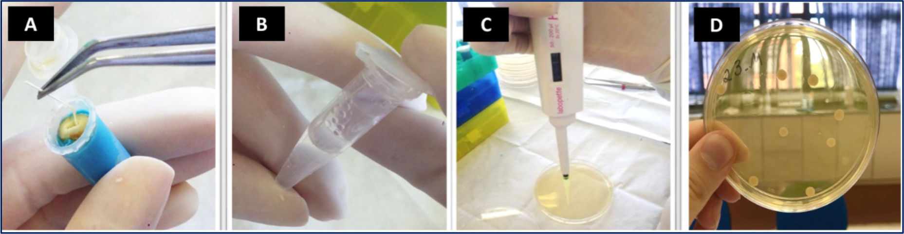
The microbiological analysis was performed in two stages: initial sample (S1) - after contamination and before filling material protocols; final sample (S2) - after filling material removal. In both stages, the root canals were irrigated with 5 mL of DW and filled with sterile saline solution. Then, a sterile K-file #30 (Dentsply-Maillefer, Ballaigues, Switzerland) was inserted to promote contact with the walls for 30 seconds. Then, a sterile absorbent paper point was inserted into the root canal for 30 seconds, and the point was transferred to a tube containing 450 μL of sterile saline solution. The material was homogenized and diluted to 1x10⁻³. Aliquots of 100 μL of the solution and each dilution were cultured on blood agar plates in duplicate; these samples were incubated for 18-24 h at 37 °C. Then, the number of colony-forming units (CFUs) was counted on the plates. These procedures are illustrated in Figure 2. The effectiveness of the tested protocols was assessed by the percentage reduction in E. faecalis from the initial (S1) to the final (S2) samples.
Illustrative images of antimicrobial action evaluation after the tested protocols: (A) - sterile absorbent paper point working into the root canal; (B) - tube containing 450μL of sterile saline solution where the content of absorbent paper point was transferred; (C) - aliquots of 100μL of each dilution being cultivated on the blood agar plate; (D) - CFUs counting on the plates.
Cytotoxicity Evaluation
L929 gingival fibroblastic cells were obtained from the Laboratory of Applied Virology of the Federal University of Santa Catarina (UFSC, Florianópolis, SC, Brazil). For cell maintenance, Minimum Essential Medium (Sigma-Aldrich Brasil Ltda., Rio de Janeiro, RJ, Brazil) supplemented with 10% fetal bovine serum (Invitrogen Thermo Fisher Scientific Inc., São Paulo, SP, Brazil) was used, and cells were kept in 75 cm2 culture flasks at 37 °C and 5% CO2.
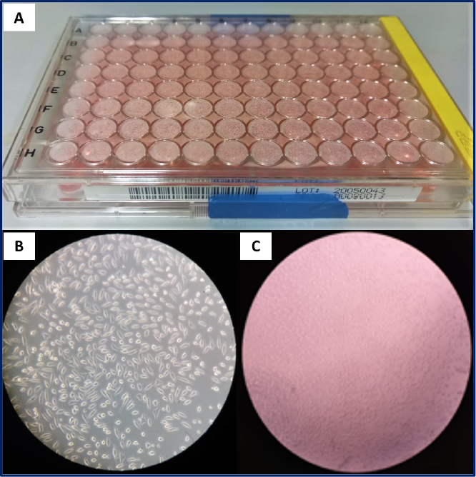
The assay was performed using 3-(4,5-dimethylthiazol-2-yl)-2,5-diphenyltetrazolium bromide (MTT; Sigma-Aldrich Brasil Ltda., Rio de Janeiro, RJ, Brazil), a colorimetric assay for evaluating cell viability. The L929 cells were trypsinized, counted, and distributed into 96-well plates at a concentration of 1 × 10⁵ cells per well (Figure 3). The Dulbecco's Modified Eagle Medium (DMEM) (Sigma-Aldrich Brasil Ltda., Rio de Janeiro, RJ, Brazil) was used, enriched with 5% fetal bovine serum. After 24 hours of incubation at 37 °C in an oven with 5% CO2, cell confluence was observed under an inverted microscope. Then, the DMEM medium was removed by aspiration, and the tested filling materials were added, being incubated for 1 hour. In the negative control group 1 (DW), 100 μL of DW was added. In groups 2 (ZOE paste), 3 (Ca(OH)2 paste), 4 (Iodoform paste), and 5 (experimental paste), 50 mg of the filling material was added.

Illustrative images of cytotoxicity evaluation: (A) - box containing the well plates where the L929 fibroblastic cells were distributed at a concentration of 1 × 10⁵ cells per well; (B) - viable L929 fibroblastic cells before the tested protocols; (C) - nonviable L929 fibroblastic cells after the tested protocols.
After 1 hour, the wells were washed with 200 μL of sterile PBS, and 50 mL of MTT solution (1 mg/mL in DMEM) was added, followed by incubation for 4 hours. MTT was carefully removed to avoid cell damage, and 100 mL of dimethylsulfoxide (DMSO) was added to solubilize the formazan crystals. DMSO was also added to empty wells for white colorimetric calculation. The plate was placed in the mixer for 10 min, and absorbance was measured using a 490 nm filter.
All experiments were performed in triplicate, and cell viability percentages were calculated relative to the negative control group using the following formula: viability (%) = (sample absorbance - mean blank absorbance) × 100 / (control absorbance - mean blank absorbance).
pH Evaluation
Colorimetric pH determination was used to measure the pH of each filling paste. This technique is based on the use of paper strips that change color according to the pH, associating the color with a pH value that can be acidic (0-6), neutral (=7), or alkaline (8-14), when these paper strips come into contact with the tested filling pastes (Figure 4 - C). In this way, the color change is observed, and the pH measurement is performed.

Illustrative images of (A) and (B) - flow evaluation by using the mold plate and measurement on graph paper; (C) - pH evaluation by using the colorimetric paper strips; (D) - radiopacity evaluation by using the aluminum stepwedge.
The filling pastes were prepared as previously described in the evaluation of antimicrobial action. Suspensions of tested filling pastes in sterile water were prepared at a concentration of 100 mg/mL as described by Zhang et al. [11]. The pH was determined in 4 mL of each suspension, stored in closed 10-mL BD Falcon test tubes, and deionized water (Nalgene, Rochester, NY, USA), thereby eliminating the effects of environmental factors until the measurement time throughout the experimental period. Before measurements, the suspensions were vortexed for 30 seconds. The pH was recorded immediately after mixing for 30 seconds and after 14 days.
The colorimetric paper strips were placed in contact with the homogenized mixture from each group for 30 seconds. After this period, the paper strip was removed, and the pH value result was immediately recorded for each group. After 14 days of storage at room temperature in closed cabinets and without exposure to light, the mixture was homogenized again by vortexing for 30 seconds, and a new pH measurement was performed. Five samples per group were prepared, and measurements were performed in triplicate in each evaluation period. The pH values of each tested filling paste were recorded immediately and after 14 days, and the mean pH for each group was calculated and subjected to statistical analysis.
Flow Evaluation
The flow evaluation was performed immediately after preparation of the tested filling pastes and again after 14 days, for both the commercially available and prepared pastes. The prepared pastes were stored in hermetic, double-walled polyethylene bottles. In the device used to determine the flow, a circular glass mold plate (diameter = 20 cm; thickness = 0.2 cm) with a central hole of 1.2 cm in diameter was placed on a glass support plate (20 cm x 20 cm), positioned on a millimeter scale, and a light source. The tested filling pastes were introduced into the hole of the mold plate, and the surface was leveled with a spatula.
The mold plate was carefully removed, and a 1000 g glass plate was placed over the tested filling pastes. After 1 minute, the diameters of the filling paste were measured at two opposite positions using the scale on the millimeter paper. Subsequently, the average diameter was calculated. After 1 minute, a second 1000 g glass plate was placed over the first, and a second measurement of the mean diameter was performed. After 1 minute, a third 1000 g glass plate was placed over the previously positioned plates containing the tested filling paste, and a third measurement was performed. The three measurements were recorded (in mm) (Figure 4 - A and B). Five samples per group were evaluated.
After 14 days of storage at room temperature in closed cabinets, without exposure to light, all procedures were repeated, yielding a second average of the three measurements (in mm) for each sample of each filling paste. The descriptive analysis of the flow of each tested filling paste was performed immediately and after 14 days, and a mean flow value was assigned to each group, which was then submitted to statistical analysis.
Radiopacity Evaluation
This evaluation was performed in accordance with the ISO 6876/2001 specifications. After dispensing onto a glass plate or handling the tested filling pastes as described in the previous evaluations, the filling pastes were inserted into a polycarbonate mold with a 10 mm diameter and 1 mm thickness, forming a specimen of the respective tested filling paste. In total, 5 specimens were fabricated for each group. All specimens from each group were positioned on an intraoral occlusal phosphor plate measuring 5.7 × 7.6 cm (Dürr Dental, Bietigheim-Bissingen, Germany), next to a graduated aluminum step wedge with thicknesses ranging from 0.5 to 5 mm (in 0.5 mm increments) (Figure 4 - D).
The radiographic image was obtained with a conventional X-ray machine (Dabi Atlante, Ribeirão Preto, SP, Brazil) at 70 kVp and 8 mA. The radiographic recording was performed with an exposure time of 0.3 seconds and a focal length of 30 cm. After radiographic imaging, the intraoral occlusal phosphor plate for each specimen in each group was scanned with a digital scanner (VistScan Mini View - Dürr Dental, Bietigheim-Bissingen, Germany), and the resulting images were imported into Viewbox Studio (Dürr Dental, Bietigheim-Bissingen, Germany).
The radiographic density values obtained were converted into mm Al. The software's equal-density tool was used to identify areas of equal density. The means and standard deviations of the grey levels (pixel density) of the aluminum stepwedge and the specimens were obtained in a standardized central area of 1.5 mm2. Five samples per group were evaluated, and the results were analyzed by averaging the five measurements per sample.
Statistical Analysis
The Kolmogorov-Smirnov test was used to assess the normality of the data distribution. The one-way ANOVA followed by Tukey post-hoc tests was used for intergroup comparison in all evaluations (α=5%). All statistical analyses were performed with Stat Plus software, version 6.0 (Stat Plus Analyst Soft Inc., Vancouver, BC, Canada).
Results
The mean ± standard deviation of antimicrobial activity (% of bacterial reduction), cytotoxicity (% of cell viability), pH values, flow (mm), and radiopacity (mm Al) for each group are expressed in Table 1.
Mean ± standard deviation of antimicrobial activity (% of bacterial reduction), cytotoxicity (% of cell viability), pH values, flow (mm), and radiopacity (mm Al) for each group.
In the antimicrobial action evaluation, no statistically significant differences were found among groups 2 (ZOE paste), 3 (Ca(OH)2 paste), 4 (Iodoform paste), and 5 (Experimental paste) (p>0.05). At the same time, all experimental groups were significantly different from the negative control group (p<0.05). In the cytotoxicity evaluation, the highest percentage of viable cells was observed in groups 1 (negative control), 4 (Iodoform paste), and 5 (Experimental paste), with no statistically significant differences among them (p>0.05). The lowest percentage of viable cells was observed in group 2 (ZOE paste), followed by group 3 (Ca(OH)2 paste), and was statistically different from all other groups (p<0.05).
In the pH evaluation, the highest pH values were observed for groups 3 (Ca(OH)2 paste) and 5 (Experimental paste), with no statistically significant differences between them (p>0.05). Both groups differed significantly from all other groups (p<0.05). In the flow evaluation, the highest flow was observed for groups 3 (Ca(OH)2 paste), 4 (Iodoform paste), and 5 (Experimental paste), with no statistically significant differences between them (p>0.05). These groups differed significantly from all other groups (p<0.05).
In the radiopacity evaluation, no statistically significant differences were found among groups 2 (ZOE paste), 3 (Ca(OH)2 paste), 4 (Iodoform paste), and 5 (Experimental paste) (p>0.05). At the same time, all experimental groups were significantly different from the negative control group (p<0.05).
Discussion
The antimicrobial activity of filling materials used in endodontic treatment of primary teeth plays a significant role in reducing the number of microorganisms remaining in the endodontic space. Moreover, these materials should be harmless to the periapical tissues and permanent tooth germs, given the physiological process of root resorption [1]. Despite significant advances in techniques, resources, and materials, endodontic treatment of primary teeth still presents several challenges. This can be explained by the anatomical characteristics of the root canal, the importance of maintaining the primary tooth until complete exfoliation, and the lack of a standardized root canal filling material for primary teeth. The ZOE paste has been used for a long time in the endodontics of primary teeth [12]. However, due to its irritating potential and low resorption ability, materials containing (Ca(OH)2) or iodoform have been recommended, especially due to their antimicrobial activity, biocompatibility, and easy resorption [13]. At the same time, these pastes also presented limitations in the endodontic therapy of primary teeth [3,4]. In this scenario, the present study proposed a new filling paste for primary teeth to meet some of the requirements for clinical use.
According to literature, the mineral density for dentin of primary teeth is significantly lower when compared to dentin of permanent teeth [14]. At the same time, collagen degradation is faster and more substantial in primary teeth [15]. The primary dentin also exhibits a greater number of dentin tubules, leading to a reduced intertubular dentin area, reduced buffering capacity, and increased diffusion of antimicrobial agents [16]. Despite these differences, it is very difficult to use primary teeth in the in vitro studies. This is because when a primary tooth is removed or lost, the root is almost completely resorbed. Therefore, it is not possible to evaluate intracanal decontamination protocols on the roots of primary teeth. For this reason, permanent teeth were used in the antimicrobial evaluation of the present study. Furthermore, similarities between the dentin of primary and permanent teeth have been reported, including demineralization rates, organic and inorganic content, and microhardness [15,17]. Thus, the root canal of permanent teeth serves as an alternative in antimicrobial evaluations like this.
The cytotoxicity evaluation was performed in accordance with previous studies on intracanal decontamination protocols [9,10]. The MTT assay is a simple, reproducible, and widely used method to assess the cytotoxicity of decontamination protocols commonly used in endodontic therapy [18]. The fibroblastic cells used in this evaluation are diploid, continuous host cells, which are more likely to exhibit cytotoxic effects in response to dental materials or chemical substances [19]. In your turn, the use of paper strips that change color with the pH of the tested substance is well recommended in the literature, as a practical, fast, and easy-to-reproduce method [20]. The flow evaluation was performed according to the principles of a previous study [21], enabling assessment of the ability of the tested filling pastes to flow under significant stress over time, thereby reproducing the conditions necessary for flow. Finally, the radiopacity evaluation was performed in accordance with the ISO 6876/2001 specifications. This is a regulatory standard for evaluating the radiopacity of dental materials. Considering these points, the present study carried out this set of evaluations.
E. faecalis is a highly resistant, facultatively anaerobic microorganism. It shows virulence factors, the ability to persist under nutrient limitation, and the ability to colonize root canals in a biofilm, making its removal difficult [22]. For these reasons, E. faecalis was chosen as the sample contaminant in the present study, posing a significant challenge to the decontamination process using the tested protocols. In this proposal, the experimental paste demonstrated effectiveness in eliminating E. faecalis from root canals, confirming the first hypothesis of the present study. The composition of the experimental paste was responsible for this high antimicrobial activity. The CHX induces changes in cellular osmotic equilibrium, resulting in the precipitation of cytoplasmic content and the death of microorganisms [5]. Neomycin sulfate penetrates microbial cells via active oxygen transport and inhibits microbial protein synthesis, leading to cell death [6]. Finally, Ca(OH)2 becomes alkaline in the root canal space, denatures microbial proteins, and damages bacterial cell membranes [7]. In this way, the combination of the distinct mechanisms of action of the components resulted in effective antimicrobial activity of the experimental paste against E. faecalis.
Ca(OH)2 is recognized for its ability to promote tissue necrosis, which is why it is recommended in cases of root resorption [23]. At the same time, it induces chronic inflammation and reduces cell viability when it comes into contact with connective tissue [24]. This was observed in the present study, where the use of calcium hydroxide paste significantly reduced the number of viable fibroblast cells. Despite the presence of Ca(OH)2 in their composition, the iodoformed and experimental pastes exhibited the highest cell viability. It confirms the second hypothesis of the present study. This can be explained by a lower Ca(OH)2 concentration and its dilution by the other components in the tested pastes. In your turn, the ZOE paste showed the lowest cell viability levels, consistent with findings in the literature, which also reported its irritating and cytotoxic effects [2].
Given that the acidic environment is most conducive to microbial proliferation, it is essential to alkalinize the root canal space to neutralize microbial content [22]. According to the results of the present study, the experimental paste yielded the highest pH values, similar to those of the group treated with the Ca(OH)2 paste. It confirms the third hypothesis of the present study. The experimental paste contains Ca(OH)2 and releases hydroxyl ions (OH-). As a consequence of prolonged release, the environment of the root canal space becomes significantly alkaline [7]. In this way, the pH increases, representing an important mechanism of antimicrobial action of the tested experimental paste.
On the other hand, the tested iodoform paste showed a slight increase in pH, despite the presence of Ca(OH)2 in its composition. The tested iodoform paste presents silicone oil as a vehicle. It is a very viscous vehicle, and it may contribute to a slower, more gradual release of hydroxyl ions from Ca(OH)2, necessitating a longer time to achieve a satisfactory elevation of pH. Perhaps in periods longer than 14 days of observation, the pH levels could be higher.
Flowability is a physical property related to a material's ability to flow under pressure and is a basic requirement for filling materials in endodontics [1]. The experimental paste demonstrated the highest flow values, similar to those of Ca(OH)2 and iodoform pastes. The CHX has high diffusion as a vehicle for the experimental paste [25]. It confirms the fourth hypothesis of the present study. It ensures that the experimental paste can penetrate the depths of dentinal tubules and anatomical complexities, adhere to the root canal walls, and provide satisfactory sealing of the root canal space.
On the other hand, radiopacity is also a basic requirement of any filling material, allowing assessment of whether the canal has been adequately filled and distinguishing it from anatomical structures [1]. According to the results of the present study, the radiopacity of the experimental paste is in accordance with the requirements of ISO 6876/2001, which specifies a minimum of 3 mm Al for root canal filling materials. It confirms the fifth hypothesis of the present study. Barium sulfate is a radiopacifier commonly added to filling material formulations. Similarly, this substance was added to the experimental paste, ensuring a radiopacity value greater than 9 mm Al in the radiopacity test. Therefore, the experimental paste presented satisfactory flow and radiopacity in the present study.
The primary limitation of this study was the inability to use primary teeth in in vitro assays due to physiological root resorption and other intrinsic factors, which necessitated the use of permanent teeth as experimental substitutes. Despite this methodological constraint, the experimental paste demonstrated relevant clinical properties, including antimicrobial efficacy, low cytotoxicity, alkaline pH, adequate flowability, and satisfactory radiopacity, indicating its potential for safe and effective application in endodontic procedures on primary dentition. Then, a ready-to-use paste in a plastic tube could be used, with an injection tip compatible with the root canal diameter, if the experimental paste is commercialized. Additionally, the study highlights a conceptual mismatch between endodontics and pediatric dentistry, as endodontics has traditionally evaluated filling materials through multidimensional analyses of physicochemical and biological properties. In contrast, pediatric dentistry often limits its investigations to isolated properties such as biocompatibility, radiopacity, resorption capacity, and antimicrobial activity of filling pastes. This gap underscores the urgent need to expand the scope of investigation in primary dentition, focusing on integrated evaluations that account for their histological and functional specificities, thereby promoting the development of endodontic materials optimized for pediatric use. Furthermore, investigations into the physicochemical stability of the material, its interactions with the endodontic microbiota characteristic of the permanent dentition, the resorption of the material in in vivo and clinical studies, and the development of laboratory models that more accurately replicate the biological conditions of the primary dentition are suggested. Finally, randomized controlled clinical trials in pediatric populations are recommended to validate the efficacy and safety of the experimental paste under real clinical conditions.
Conclusion
Within the limitations of the present study, the new filling paste for primary teeth demonstrates satisfactory antimicrobial activity, cytotoxicity, pH, flow, and radiopacity.
-
Financial Support
None.
Data Availability
The data used to support the findings of this study can be made available upon request to the corresponding author.
ν References
-
[1] Mortazavi M, Mesbahi M. Comparison of zinc oxide and eugenol, and Vitapex for root canal treatment of necrotic primary teeth. Int J Paediatr Dent 2004; 14(6):417-424. https://doi.org/10.1111/j.1365-263X.2004.00544.x
» https://doi.org/10.1111/j.1365-263X.2004.00544.x -
[2] Rhaiem M, Elelmi Y, Baaziz A, Chatti M, Maatouk F, Ghedira H. Alternative root canal filling materials to zinc oxide eugenol in primary teeth: A systematic review of the literature. Eur Arch Paediatr Dent 2023; 24(5):533-547. https://doi.org/10.1007/s40368-023-00839-6
» https://doi.org/10.1007/s40368-023-00839-6 -
[3] Cassol DV, Duarte ML, Pintor AVB, Barcelos R, Primo LG. Iodoform vs calcium hydroxide/zinc oxide based pastes: 12-month findings of a randomized controlled trial. Braz Oral Res 2019; 33:e002. https://doi.org/10.1590/1807-3107bor-2019.vol33.0002
» https://doi.org/10.1590/1807-3107bor-2019.vol33.0002 -
[4] Najjar RS, Alamoudi NM, El-Housseiny AA, Al Tuwirqi AA, Sabbagh HJ. A comparison of calcium hydroxide/iodoform paste and zinc oxide eugenol as root filling materials for pulpectomy in primary teeth: A systematic review and meta-analysis. Clin Exp Dent Res 2019; 5(3):294-310. https://doi.org/10.1002/cre2.173
» https://doi.org/10.1002/cre2.173 -
[5] Ferraz CC, Gomes BP, Zaia AA, Teixeira FB, Souza-Filho FJ. In vitro assessment of the antimicrobial action and the mechanical ability of chlorhexidine gel as an endodontic irrigant. J Endod 2001; 27(7):452-455. https://doi.org/10.1097/00004770-200107000-00004
» https://doi.org/10.1097/00004770-200107000-00004 -
[6] da Silva Magalhães K, Kuerten Gil AC, Goulart TS, Schuldt DPV, Coelho BS, Figueiredo D de R, et al. Efficacy of disinfection procedures performed prior to regenerative endodontic therapy: An integrative review. Aust Endod J 2023; 49(2):418-427. https://doi.org/10.1111/aej.12670
» https://doi.org/10.1111/aej.12670 -
[7] Leonardo MR, da Silva LA, Tanomaru Filho M, Bonifácio KC, Ito IY. In vitro evaluation of antimicrobial activity of sealers and pastes used in endodontics. J Endod 2000; 26(7):391-394. https://doi.org/10.1097/00004770-200007000-00003
» https://doi.org/10.1097/00004770-200007000-00003 -
[8] Verma R, Fischer BI, Gregory RL, Yassen GH. The radiopacity and antimicrobial properties of different radiopaque double antibiotic pastes used in regenerative endodontics. J Endod 2018; 44(9):1376-1380. https://doi.org/10.1016/j.joen.2018.06.010
» https://doi.org/10.1016/j.joen.2018.06.010 -
[9] Souza MA, Zanella ML, Vanin GN, Dallepiane FG, Pizzi CYM, Ferreira ER, et al. Antimicrobial action and cytotoxicity of hypochlorous acid obtained from an innovative electrolytic device - An in vitro study. Arch Oral Biol 2024; 163:105966. https://doi.org/10.1016/j.archoralbio.2024.105966
» https://doi.org/10.1016/j.archoralbio.2024.105966 -
[10] Souza MA, Steier L, Vanin GN, Zanella ML, Pizzi CM, Ferreira ER, et al. Antimicrobial action, cytotoxicity and erosive potential of hypochlorous acid obtained from an electrolytic device compared with sodium hypochlorite. Clin Oral Investig 2024; 28(5):282. https://doi.org/10.1007/s00784-024-05675-6
» https://doi.org/10.1007/s00784-024-05675-6 -
[11] Zhang H, Pappen FG, Haapasalo M. Dentin enhances the antibacterial effect of mineral trioxide aggregate and bioaggregate. J Endod 2009; 35(2):221-224. https://doi.org/10.1016/j.joen.2008.11.001
» https://doi.org/10.1016/j.joen.2008.11.001 -
[12] Reddy S, Ramakrishna Y. Evaluation of antimicrobial efficacy of various root canal filling materials used in primary teeth: A microbiological study. J Clin Pediatr Dent 2007; 31(3):193-198. https://doi.org/10.17796/jcpd.31.3.t73r4061424j2578
» https://doi.org/10.17796/jcpd.31.3.t73r4061424j2578 -
[13] Ding SJ, Kao CT, Shie MY, Hung C, Huang TH. The physical and cytological properties of white MTA mixed with Na2HPO4 as an accelerant. J Endod 2008; 34(6):748-751. https://doi.org/10.1016/j.joen.2008.02.041
» https://doi.org/10.1016/j.joen.2008.02.041 -
[14] Hayashi-Sakai S, Sakamoto M, Hayashi T, Kondo T, Sugita K, Sakai J, et al. Evaluation of permanent and primary enamel and dentin mineral density using micro-computed tomography. Oral Radiol 2019; 35(1):29-34. https://doi.org/10.1007/s11282-018-0315-2
» https://doi.org/10.1007/s11282-018-0315-2 -
[15] Scheffel DLS, Cury JA, Tenuta LMA, Scheffel RH, Perez C, Soares DG, et al. Proteolytic activity, degradation, and dissolution of primary and permanent teeth. Int J Paediatr Dent 2020; 30(5):650-659. https://doi.org/10.1111/ipd.12632
» https://doi.org/10.1111/ipd.12632 -
[16] Ruschel HC, Chevitarese O. Density and diameter of dentinal tubules of first and second primary human molars--Comparative scanning electron microscopy study. J Clin Pediatr Dent 2002; 26(3):297-304. https://doi.org/10.17796/jcpd.26.3.t5344598326r5687
» https://doi.org/10.17796/jcpd.26.3.t5344598326r5687 -
[17] Torres CP, Miranda Gomes-Silva J, Menezes-Oliveira MAH, Silva Soares LE, Palma-Dibb RG, Borsatto MC. FT-Raman spectroscopy, µ-EDXRF spectrometry, and microhardness analysis of the dentin of primary and permanent teeth. Microsc Res Tech 2018; 81(5):509-514. https://doi.org/10.1002/jemt.23005
» https://doi.org/10.1002/jemt.23005 -
[18] Coaguila-Llerena H, Ochoa-Rodríguez VM, Passos Barbieri I, Ramos SG, Faria G. Calcium hypochlorite cytotoxicity mechanism in fibroblasts and effect on osteoblast mineralization. Int Endod J 2024; 57(1):64-77. https://doi.org/10.1111/iej.13983
» https://doi.org/10.1111/iej.13983 -
[19] Eldeniz AU, Mustafa K, Ørstavik D, Dahl JE. Cytotoxicity of new resin-, calcium hydroxideand silicone-based root canal sealers on fibroblasts derived from human gingiva and L929 cell lines. Int Endod J 2007; 40(5):329-237. https://doi.org/10.1111/j.1365-2591.2007.01211.x
» https://doi.org/10.1111/j.1365-2591.2007.01211.x -
[20] Hauss Monteiro DD, Elias DC, Costa R, Carvalho M, Ferreira RC, Moreira AN, et al. Effect of salivary flow on bleached enamel roughness and mineral content: An in situ and in vitro study. Oper Dent 2023; 48(2):155-165. https://doi.org/10.2341/21-062-L
» https://doi.org/10.2341/21-062-L -
[21] Cefali LC, Souza-Moreira TM, Corrêa MA, Salgado HRN, Isaac VLB. Development and evaluation of an emulsion containing lycopene for combating acceleration of skin aging. Braz J Pharm Sci 2015; 51(3):579-590. https://doi.org/10.1590/S1984-82502015000300010
» https://doi.org/10.1590/S1984-82502015000300010 -
[22] Guerreiro-Tanomaru JM, de Faria-Júnior NB, Duarte MAH, Ordinola-Zapata R, Graeff MSZ, Tanomaru-Filho M. Comparative analysis of Enterococcus faecalis biofilm formation on different substrates. J Endod 2013; 39(3):346-350. https://doi.org/10.1016/j.joen.2012.09.027
» https://doi.org/10.1016/j.joen.2012.09.027 -
[23] Lin S, Moreinos D, Wisblech D, Rotstein I. Regenerative endodontic therapy for external inflammatory lateral resorption following traumatic dental injuries: Evidence assessment of best practices. Int Endod J 2022; 55(11):1165-1176. https://doi.org/10.1111/iej.13811
» https://doi.org/10.1111/iej.13811 -
[24] Poggio C, Ceci M, Dagna A, Beltrami R, Colombo M, Chiesa M. In vitro cytotoxicity evaluation of different pulp capping materials: A comparative study. Arh Hig Rada Toksikol 2015; 66(3):181-188. https://doi.org/10.1515/aiht-2015-66-2589
» https://doi.org/10.1515/aiht-2015-66-2589 -
[25] Gomes BPFA, Souza SFC, Ferraz CCR, Teixeira FB, Zaia AA, Valdrighi L, et al. Effectiveness of 2% chlorhexidine gel and calcium hydroxide against Enterococcus faecalis in bovine root dentine in vitro. Int Endod J 2003; 36(4):267-275. https://doi.org/10.1046/j.1365-2591.2003.00634.x
» https://doi.org/10.1046/j.1365-2591.2003.00634.x
Edited by
-
Academic Editor:
Carina Silva-Boghossian