Increasing the Number of Osteoblasts and Decreasing RANKL Expression on Diabetic Periodontitis in Rats Post-Administration of Nanoliposome Papaya Seed Extract
ABSTRACT
Objective: To evaluate the impact of nanoliposome papaya seed extract on the number of osteoblasts and RANKL expression in a diabetic periodontitis rat model.
Material and Methods: Thirty-six Sprague Dawley rats were randomly divided into three groups; each group was Group T0 (rats induced to become diabetic periodontitis without treatment), Group T1 (rats induced to become diabetic periodontitis and given 96% papaya seed extract) and Group T2 (rats induced to become diabetic periodontitis and given 96% nanoliposome papaya seed extract). They were assessed at three points in time: day 3, day 7, and day 14. The extracts were given orally at 0.5 ml and drops as much as 0.03 ml on the gingival sulcus, and treatments were given once a day in each group at a predetermined time. Osteoblast cells were counted by hematoxylin-eosin (HE) staining, and RANKL expression was counted by immunohistochemistry (IHC) staining. The difference in osteoblast cells and RANKL expression between groups were analysed by one-way ANOVA and post hoc tests.
Results: There was a significant difference in the number of osteoblasts on day 14 in the groups; a significant difference was also seen in RANKL expression on day 7 and day 14 in the groups.
Conclusion: Administration of nanoliposome papaya seed extract affects the enhancement of the number of osteoblasts and reduction of RANKL expression in vivo.
Keywords:
Plant Extracts; Carica; Osteoblast; Diabetes Complications; Periodontics.
Introduction
Periodontal disease, an illness of supportive tissues of teeth, is a common oral infection suffered by humans [1]. According to the Global Burden of Disease Study [2], periodontal disease was the 11th most prevalent condition in the world. The type of periodontal disease that is known as a destructive and irreversible stage is periodontitis. Periodontitis causes destruction in the periodontal ligament, cementum, alveolar bone, and gingiva, and it influences quality of life. Periodontitis is a multifactorial and highly prevalent progressive disease; it was reported to affect 10.5%-12% of the world’s population. Chronic periodontitis is the most prevalent form of periodontitis, it is usually accompanied by the enhancement in the proportion of gram-negative organisms in subgingival biofilm like Porphromonas gingivalis, Tannerella forsythia, and Treponema denticola. The predominant bacterial pathogen in chronic periodontitis is Porphyromonas gingivalis, which has virulence factors that can invade periodontal tissue and also induce further immune responses with an increasing concentration of proinflammatory mediators that may increase periodontal breakdown [1,3,4].
Epidemiological data proves that diabetes is a major risk factor for periodontitis, and periodontitis can cause various complications, one of which is worsening glycaemic control. People with diabetes mellitus have a high prevalence of periodontitis [5]. In Indonesia, the prevalence of periodontitis in diabetes mellitus patients reaches 75% [6]. Diabetic patients were three times more likely to develop alveolar bone loss and loss of attachment than patients who did not experience diabetes mellitus [7]. A previous study by Tse stated that diabetes mellitus has an interrelation with periodontitis; uncontrolled diabetes mellitus can increase the periodontal tissue inflammatory process, which aggravates alveolar bone destruction, and periodontitis can increase cytokine production, which worsens glycaemic control in diabetes mellitus [8]. Periodontal tissue condition may be exacerbated by impaired glycaemic control, immune dysfunction of bacterial invasion, oxidative stress, and increased inflammatory cytokine presence in diabetes. In the gingival sulcular fluid, several inflammatory cytokines can be triggered by hyperglycaemia. On the other hand, periodontitis enhances systemic oxidative stress and acute phase reactants such as C-reactive protein. So persistent chronic inflammation in periodontal tissue can affect the control of diabetes [9].
The main treatment in periodontitis is removing local factors as initial therapy with supporting therapy, such as using antibiotics and non-steroidal anti-inflammatory drugs if needed. Regenerative surgery is the gold standard in stimulating bone regeneration [10]. However, treatment with antibiotics may cause shortand long-term side effects, for example, bacterial resistance, infections, allergies, and diseases in the kidneys, heart, and liver [11].
Recently, people focused on traditional herbs with natural compounds as an alternative treatment. It showed fewer side effects, low cost, high availability, and ability as a treatment and therapy agent. One of the potential traditional herbs is papaya seeds (Carica papaya Linn); it contains various bioactive compounds, namely alkaloids, flavonoids, saponins and phenols, which act as antibacterial and anti-inflammatory agents that promote wound healing [12]. The active compounds found in papaya seed extract that play a role in wound healing, especially flavonoids, have secondary compounds, such as quercetin, that have anti-inflammatory properties capable of increasing the production of osteoblasts in the osteogenesis process, and phenols, which have bioactive components, such as chlorogenic acid, that are capable of inhibiting osteoclast production [13].
Remodelling alveolar bone depends on the balance of bone deposition by osteoblasts and bone resorption by osteoclasts. Osteoblasts are a type of mesenchymal cell that play a role in producing the organic matrix of bone in the process of bone formation and development [14]. Receptor activator of nuclear factor-kappa B ligand (RANKL), its receptor activator of nuclear factor-kappa B (RANK) and osteoprotegerin (OPG) are key molecules in regulating osteoclast differentiation, recruitment and function. RANKL is essential for the complete differentiation of osteoclast precursor cells, so it plays an important role in periodontal bone resorption [15].
Although natural compounds showed high potency as treatment and therapy agents, they generally have a large molecular size and low water solubility leading to poor absorption and bioavailability [16,17]. Nanoliposomes are one way to modify drug delivery systems. They can optimise the therapeutic effect by providing specific targets in action. Liposomes are artificial vesicles made of phospholipids and cholesterol. They have been widely used as a carrier of active substances in medicine because of their ability to increase drug stability and are easily absorbed by the membrane [18]. This study aimed to evaluate the effectiveness of nanoliposome papaya seed extract on the number of osteoblasts and RANKL expression in a diabetic periodontitis rat model.
Material and Methods
Ethical Clearance
This research was evaluated and approved by the Ethical Committee of The Institute of Bioscience, Universitas Brawijaya (Certificate number 1198-KEP-UB). This was true experimental design research and randomised post-test-only method.
Papaya Seeds Extract Nanoliposome Synthesis
Extraction of papaya seeds with 96% ethanol solvent and synthesis of nanoliposomes was accomplished using the same method as previous research done by Pusporini et al. [13,19]. Papaya seeds were extracted using the maceration method and then synthesised into nanoliposome by combining Mozafari and sonication methods to reduce the particle size by 1-1000 nm [20,21].
Experimental Animals
Young, male and healthy Sprague Dawley rats (± 250-300 g) were acclimatised for one week at Animal House, Faculty of Dentistry, Universitas Brawijaya. All animals were allowed free access to water and fed with standard rat pellets. Thirty-six Sprague Dawley rats were randomly divided into three groups: group T0 (rats induced to become diabetic periodontitis without treatment), group T1 (rats induced to become diabetic periodontitis and given 96% papaya seed extract), and group T2 (rats induced to become diabetic periodontitis and given 96% nanoliposome papaya seed extract). Observations were assessed at three points in time: day 3, day 7, and day 14, so each group consisted of four rats.
After the rats had fasted for at least eight hours, they were induced to be in a diabetic condition by injection of nicotinamide 150 mg/kgBW dissolved with normal saline intraperitoneally, followed 15 minutes later by an injection of STZ 50 mg/kgBW dissolved with a citrate buffer (pH 4.5). This induction was repeated 24 hours later with the same dose. After induction, blood was collected from rats, and glucose levels were checked using a commercial glucometer. Rats reached diabetes mellitus if fasting blood glucose > 126 mg/dL [22,23]. For the induction of periodontitis, previously rats were anaesthetised by injecting ketamine HCL intramuscularly into the hamstring at a dose of 0.2ml/200grBW; further induction was done by tying a silk ligature of 3.0 size to sub gingiva around central incisor of lower jaw and dropping 5 µg LPS P. gingivalis in 0.05 ml of PBS once a day on the central labial incisors of the lower jaw for seven days [19,24,25]. The examination results of periodontitis condition are the gingiva looks redness and swelling, a periodontal pocket exists, and BOP (+) [26]. Both the papaya seed extract and nanoliposomes papaya seed extract were administered orally at 0.5 ml and locally at 0.03 ml on the pocket. The treatments were given once daily in each group at a predetermined time [19,27]. All samples were sacrificed with the cervical dislocation method under anaesthesia on days 4, 8, and 15. After being sacrificed, the lower jaw was cleaned with NaCl 0.9%, then placed in a closed container containing a 10% formalin buffer for fixation and sent to the anatomical pathology laboratory for histological preparation. Rats were properly buried.
Histopathological analysis used HE staining to calculate the number of osteoblast cells and IHC staining to calculate the RANKL expression number. Identification of osteoblast cells on preparation with HE staining have the form; cuboidal or flat with a single dark purple nucleus [28]. Identification of RANKL expression on preparation with IHC staining was determined through osteoblast cells; osteoblasts that positively expressed RANKL were marked by dark brown cytoplasms [29]. Calculation of osteoblast cells and RANKL expression used a light microscope with 400x magnification for five fields of view; it was done in the alveolar margin of the mesiolabial region of the incisors.
Statistical Analysis
The data was analysed using IBM SPSS statistics 22 with a significance level or probability value of 0.05 (p=0.05) and a confidence level of 95% (α=0.05). The data analysis was performed using a normality test with the Shapiro-Wilks test. The homogeneity test was done using the Levene test (p>0.05). One-way ANOVA tests and post hoc tests were performed to determine the significance of differences between groups (p<0.05).
Results
The clinical presentation of periodontitis condition and characteristics of animal model of diabetic periodontitis can be seen in Figure 1.

Periodontitis model in rat. A. Normal; B. Periodontitis; C. Characteristics of diabetic periodontitis model.
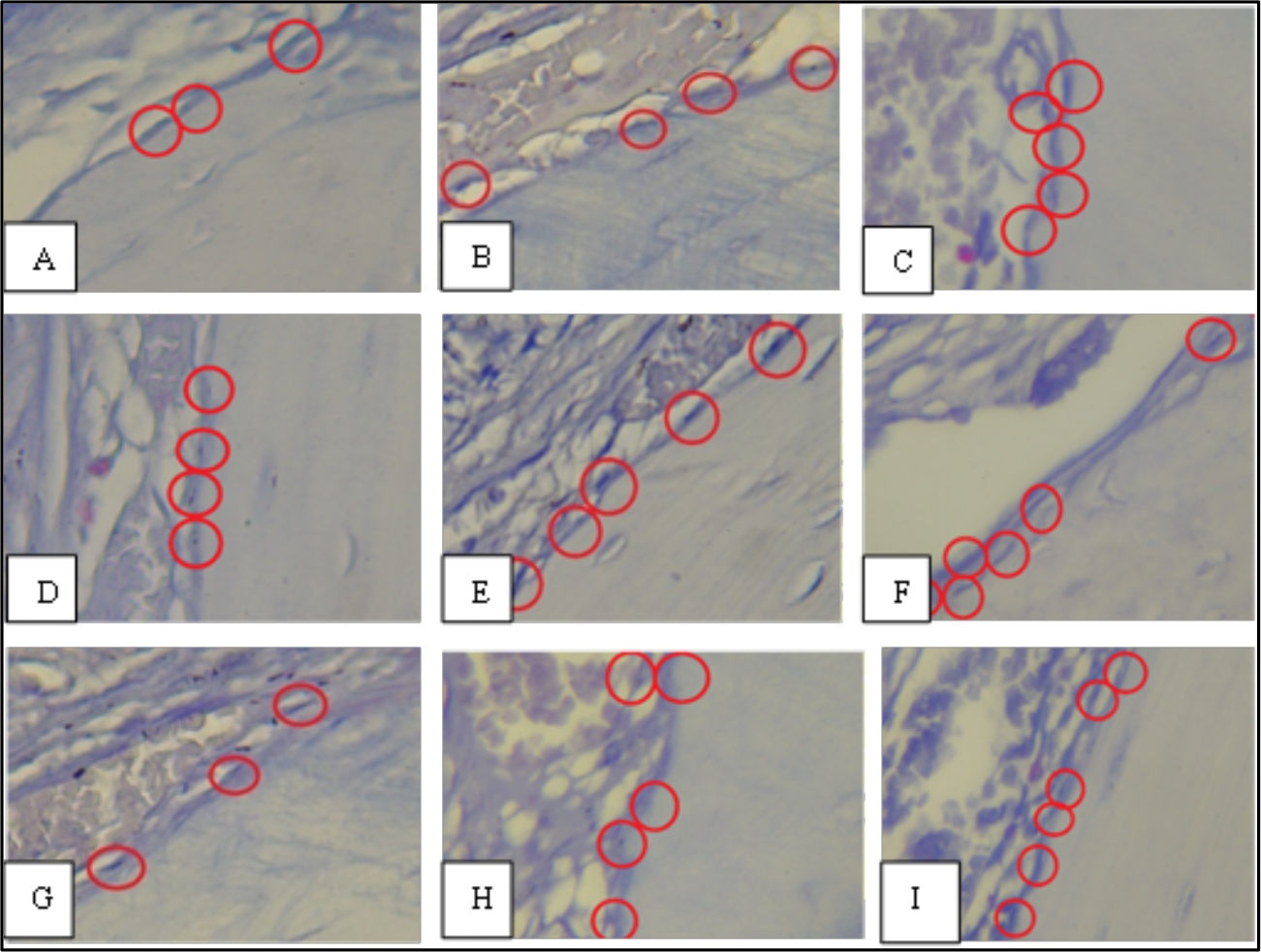
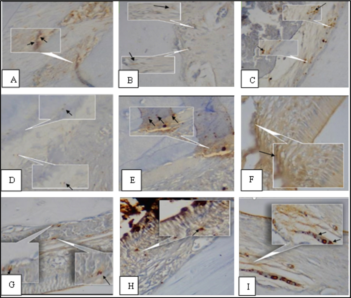
Histopathological observation on the alveolar bone around the central incisor of the lower jaw was carried out on days 3, 7, and 14. The mean and standard deviation of osteoblasts and RANKL expression from each group were presented in Figures 2 and 3. There were patterns of decreased numbers of osteoblasts in group T0 and an enhanced number of osteoblasts in groups T1 and T2 at three time-point evaluations. The highest average number of osteoblasts was found in the nanoliposome papaya seed extract group (group T2) on day 14. There were patterns of enhanced numbers of RANKL expression in group T0 and decreased numbers of RANKL expression in groups T1 and T2 at three time-point evaluations. The lowest average number of RANKL expression is found in the nanoliposome papaya seed extract (group T2) on day 14. The fluctuation is described in Figures 2 and 3, and the histopathological image of osteoblasts and RANKL expression is described in Figures 4 and 5.


Overview of osteoblast cells using HE staining. The red round mark shows osteoblast cells in the alveolar bone that were observed using a 400x magnification Olympus digital microscope. A) T0-3; B) T1-3; C) T2-3; D) T0-7; E) T1-7; F) T2-7; G) T0-14; H) T1-14; I) T2-14.
Overview of RANKL expression using IHC staining. Arrows and inserts indicate RANKL expression in the alveolar bone that was observed using a 400x magnification Olympus digital microscope. A) T0-3; B) T1-3; C) T2-3; D) T0-7; E) T1-7; F) T2-7; G) T0-14; H) T1-14; I) T2-14.
According to the normality and homogeneity test, it showed that the data is normal and homogenous. The one-way ANOVA test showed a significant difference in the number of osteoblasts on day 14 (p=0.002); a significant difference was also seen in RANKL expression on day 7 (p=0.002) and day 14 (p=0.000). Post hoc tests showed a significant difference in the number of osteoblasts in group T0 compared with group T2 and group T1 compared with group T2 on day 14. Post hoc tests showed a significant difference in the number of RANKL expression in each group on day 7, also group T0 compared with group T1, and group T0 compared with group T2 on day 14 (Table 1).
Discussion
There is a reciprocal relationship between diabetes mellitus and periodontitis. Periodontitis can increase the production of cytokines that worsens glycaemic control; conversely, high blood sugar levels can increase the inflammatory process of the periodontal tissue [30]. The bone remodelling cycle operates continually as osteoclasts constantly remove mature bone with new bone simultaneously formed by osteoblasts. A balance between bone resorption by osteoclast and bone formation by osteoblast determines the level of bone mass. Osteoblast cells influence the regulation of osteoclastogenesis through the regulatory system of OPG/RANKL/RANK. RANKL/OPG ratio is a determinant of the bone resorption process. RANKL is a member of TNF, it will bind to its receptor RANK to stimulate osteoclast differentiation and activation, they play a role in the formation and function of osteoclasts. OPG is a membrane that surrounds and secretes proteins attached to RANKL to inhibit its role on RANK receptors [15]. Hyperglycaemia, a condition related to diabetes mellitus, can increase the production of advanced glycation end products (AGEs), receptor of AGEs (RAGE), and reactive oxygen species (ROS). Interaction between AGEs and RAGE causes immune dysfunction and enhancement of proinflammatory cytokine expression. ROS can stimulate the enhancement of proinflammatory cytokine by activating intercellular signalling such as mitogen-activated protein kinase (MAPKs) and nuclear factor kappa B (NF-kB); it can damage tissue repair [31].
In this study, the condition of the experimental animals was made periodontitis condition accompanied by diabetes mellitus; in addition, they received different treatments as already mentioned in the method. The clinical signs that were seen in the rat models were bleeding on probing, redness, swelling, periodontal pocket, and fasting blood glucose >126 mg/dl. In group T0, which did not receive any treatment, there was a pattern of decreased number of osteoblasts and an increased number of RANKL expression on the predetermined time series, namely days 3, 7, and 14. In each time series, the lowest number of osteoblasts and the highest number of RANKL expression were found in group T0. This shows there is an inflammatory process in the group. Statistically, on day three, there were no significant differences in osteoblast number and RANKL expression between groups. The reduction of osteoblasts in periodontitis rats in this research was caused by inflammation triggered by LPS P. gingivalis that can activate multiple cell types via the production of proinflammatory cytokines [26,32].
Histopathologically periodontitis was signalled by the increase of polymorphonuclear leukocyte infiltration, production of proinflammatory cytokines and prostaglandins, amplification of lytic enzyme, and activation of osteoclasts along with a high level of ROS [19,33]. This condition is exacerbated by the presence of high blood sugar levels; hyperglycaemia can lead to the regulation of the immune response through direct influence on the function of immune cells, and it can worsen inflammation. Some research show there is an increase of inflammatory mediators such as expression of TNF-α, IL-β, IL-6, and PGE2 in chronic periodontitis with diabetes mellitus. Enhancing inflammatory mediators can increase the expression of RANKL, which will bind to its receptor RANK as the initial process of alveolar bone destruction. This condition will stimulate the osteoclast precursor to differentiate and induce TNF receptor-associated factor 6 (TRAF 6), which activates NF-kB and MAPKs, which are key to osteoclast formation; it will be in line with the reduction of the number of osteoblast cells. It leads to the destruction of inorganic minerals from the alveolar bone; then, bone resorption could occur. The uncontrollable destruction will cause high severity [31,34].
In groups T1 (given 96% papaya seed extract) and T2 (given 96% nanoliposome papaya seed extract), there was a pattern of an increased number of osteoblasts and a decreased number of RANKL expression on the predetermined time series, namely days 3, 7 and 14. In each time series, the highest number of osteoblasts and the lowest number of RANKL expression was found in group T2. Statistically, there is a significant difference between groups in RANKL expression on days 7 and 14 and the number of osteoblasts on day 14. It indicated that treatment with papaya seed extract has the potential to reduce periodontitis via an anti-inflammatory mechanism. Based on the previous findings, papaya seed extract contains active compounds such as flavonoids and phenols that promote alveolar bone recovery. Flavonoids in papaya seed have antibacterial, antifungal, and antioxidant properties that are able to restore alveolar bone density by preventing bone resorption. Flavonoids have a secondary flavonol compound, namely quercetin which has antioxidant and anti-inflammatory activity that can reduce the inflammatory production of IL-1, IL-6 and, TNF-α; quercetin is also capable of inhibiting intracellular signalling pathways such as MAPKs and NF-kB resulting in reduced inflammation and oxidative stress [19]. Chlorogenic acid is a phenolic compound in papaya seed that acts as an antioxidant and anti-inflammatory agent that could reduce RANKL production, inhibit osteoclast activity and bone resorption resulting in increasing the number of osteoblast cells [35].
Based on a post hoc test on day 14, there is no significant difference between groups T0 and T1 in the number of osteoblasts; however, in RANKL expression, that significant difference exists. There is no significant difference between groups T1 and T2 in the number of RANKL expression; however, that significant difference exists in the number of osteoblasts. This condition can be caused by several possibilities. The use of herbal medicine has several disadvantages: low solubility, lack of permeability to penetrate the absorption barrier, low bioavailability, and unpredictable toxicity [16,17]. Efforts to optimise the active compounds of papaya seed extract can be carried out in the form of liposomes. In this research, the treatment with papaya seed extract administered with nanoliposome showed the best result.
Various types of nanoliposomes are currently used clinically as delivery systems for drugs. The nanoliposomal system is a potential drug delivery system because nanoliposomes incorporate hydrophilic and lipophilic therapeutic agents relatively easily and can be delivered directly to an accessible body site such as the periodontium. With relative non-immunogenicity and low toxicity, nanoliposomes also naturally target macrophages [36]. The process of alveolar bone repair includes two stages of cellular activity, i.e. the resorption of old bone by osteoclasts through the process of acidification and proteolytic digestion and the formation of new bone by osteoblasts; osteoblast cells will invade the area, and formation occurs by secreting osteoid (collagen and protein matrix) which then mineralises. This phase lasts for 7 to 14 days, and after that, there is suppression of osteoclast formation and stimulation of osteoblastogenesis. During the repair process, differentiation occurs, including chemotaxis, cell attachment, mitosis, and osteoblast differentiation leading to deposition of new alveolar bone. The limitation of this study is that observations were made only up to day 14; more time is necessary to observe the process of osteoblastogenesis in more detail [37]. In addition, problems often arise in the preparation of nanoparticles, the occurrence of rapid aggregation, and uneven particle sizes, so the stability of the dispersion system becomes difficult to control. It is important to examine particle size, particle morphological characters, and potential zeta values as a solution to these problems [38].
Conclusion
There is an effect of the administration of nanoliposome papaya seed extract on a diabetic periodontitis rat model; it can increase the number of osteoblasts and decrease the number of RANKL expression in vivo. Comparing the different doses and observation times, as well as toxicity and material characterisation tests, is necessary to do for further research.
Data Availability
The data used to support the findings of this study can be made available upon request to the corresponding author.
Funding
References
- [1] Newman MG, Takei HH, Klokkevold PR, Carranza FA. Clinical Periodontology. Thirteenth. Carranza F, editor. Philadelphia: Elsevier; 2019. 602 p.
-
[2] Vos T, Abajobir AA, Abbafati C, Abbas KM, Abate KH, Abd-Allah F, et al. Global, regional, and national incidence, prevalence, and years lived with disability for 328 diseases and injuries for 195 countries, 1990-2016: A systematic analysis for the Global Burden of Disease Study 2016. Lancet 2017; 390(10100):1211-1259. https://doi.org/10.1016/S0140-6736(17)32154-2
» https://doi.org/10.1016/S0140-6736(17)32154-2 -
[3] Yamasaki Y, Nomura R, Nakano K, Naka S, Matsumoto-Nakano M, Asai F, et al. Distribution of periodontopathic bacterial species in dogs and their owners. Arch Oral Biol 2012; 57(9):1183-1188. https://doi.org/10.1016/j.archoralbio.2012.02.015
» https://doi.org/10.1016/j.archoralbio.2012.02.015 -
[4] Song J, Gao Z, Li M. Efficacy of adjunctive photodynamic therapy for chronic periodontitis undergoing scaling and root Planing: A Meta-analysis of randomized controlled trials. Opt Laser Technol 2022; 149(December 2021):107808. https://doi.org/10.1016/j.optlastec.2021.10780
» https://doi.org/10.1016/j.optlastec.2021.10780 -
[5] Zhou X, Zhang W, Liu X, Zhang W, Li Y. Interrelationship between diabetes and periodontitis: Role of hyperlipidemia. Arch Oral Biol 2015; 60(4):667-674. https://doi.org/10.1016/j.archoralbio.2014.11.008
» https://doi.org/10.1016/j.archoralbio.2014.11.008 -
[6] Rikawarastuti R, Anggreni E, Ngatemi N. Diabetes melitus dan tingkat keparahan jaringan periodontal. Kesmas Natl Public Heal J 2015; 9(3):277-281. https://doi.org/10.21109/kesmas.v9i3.693 [In Indonesian].
» https://doi.org/10.21109/kesmas.v9i3.693 -
[7] Sari R, Herawati D, Nurcahyanti R, Wardani PK. Prevalensi periodontitis pada pasien diabetes mellitus (Studi observasional di poliklinik penyakit dalam RSUP Dr. Sardjito). Maj Kedokt Gigi Indones 2017; 3(2):98-104. https://doi.org/10.22146/majkedgiind.11241 [In Indonesian].
» https://doi.org/10.22146/majkedgiind.11241 -
[8] Tse SY. Diabetes mellitus and periodontal disease: awareness and practice among doctors working in public general out-patient clinics in Kowloon West Cluster of Hong Kong. BMC Fam Pract 2018; 19(1):199. https://doi.org/10.1186/s12875-018-0887-2
» https://doi.org/10.1186/s12875-018-0887-2 -
[9] Kolluri A, Gopalkrishna P, Josyula VR, Gatta AK, Chakravarthy KP. Comparison of Oncostatin M in patients with chronic periodontitis with and without diabetes. Pesqui Bras Odontopediatria Clín Integr 2022; 22:e210136. https://doi.org/10.1590/pboci.2022.015
» https://doi.org/10.1590/pboci.2022.015 -
[10] Nibali L, Koidou VP, Nieri M, Barbato L, Pagliaro U, Cairo F. Regenerative surgery versus access flap for the treatment of intra-bony periodontal defects: A systematic review and meta-analysis. J Clin Periodontol 2020; 47(S22):320-351. https://doi.org/10.1111/jcpe.13237
» https://doi.org/10.1111/jcpe.13237 - [11] Suardi HN. Antibiotik dalam Dunia Kedokteran Gigi. Cakradonya Dent J 2014; 6(2):678-744. [In Indonesian].
-
[12] Jaipah N, Saraswati I, Hapsari R. Uji Efektivitas Antimikroba Ekstrak Biji Pepaya (Carica Papaya L.) Terhadap Pertumbuhan Escherichia Coli Secara in Vitro. Diponegoro Med J (Jurnal Kedokt Diponegoro) 2017; 6(2):947-955. https://doi.org/10.14710/dmj.v6i2.18605 [In Indonesian].
» https://doi.org/10.14710/dmj.v6i2.18605 -
[13] Pusporini R, Baabdullah HO, Andyka V. Quantification of quercetin and chlorogenic acid in papaya seed ethanol extract. Asian J Pharm Clin Res 2019; 13(1):151-153. https://doi.org/10.22159/ajpcr.2020.v13i1.36242
» https://doi.org/10.22159/ajpcr.2020.v13i1.36242 -
[14] Berendsen AD, Olsen BR. Bone development. Bone 2015; 80:14-18. https://doi.org/10.1016/j.bone.2015.04.035
» https://doi.org/10.1016/j.bone.2015.04.035 -
[15] Chen B, Wu W, Sun W, Zhang Q, Yan F, Xiao Y. RANKL expression in periodontal disease: Where does RANKL Come from? Biomed Res Int 2014; 2014:731039. https://doi.org/10.1155/2014/731039
» https://doi.org/10.1155/2014/731039 -
[16] Ajazuddin, Saraf S. Applications of novel drug delivery system for herbal formulations. Fitoterapia 2010; 81(7):680-689. https://doi.org/10.1016/j.fitote.2010.05.001
» https://doi.org/10.1016/j.fitote.2010.05.001 -
[17] Mensah MLK, Komlaga G, Forkuo AD, Firempong C, Anning AK, Dickson RA. Toxicity and safety implications of herbal medicines used in Africa. In: Builders PF. Herbal Medicine. London: IntechOpen; 2019. https://doi.org/10.5772/intechopen.72437
» https://doi.org/10.5772/intechopen.72437 -
[18] Taléns-Visconti R, Díez-Sales O, de Julián-Ortiz JV, Nácher A. Nanoliposomes in cancer therapy: Marketed products and current clinical trials. Int J Mol Sci 2022; 23(8):4249. https://doi.org/10.3390/ijms23084249
» https://doi.org/10.3390/ijms23084249 -
[19] Pusporini R, Basori A, Krismariono A. Anti-inflammatory role of papaya seed extracts in inhibiting osteoclastogenesis of rats with Periodontitis. Maj Kedokt Gigi Indones 2019; 4(2):95-101. https://doi.org/10.22146/majkedgiind.26829
» https://doi.org/10.22146/majkedgiind.26829 - [20] Pusporini R, Baabdullah HO, Andyka V. Evaluation of synthesis method of nanoliposome papaya seed extract. Malaysian J Med Heal Sci 2020; 16(12):7.
- [21] Ramadon D, Mun’im A. Pemanfaatan nanoteknologi dalam sistem penghantaran obat baru untuk produk bahan alam. J Ilmu Kefarmasian Indones 2016; 14(2):118-127. [In Indonesian].
-
[22] Husna F, Suyatna FD, Arozal W, Purwaningsih EH. Model hewan coba pada penelitian diabetes. Pharm Sci Res 2019; 6(3):131-141. https://doi.org/10.7454/psr.v6i3.4531 [In Indonesian].
» https://doi.org/10.7454/psr.v6i3.4531 -
[23] Saputra NT, Suartha IN, Dharmayudha AAGO. Agen Diabetagonik Streptozotocin untuk Membuat Tikus Putih Jantan Diabetes Mellitus. Bul Vet Udayana 2018; 10(2):116-121. https://doi.org/10.24843/bulvet.2018.v10.i02.p02 [In Indonesian].
» https://doi.org/10.24843/bulvet.2018.v10.i02.p02 -
[24] Prasetya RC, Purwanti N, Haniastuti T. Infiltrasi Neutrofil pada Tikus dengan Periodontitis setelah Pemberian Ekstrak Etanolik Kulit Manggis. Maj Kedokt Gigi Indones 2014; 21(1):33-38. https://doi.org/10.22146/majkedgiind.8520 [In Indonesian].
» https://doi.org/10.22146/majkedgiind.8520 -
[25] Guimarães MR, de Aquino SG, Coimbra LS, Spolidorio LC, Kirkwood KL RCJ. Curcumin modulates the immune response associated with LPS-induced periodontal disease in rats. Innate Immun 2012; 18(1):155-163. https://doi.org/10.1177/1753425910392935
» https://doi.org/10.1177/1753425910392935 - [26] Ionel A, Lucaciu O, Moga M, Buhatel D, Ilea A, Tabaran F, et al. Periodontal disease induced in Wistar rats - Experimental study. Hum Vet Med 2015; 7(2):90-95.
-
[27] Krismariono A. The decreasing of NFκB level in gingival junctional epithelium of rat exposed to Porphyromonas gingivalis with application of 1% curcumin on gingival sulcus. Dent J 2015; 48(1):35-38. https://doi.org/10.20473/j.djmkg.v48.i1.p35-38
» https://doi.org/10.20473/j.djmkg.v48.i1.p35-38 -
[28] Majdina S, Mulawarmanti D, Rizka Y. Efektifitas kombinasi terapi oksigen hiperbarik dan gel teripang emas (Stichopus hermanii) terhadap peningkatan jumlah osteoblas pada tikus diabetes melitus yang diinduksi bakteri Porphyromonas gingivalis. Denta 2016; 10(1):31-41. https://doi.org/10.30649/denta.v10i1.30 [In Indonesian].
» https://doi.org/10.30649/denta.v10i1.30 -
[29] Devitaningtyas N, Syaify A, Herawati D. Combining 10% propolis with carbonated hydroxyapatite to observe the RANKL expression in a rabbit’s alveolar bone. Dent J 2020; 53(4):212-216. https://doi.org/10.20473/j.djmkg.v53.i4.p212-216
» https://doi.org/10.20473/j.djmkg.v53.i4.p212-216 -
[30] Llambés F. Relationship between diabetes and periodontal infection. World J Diabetes 2015; 6(7):927. https://doi.org/10.4239/wjd.v6.i7.927
» https://doi.org/10.4239/wjd.v6.i7.927 - [31] Hikmah N, Shita ADP. Peran RANKL pada Proses Resorpsi Tulang Alveolar Kondisi Diabetes. Stomatognatic 2013; 10(3):105-109. [In Indonesian].
-
[32] Nativel B, Couret D, Giraud P, Meilhac O, D’Hellencourt CL, Viranaïcken W, et al. Porphyromonas gingivalis lipopolysaccharides act exclusively through TLR4 with a resilience between mouse and human. Sci Rep 2017; 7(1):1-12. https://doi.org/10.1038/s41598-017-16190-y
» https://doi.org/10.1038/s41598-017-16190-y -
[33] Weitzmann MN. The role of inflammatory cytokines, the RANKL/OPG axis, and the immunoskeletal interface in physiological bone turnover and osteoporosis. Scientifica 2013; 2013:1-29. https://doi.org/10.1155/2013/125705
» https://doi.org/10.1155/2013/125705 -
[34] Usui M, Onizuka S, Sato T, Kokabu S, Ariyoshi W, Nakashima K. Mechanism of alveolar bone destruction in periodontitis - Periodontal bacteria and inflammation. Jpn Dent Sci Rev 2021; 57:201-208. https://doi.org/10.1016/j.jdsr.2021.09.005
» https://doi.org/10.1016/j.jdsr.2021.09.005 -
[35] Kwak SC, Lee C, Kim JY, Oh HM, So HS, Lee MS, et al. Chlorogenic acid inhibits osteoclast differentiation and bone resorption by down-regulation of receptor activator of nuclear factor kappa-b ligand-induced nuclear factor of activated t cells c1 expression. Biol Pharm Bull 2013; 36(11):1779-1786. https://doi.org/10.1248/bpb.b13-00430
» https://doi.org/10.1248/bpb.b13-00430 -
[36] Liu D, Yang PS. Minocycline hydrochloride nanoliposomes inhibit the production of TNF-α in LPS-stimulated macrophages. Int J Nanomedicine 2012; 7:4769-4775. https://doi.org/10.2147/IJN.S34036
» https://doi.org/10.2147/IJN.S34036 -
[37] Hienz SA, Paliwal S, Ivanovski S. Mechanisms of bone resorption in periodontitis. J Immunol Res 2015; 2015:615486. https://doi.org/10.1155/2015/615486
» https://doi.org/10.1155/2015/615486 - [38] Pusporini R, Listari KM, Nugraeni Y, Hidayat LH, Ito MEIT. The effect of papaya seeds extracts nanoliposomes administration on osteoclasts number of diabetic periodontitis animal model. J Int Dent Med Res 2021; 14(2):563-568.