Abstract
Nanocellulose (BNC) is a natural polymer produced by bacteria. Its structure has only glucose monomer, it has various properties such as high water holding capacity, unique nanostructure, high crystallinity and high mechanical strength. Pure BNC or in combination with different components can be used for a wide range of applications. Aloe vera is a medicinal plant with polysaccharides in its composition that has a potential for tissue regeneration and repair. The aim of this study was to evaluate the effect of incorporating Aloe vera (A. vera) into BNC membranes produced with three fractions of A. vera extract (BNC-Aloe) on the behavior of epithelial cells. Human fibroblasts and keratinocytes were shown to have increased metabolic activity and proliferation when cultured on BNC-Aloe membranes compared to control. Quantification of collagen biosynthesis was significantly higher in BNC-Aloe membranes. In conclusion, BNC-Aloe membranes are suggested as a material for the purpose of skin tissue repair.
Keywords:
tissue engineering; nanocellulose; Aloe vera; fibroblasts; keratinocytes
1. Introduction
The skin tissue is the largest organ in the human body. This organ is considered to be the body's first defense barrier. Skin tissue performs important functions such as homeostasis, body temperature body temperature and protection against dehydration, in addition to provide support for blood vessels and nerves[11 Lin, W., Lien, C., Yeh, H., Yu, C., & Hsu, S. (2013). Bacterial cellulose and bacterial cellulose–chitosan membranes for wound dressing applications. Carbohydrate Polymers, 94(1), 603-611. http://dx.doi.org/10.1016/j.carbpol.2013.01.076. PMid:23544580.
http://dx.doi.org/10.1016/j.carbpol.2013...
,22 Pang, M., Huang, Y., Meng, F., Zhuang, Y., Liu, H., Du, M., Ma, Q., Wang, Q., Chen, Z., Chen, L., Cai, T., & Cai, Y. (2020). Application of bacterial cellulose in skin and bone tissue engineering. European Polymer Journal, 122, 109365. http://dx.doi.org/10.1016/j.eurpolymj.2019.109365.
http://dx.doi.org/10.1016/j.eurpolymj.20...
]. Extensive and deep damage to the skin and mucous membranes can cause destruction of the dermis and epidermis. The damage of skin can be resolved using human skin grafts, autologous or not. However, this solution is limited by the scarcity of donors and the risk of donors and with the risk of graft rejection[33 Souto, L. R. M., Rehder, J., Vassallo, J., Cintra, M. L., Kraemer, M. H. S., & Puzzi, M. B. (2006). Model for human skin reconstructed in vitro composed of associated dermis and epidermis. Sao Paulo Medical Journal, 124(2), 71-76. http://dx.doi.org/10.1590/S1516-31802006000200005. PMid:16878189.
http://dx.doi.org/10.1590/S1516-31802006...
,44 Souto, L. R. M., Vassallo, J., Rehder, J., Pinto, G. A., & Puzzi, M. B. (2009). Immunoarchitectural characterization of a human skin model reconstructed in vitro. Sao Paulo Medical Journal, 127(1), 28-33. http://dx.doi.org/10.1590/S1516-31802009000100007. PMid:19466292.
http://dx.doi.org/10.1590/S1516-31802009...
].
The development of temporary and/or permanent and/or permanent replacements for injured tissue arises from this context. One approach to developing functional skin substitutes is to produce to produce three-dimensional biomaterials, which resemble the physiological microenvironment in the presence of the extracellular matrix when cultured with human epidermal cells and human dermal fibroblasts the growth of the artificial tissue[55 Bell, E., Sher, S., Hull, B., Merrill, C., Rosen, S., Chamson, A., Asselineau, D., Dubertret, L., Coulomb, B., Lapiere, C., Nusgens, B., & Neveux, Y. (1983). The reconstitution of living skin. The Journal of Investigative Dermatology, 81(Suppl. 1), 2S-10S. http://dx.doi.org/10.1111/1523-1747.ep12539993. PMid:6306115.
http://dx.doi.org/10.1111/1523-1747.ep12...
,66 Grøn, B., Stoltze, K., Andersson, A., & Dabelsteen, E. (2002). Oral fibroblasts produce more HGF and KGF than skin fibroblasts in response to co-culture with keratinocytes. Acta Pathologica, Microbiologica, et Immunologica Scandinavica, 110(12), 892-898. http://dx.doi.org/10.1034/j.1600-0463.2002.1101208.x. PMid:12645668.
http://dx.doi.org/10.1034/j.1600-0463.20...
].
The dressings that are being commercialized assist the tissue regeneration[77 Sayag, J., Lieaume, S., & Bohbot, S. (1996). Healing properties of calcium alginate dressings. Journal of Wound Care, 5(8), 357-362. http://dx.doi.org/10.12968/jowc.1996.5.8.357. PMid:27935753.
http://dx.doi.org/10.12968/jowc.1996.5.8...
8 Koide, M., Osaki, K., Konishi, J., Oyamada, K., Katakura, T., Takahashi, A., & Yoshizato, K. (1993). A new type of biomaterial for artificial skin: dehydrothermally cross-linked composites of fibrillar and denatured collagens. Journal of Biomedical Materials Research, 27(1), 79-87. http://dx.doi.org/10.1002/jbm.820270111. PMid:8421002.
http://dx.doi.org/10.1002/jbm.820270111...
-99 Badylak, S. F. (2007). The extracellular matrix as a biologic scaffold material. Biomaterials, 28(25), 3587-3593. http://dx.doi.org/10.1016/j.biomaterials.2007.04.043. PMid:17524477.
http://dx.doi.org/10.1016/j.biomaterials...
]. As these treatments are costly and often do not induce and often do not induce the effective cure of the lesion, emphasizing the need to invest in the development of new treatments and devices treatments and devices that present a better cost-benefit.
In the development of a biomaterial with active substances was used Bacterial Cellulose. It is a natural polymer synthesized by various bacteria, including those from the genus Komagataeibacter, formerly classified in genus Gluconacetobacter[1010 Brown, R. M., Jr., & Montezinos, D. (1976). Cellulose microfibrils: visualization of biosynthetic and orienting complexes in association with the plasma membrane. Proceedings of the National Academy of Sciences of the United States of America, 73(1), 143-147. http://dx.doi.org/10.1073/pnas.73.1.143. PMid:1061108.
http://dx.doi.org/10.1073/pnas.73.1.143...
]. Cellulose-producing bacteria under specific conditions synthesizes a nanofiber network with excellent properties, such as biocompatibility, elasticity, transparency, purity and mechanical stability, which we call bacterial nanocellulose (BNC)[22 Pang, M., Huang, Y., Meng, F., Zhuang, Y., Liu, H., Du, M., Ma, Q., Wang, Q., Chen, Z., Chen, L., Cai, T., & Cai, Y. (2020). Application of bacterial cellulose in skin and bone tissue engineering. European Polymer Journal, 122, 109365. http://dx.doi.org/10.1016/j.eurpolymj.2019.109365.
http://dx.doi.org/10.1016/j.eurpolymj.20...
,1111 Rambo, C. R., Recouvreux, D. O. S., Carminatti, C. A., Pitlovanciv, A. K., Antônio, R. V., & Porto, L. M. (2008). Template assisted synthesis of porous nanofibrous cellulose membranes for tissue engineering. Materials Science and Engineering C, 28(4), 549-554. http://dx.doi.org/10.1016/j.msec.2007.11.011.
http://dx.doi.org/10.1016/j.msec.2007.11...
12 Recouvreux, D. O. S., Carminatti, C. A., Pitlovanciv, A. K., Rambo, C. R., Porto, L. M., & Antônio, R. V. (2008). Cellulose biosynthesis by the beta-proteobacterium, Chromobacterium violaceum. Current Microbiology, 57(5), 469-476. http://dx.doi.org/10.1007/s00284-008-9271-0. PMid:18820969.
http://dx.doi.org/10.1007/s00284-008-927...
13 Souza, S. S., Berti, F. V., Oliveira, K. P. V., Pittella, C. Q. P., Castro, J. V., Pelissari, C., Rambo, C. R., & Porto, L. M. (2019). Nanocellulose biosynthesis by Komagataeibacter hansenii in a defined minimal culture medium. Cellulose, 26(3), 1641-1655. http://dx.doi.org/10.1007/s10570-018-2178-4.
http://dx.doi.org/10.1007/s10570-018-217...
14 Sperotto, G., Stasiak, L. G., Godoi, J. P. M. G., Gabiatti, N. C., & Souza, S. S. (2021). A review of culture media for bacterial cellulose production: complex, chemically defined and minimal media modulations. Cellulose, 28(5), 2649-2673. http://dx.doi.org/10.1007/s10570-021-03754-5.
http://dx.doi.org/10.1007/s10570-021-037...
-1515 Anton-Sales, I., Beekmann, U., Laromaine, A., Roig, A., & Kralisch, D. (2019). Opportunities of bacterial cellulose to treat epithelial tissues. Current Drug Targets, 20(8), 808-822. http://dx.doi.org/10.2174/1389450120666181129092144. PMid:30488795.
http://dx.doi.org/10.2174/13894501206661...
]. Along with fractions of Aloe vera (A. vera). The extract of parenchymal tissue of A. vera contains polysaccharides, sugars, minerals, proteins, lipids, phenolic compounds[1616 Rahman, S., Carter, P., & Bhattarai, N. (2017). Aloe vera for tissue engineering applications. Journal of Functional Biomaterials, 8(1), 6. http://dx.doi.org/10.3390/jfb8010006. PMid:28216559.
http://dx.doi.org/10.3390/jfb8010006...
]. A. vera has long been considered as a safe functional food material that can be used orally and topically[1717 Tanaka, M., Yamada, M., Toida, T., & Iwatsuki, K. (2012). Safety evaluation of supercritical carbon dioxide extract of Aloe vera gel. Journal of Food Science, 77(1), T2-T9. http://dx.doi.org/10.1111/j.1750-3841.2011.02452.x. PMid:22260137.
http://dx.doi.org/10.1111/j.1750-3841.20...
]. The extracted material generated three distinct fractions: T, G and F. The T fraction contains all the components present in the parenchymal tissue, synergy with all natural components. A portion of this fraction was centrifuged, producing G fraction containing the components of the parenchymal tissue, except the fibers. F fraction contained only the polysaccharides of the parenchymal tissue of the A. vera[1818 Godinho, J. F., Berti, F. V., Müller, D., Rambo, C. R., & Porto, L. M. (2016). Incorporation of Aloe vera extracts into nanocellulose during biosynthesis. Cellulose(1), 23, 545-555. http://dx.doi.org/10.1007/s10570-015-0844-3.
http://dx.doi.org/10.1007/s10570-015-084...
]. It is a plant that has immune-active properties with therapeutic functions that have been popularly used in a number of applications, such as anti-inflammatory and wound healing purposes[1919 Davis, R. H., Donato, J. J., Hartman, G. M., & Haas, R. C. (1994). Anti-inflammatory and wound healing activity of a growth substance in Aloe vera. Journal of the American Podiatric Medical Association, 84(2), 77-81. http://dx.doi.org/10.7547/87507315-84-2-77. PMid:8169808.
http://dx.doi.org/10.7547/87507315-84-2-...
20 Kang, M., Kim, S. Y., Kim, Y. T., Kim, E., Lee, S., Ko, S., Wijesinghe, W. A. J. P., Samarakoon, K. W., Kim, Y., Cho, J. H., Jang, H., & Jeon, Y. (2014). In vitro and in vivo antioxidant activities of polysaccharide purified from aloe vera (Aloe barbadensis) gel. Carbohydrate Polymers, 99, 365-371. http://dx.doi.org/10.1016/j.carbpol.2013.07.091. PMid:24274519.
http://dx.doi.org/10.1016/j.carbpol.2013...
-2121 Sierra-García, G. D., Castro-Ríos, R., González-Horta, A., Lara-Arias, J., & Chávez-Montes, A. (2014). Acemannan, an extracted polysaccharide from Aloe vera: a literature review. Natural Product Communications, 9(8), 1217-1221. http://dx.doi.org/10.1177/1934578X1400900836. PMid:25233608.
http://dx.doi.org/10.1177/1934578X140090...
] According to some studies, A. vera interacts with fibroblast growth factors stimulating proliferation and increasing collagen[2222 Chithra, P., Sajithlal, G. B., & Chandrakasan, G. (1998). Influence of Aloe vera on the glycosaminoglycans in the matrix of healing dermal wounds in rats. Journal of Ethnopharmacology, 59(3), 179-186. http://dx.doi.org/10.1016/S0378-8741(97)00112-8. PMid:9507902.
http://dx.doi.org/10.1016/S0378-8741(97)...
,2323 Boudreau, M. D., & Beland, F. A. (2006). An evaluation of the biological and toxicological properties of Aloe barbadensis (miller), Aloe vera. Journal of Environmental Science and Health. Part C, Environmental Carcinogenesis & Ecotoxicology Reviews, 24(1), 103-154. http://dx.doi.org/10.1080/10590500600614303. PMid:16690538.
http://dx.doi.org/10.1080/10590500600614...
].
The incorporation of other components into the nanocellulose matrix during the synthesis can increase its applications by improving their physicochemical properties[1111 Rambo, C. R., Recouvreux, D. O. S., Carminatti, C. A., Pitlovanciv, A. K., Antônio, R. V., & Porto, L. M. (2008). Template assisted synthesis of porous nanofibrous cellulose membranes for tissue engineering. Materials Science and Engineering C, 28(4), 549-554. http://dx.doi.org/10.1016/j.msec.2007.11.011.
http://dx.doi.org/10.1016/j.msec.2007.11...
,1212 Recouvreux, D. O. S., Carminatti, C. A., Pitlovanciv, A. K., Rambo, C. R., Porto, L. M., & Antônio, R. V. (2008). Cellulose biosynthesis by the beta-proteobacterium, Chromobacterium violaceum. Current Microbiology, 57(5), 469-476. http://dx.doi.org/10.1007/s00284-008-9271-0. PMid:18820969.
http://dx.doi.org/10.1007/s00284-008-927...
,2424 Saibuatong, O., & Phisalaphong, M. (2010). Novo Aloe vera-bacterial cellulose composite film from biosynthesis. Carbohydrate Polymers, 79(2), 455-460. http://dx.doi.org/10.1016/j.carbpol.2009.08.039.
http://dx.doi.org/10.1016/j.carbpol.2009...
25 Stumpf, T. R., Pértile, R. A. N., Rambo, C. R., & Porto, L. M. (2013). Enriched glucose and dextrin mannitol-based media modulates fibroblast behavior on bacterial cellulose membranes. Materials Science and Engineering C, 33(8), 4739-4745. http://dx.doi.org/10.1016/j.msec.2013.07.035. PMid:24094182.
http://dx.doi.org/10.1016/j.msec.2013.07...
-2626 Moniri, M., Moghaddam, A. B., Azizi, S., Rahim, R. A., Ariff, A. B., Saad, W. Z., Navaderi, M., & Mohamad, R. (2017). Production and status of bacterial cellulose in biomedical engineering. Nanomaterials, 7(9), 257. http://dx.doi.org/10.3390/nano7090257. PMid:32962322.
http://dx.doi.org/10.3390/nano7090257...
]. In most cases, considering the main characteristics of BNC, a modification is suitable, since the nanomaterials based on BNC generally present high value-added with great potential of applications. In this perspective BNC membranes incorporated with A. vera extracts were previously developed[1818 Godinho, J. F., Berti, F. V., Müller, D., Rambo, C. R., & Porto, L. M. (2016). Incorporation of Aloe vera extracts into nanocellulose during biosynthesis. Cellulose(1), 23, 545-555. http://dx.doi.org/10.1007/s10570-015-0844-3.
http://dx.doi.org/10.1007/s10570-015-084...
] but the potential application for skin regeneration remains to be evaluated. To widen the applicability of BNC-Aloe membranes, this study presents an in vitro analysis of the behavior of human epithelial cells grown in the porous surface (porous side) of the membranes to provide evidence on the biocompatibility, efficacy of the BNC-Aloe on epithelial tissue repair, in addition to quantifying collagen synthesis.
2. Materials and Methods
2.1 BNC and BNC-Aloe membranes
A. vera leaves were used to obtain three different polysaccharide portions following the same procedure previously developed by Godinho[1818 Godinho, J. F., Berti, F. V., Müller, D., Rambo, C. R., & Porto, L. M. (2016). Incorporation of Aloe vera extracts into nanocellulose during biosynthesis. Cellulose(1), 23, 545-555. http://dx.doi.org/10.1007/s10570-015-0844-3.
http://dx.doi.org/10.1007/s10570-015-084...
]. A. vera gel pulp – GP, A. vera gel extract – GE and polysaccharide fraction – PF were used as a supplement of mannitol-based bacterial culture medium[1818 Godinho, J. F., Berti, F. V., Müller, D., Rambo, C. R., & Porto, L. M. (2016). Incorporation of Aloe vera extracts into nanocellulose during biosynthesis. Cellulose(1), 23, 545-555. http://dx.doi.org/10.1007/s10570-015-0844-3.
http://dx.doi.org/10.1007/s10570-015-084...
].
The bacteria Gluconacteobacter hansenii (ATCC 23769) were cultured in mannitol medium containing 60% of GE, GP and GF for 10 days. After 10 days, BNC-Aloe membranes were purified with 0.1 M NaOH for 24 h at 50 °C and finally rinsed with distilled water until reach pH 6.5. BNC-Aloe membranes were sterilized by autoclaving at 121 °C for 20 minutes before using. As a control, G. hansenii were cultured with mannitol-based medium without addition of A. vera to obtain pure BNC membranes, that were purified and sterilized following the same procedure described above. BNC and BNC-Aloe were stored in sterile conditions at room temperature until use.
2.2 Characterization of BNC membranes
The porous surface of BNC and BNC-Aloe were used to perform in vitro assays. Scanning Electron Microscopy (SEM) was performed using a JEOL JSM‒6390LV microscope (Jeol, Japan) in order to characterize the surface of BNC and BNC-Aloe. BNC membranes were freeze-dried by lyophilization as described by Berti et al.[2727 Berti, F. V., Rambo, C. R., Dias, P. F., & Porto, L. M. (2013). Nanofiber density determines endothelial cell behavior on hydrogel matrix. Materials Science and Engineering C, 33(8), 4684-4691. http://dx.doi.org/10.1016/j.msec.2013.07.029. PMid:24094176.
http://dx.doi.org/10.1016/j.msec.2013.07...
]. After drying, samples were distributed on stubs and then coated with a double gold layer.
2.3 Cell culture
Primary human fibroblasts that were extracted of eyelid skin (HDFa)[2828 Silva, A. R. P., Paula, A. C. C., Martins, T. M. M., Goes, A. M., & Pereria, M. M. (2014). Synergistic effect between bioactive glass foam and a perfusion bioreactor on osteogenic differentiation of human adipose stem cells. Journal of Biomedical Materials Research, Part A, 102(3), 818-827. http://dx.doi.org/10.1002/jbm.a.34758. PMid:23625853.
http://dx.doi.org/10.1002/jbm.a.34758...
] and immortalized keratinocyte cell line (HaCat) (ATCC) were cultured in Dulbecco's Modified Eagle's medium (DMEM) (Gibco®, USA) supplemented with 10% of fetal bovine serum (Gibco®, USA) and 1% penicillin/streptomycin (Gibco®, USA). Cell cultures were maintained in a humidified CO2 (5% in air) incubator at 37°C. Primary human fibroblast cells were used in a passage of 5 to 9 to perform all in vitro assays, cultured on petri dishes. Samples were supported on glass rings before seeding. The density of cells/samples were in volume of medium for test, before time of cultured the samples were taken and added to new plates to perform the quantitative testes.
Cellular metabolic activity- Metabolic activity was determined by mitochondrial activity through MTS [3‐(4,5‐dimethylthiazol‐2‐yl)‐5‐(3‐carboxymethoxyphenyl)‐2‐(4‐sulfophenyl)‐2H‐tetrazolium] colorimetric assay using MTS assay kit purchased from Promega Biotecnologia do Brasil Ltda. (São Paulo, Brazil). MTS assay was performed according to the manufacturer's instructions. HDFa and HaCat cells were seeded on the porous surface of BNC AND BNC-Aloe membranes with 15 mm diameter, in a density of 105 cells/sample. At the end of 1, 3, and 7 days, samples and culture medium were removed, and adhered cells were rinsed with PBS three times. Culture plates were kept in a humidified incubator at 37°C and 5% CO2, protected from light during 2 h for the MTS reaction. Metabolic activity of HDFa and HaCat cells were quantified by Micro ELISA reader (SpectraMaxPlus 384, Molecular Devices, USA) at a wavelength of 490 nm.
Cell proliferation - The PicoGreen dsDNA Quantification kit (Molecular Probes, USA) was used to quantify epithelial cells proliferation when cultured on BNC and BNC-Aloe membranes with 15 mm diameter. Epithelial cells were seeded in a density of 105 cells/ sample during 1, 3 and 7 days. To quantify dsDNA of HDFa and HaCat cell, samples were washed with PBS and submersed in 1 mL of ultrapure water for 1 hour in a humidified atmosphere at 37°C and 5% CO2. Thereafter, the plate was removed and stored in a freezer at -80 °C until analysis. Samples were incubated for 2–5 min at room temperature, protected from light and subsequently reacted with Pico Green® following the manufacturer’s protocol. A standard curve was constructed by the quantification of the λDNA provided by the Pico Green® kit. Analysis was performed using Microplate Infinite (model M200 TECAN / LAMEB 1 – UFSC) with excitation filter at 480 nm and emission filter at 530 nm.
Cell viability - Live/Dead® Viability/Cytotoxicity kit (Invitrogen, USA) was used to evaluate cell viability. It measures the intracellular esterase activity (calcein) and plasma membrane integrity (ethidium homodimer). HDFa and HaCat cells were seeded on the porous surface of the BNC and BNC-Aloe membranes with 15 mm diameter in a density of 105 cells/sample. A solution of ethidium homodimer and calcein (4:1) was prepared in PBS, and 100 µL of this solution was added on each sample following the manufacturer's protocol. Afterwards, the culture plate was incubated for 30 minutes at 37°C and 5% CO2 atmosphere. After incubation, samples were mounted on slides and observed using a fluorescence microscope (Eclipse C-L, Nikon, Japan).
2.4 Collagen biosynthesis by fibroblasts (HDFa)
HDFa were grown on the surface of BNC and BNC-Aloe membranes with 15 mm diameter for a culturing period of 7 days. Cells were then washed with PBS, fixed with 3.7% formaldehyde for 1 h at 25°C, washed again with PBS and left in a laminar flow hood to dry for 45 min. Samples were then stained with 200 µL of Sirius red solution 0.5 g (Direct Red 80% – Dye content 25%, Sigma, USA) in 500 mL of saturated aqueous picric acid solution for one hour at a temperature of 25°C, protected from light. The supernatant was removed, and samples were washed with 250 µL of 0.01 M hydrochloric acid to remove unfixed dye. Stained collagen fibers were solubilized by adding 150 µL of 0.01 M sodium hydroxide solution and left for one hour. Results of collagen biosynthesis were quantified by Micro ELISA reader (SpectraMaxPlus 384, Molecular Devices, USA) at a wavelength of 535 nm and data were expressed as mean percentage of collagen synthesis. Results were compared with the standard curve of collagen connective tissue of bovine skin, kindly provided by Dr. Durvanei Augusto Maria from Butantan Institute (São Paulo).
2.5 Statistical analysis
Statistical analyses were performed using GraphPad-Prism® version 5.0 (GraphPad Software Inc., USA). Comparisons between groups were evaluated by one-way analysis of variance (ANOVA), followed by the Bonferroni test. Differences were considered significant when p < 0.05.
3. Results and Discussions
In this study we have investigated whether the presence of A. vera fractions incorporated into BNC membranes would stimulate proliferation, metabolic activity, and collagen synthesis of epithelial cells. To the best of our knowledge this study showed for the first time that BNC membranes incorporated with A. vera could stimulate collagen synthesis of epithelial cells.
BNC and BNC-Aloe membranes were successfully synthesized following the previous procedure standardized by our research group[2929 Godinho, J. (2014). Hidrogéis de celulose bacteriana incorporados com frações de Aloe vera (Dissertação de Mestrado). Universidade Federal de Santa Catarina, Santa Catarina, Brasil.,3030 Piaia, L., Paes, C. Q., & Porto, L. M. (2014). Viability of human dermal fibroblasts cultured on bacterial cellulose and Aloe vera composites. BMC Proceedings, 8(4), 61. http://dx.doi.org/10.1186/1753-6561-8-S4-P61.
http://dx.doi.org/10.1186/1753-6561-8-S4...
]. BNC and BNC-Aloe membranes were micro structurally characterized by SEM. Figure 1 shows the porous surface of BNC and BNC-Aloe membranes that is corresponding to the surface used to culture epithelial cells. The porous surface of BNC and BNC-Aloe membranes showed similar microstructure after incorporation with A. vera fractions, which resulted in a non-homogeneous surface, as shown on Figure 2. Unlike, BNC-PF membranes showed a thinner fiber network when compared to BNC, BNC-GE and BNC-GP.


Micrographs of BNC and BNC-Aloe membranes obtained by SEM. The BNC-Aloe membranes were produced by the addition of 60% of A. vera fractions (BNC-GE, BNC-GP and BNC-PF). BNC (nanocellulose); BNC-GE (nanocellulose/ A. vera gel extract); BNC-GP (nanocellulose/ A. vera gel pulp); BNC-PF (nanocellulose/ polysaccharide fraction). The magnification used to obtain the micrographs was 3000×.
The porous surface of BNC incorporated with A. vera fractions could determine physical and functional properties of the membranes, proposing a potential biomaterial for tissue engineering applications[2525 Stumpf, T. R., Pértile, R. A. N., Rambo, C. R., & Porto, L. M. (2013). Enriched glucose and dextrin mannitol-based media modulates fibroblast behavior on bacterial cellulose membranes. Materials Science and Engineering C, 33(8), 4739-4745. http://dx.doi.org/10.1016/j.msec.2013.07.035. PMid:24094182.
http://dx.doi.org/10.1016/j.msec.2013.07...
] due their similar microstructure to the extracellular matrix[2727 Berti, F. V., Rambo, C. R., Dias, P. F., & Porto, L. M. (2013). Nanofiber density determines endothelial cell behavior on hydrogel matrix. Materials Science and Engineering C, 33(8), 4684-4691. http://dx.doi.org/10.1016/j.msec.2013.07.029. PMid:24094176.
http://dx.doi.org/10.1016/j.msec.2013.07...
,3131 Dayal, M. S., & Catchmark, J. M. (2016). Mechanical and structural property analysis of bacterial cellulose composites. Carbohydrate Polymers, 144, 447-453. http://dx.doi.org/10.1016/j.carbpol.2016.02.055. PMid:27083837.
http://dx.doi.org/10.1016/j.carbpol.2016...
32 Murphy, C. M., & O’Brien, F. J. (2010). Understanding the effect of mean pore size on cell activity in collagen-glycosaminoglycan scaffolds. Cell Adhesion & Migration, 4(3), 377-381. http://dx.doi.org/10.4161/cam.4.3.11747. PMid:20421733.
http://dx.doi.org/10.4161/cam.4.3.11747...
-3333 Fu, L., Zhang, J., & Yang, G. (2013). Present status and applications of bacterial cellulose-based materials for skin tissue repair. Carbohydrate Polymers, 92(2), 1432-1442. http://dx.doi.org/10.1016/j.carbpol.2012.10.071. PMid:23399174.
http://dx.doi.org/10.1016/j.carbpol.2012...
].
Figure 3 shows the metabolic activity profile of epithelial cells cultured on BNC and BNC-Aloe membranes cultured for 1, 3 and 7 days. At the end of 7 days of culture, it is possible to observe an increase of metabolic activity behavior of HDFa cells when cultured on the porous surface of BNC, BNC-GE, BNC-GP, BNC-PF (Figure 2a), with significant differences observed in HDFa cells cultured on BNC-PF membrane in comparison to BNC. In the first day, metabolic activity of HDFa cells cultured on BNC-GE, BNC-GP and BNC-PF decreased approximately 4%, 19% and 21%, respectively. However, significant differences were observed only for cells cultured on BNC-GP and BNC-PF. In the third day of culture, metabolic activity of HDFa cells decreased 15% in BNC-GE, 9% in BNC-GP and increased 23% in BNC-PF (considering BNC as 100%). In the last day of culture (7 days), metabolic activity of HDFa decreased 6% in BNC-GE, 2% in BNC-GP and 7% in BNC-PF.

Metabolic activity of epithelial cells cultured on the porous surface of BNC and BNC-Aloe membranes for 7 days. (a) HDFa (primary human fibroblast) cells and (b) HaCat (immortalized keratinocytes cell line). Bars represent the average standard deviation and * represents significant differences at p<0.05 comparing BNC and BNC-Aloe biomembranes.
The metabolic activity of HaCat cells cultured during 1, 3 and 7 days on BNC and BNC-Aloe membranes are shown in Figure 2b. The metabolic activity profile of HaCaT cells increased with the time when the cells were cultured on the porous surface of BNC-GE and BNC-GP membranes. When HaCaT cells were cultured on BNC, BNC-GE, BNC-GP and BNC-PF it was not possible to observe significant differences after 1 day of culture. Thus, in the third day of culture the metabolic activity of HaCaT significantly increased when cultured in BNC-GE (9%) and BNC-GP (29%) membranes. After 7 days of culture, a significant increase was observed in HaCaT cells in BNC-GE (126%), BNC-GP (141%) and BNC-PF (28%) membranes, considering BNC as 100%. The metabolic activity of HaCaT showed a linear increase when cultured on BNC-GP, for 1 day (5%), 3 days (29%) and in 7 days (141%).
The incorporation of A. vera fractions into BNC membranes was extremely important to promote significant differences on cell viability and proliferation of epithelial cells. The behaviors of fibroblasts and keratinocytes cells were differently influenced by the composition of A. vera fractions (GP, GE and PF).
The metabolic activity and proliferation of HaCat cells increased when they were cultured on BNC-GP and BNC-GE compared to BNC and BNC-PF. On the other hand, after 7 days of culture, the metabolic activity of HDFa cells was similar when compared BNC to BNC-GP.
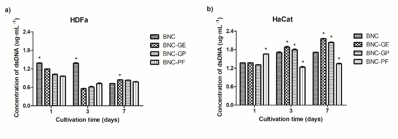
The result obtained by dsDNA quantification is showed in Figure 4. HDFa (Figure 4a) and HaCat (Figure 4b) were cultured on BNC and BNC-Aloe membranes during 1, 3 and 7 days. Significant differences were observed throughout all experimental time when HDFa cells were cultured on BNC compared to BNC-Aloe membranes. In the first day of culture, the number of proliferative HDFa cells cultured on BNC-Aloe membranes decreased almost 14% for BNC-GE, 26% for BNC-GP and 31% for BNC-PF. In the third day of culture, this number decreased almost 60%, 56% and 48% for BNC-GE, BNC-GP and BNC-PF, respectively. In the end of 7 days of in vitro culture, HDFa cells increased the proliferative profile when cultured on BNC-Aloe membranes. Unlike, the number of proliferative HDFa cells drastically decreased when they were cultured on BNC during 7 days of culture, (Figure 4a). After 7 days of culture, there were significant differences between the number of proliferative HDFa cells cultured on BNC and BNC-Aloe membranes. HDFa cells were 17% (BNC-GE), 15% (BNC-GP) and 8% (BNC-PF) more proliferative when they were cultured on BNC-Aloe membranes compared to BNC.

Proliferation of epithelial cells cultured on BNC and BNC-Aloe membranes during 1, 3 and 7 days of in vitro culture. The bars represent the average standard deviation and * represents significant differences at p<0.05 comparing BNC and BNC-Aloe membranes. Proliferation of (a) HDFa and (b) HaCat cells cultured on the porous surface of BNC, BNC-GE, BNC-GP, and BNC-PF membranes.
HaCaT cells were also cultured on BNC and BNC-Aloe membranes to determine if the incorporation of A. vera affects the HaCaT proliferation (Figure 4b). Significant differences in the number of proliferative cells cultured on BNC and BNC-Aloe membranes were observed throughout the total experimental time evaluated, except on the first day of culture when the results observed for BNC-GE were similar to BNC membranes (100%). BNC-GE and BNC-GP increased the number of proliferative HaCaT cells over the 7 days of culture, in 26% and 19% respectively (considering BNC as 100%). In the third day of culture, the number of proliferative HaCat cells increased in 10% and 5% when cultured on BNC-GE and BNC-GP, respectively. On the other hand, the HaCat cells decreased by 28% when cultured on BNC-PF membranes. At the seventh day of experiment, the number of proliferative HaCat cells decreased by 22% when cultured on BNC-PF membranes.
In addition, the proliferation of HDFa cells was stimulated by BNC-GP membranes in comparison to the BNC membranes without the addition of A. vera. The metabolic activity of HDFa decreased after three days of culture on BNC, BNC-GE and BNC-GP. A similar decrease on metabolic activity values were observed when HUVECs were cultured on the porous surface of BNC after 3 days of in vitro culture[2727 Berti, F. V., Rambo, C. R., Dias, P. F., & Porto, L. M. (2013). Nanofiber density determines endothelial cell behavior on hydrogel matrix. Materials Science and Engineering C, 33(8), 4684-4691. http://dx.doi.org/10.1016/j.msec.2013.07.029. PMid:24094176.
http://dx.doi.org/10.1016/j.msec.2013.07...
]. According to the referred authors, the cellular adaptation into BNC microarchitecture might have influenced the decrease on metabolic activity[2727 Berti, F. V., Rambo, C. R., Dias, P. F., & Porto, L. M. (2013). Nanofiber density determines endothelial cell behavior on hydrogel matrix. Materials Science and Engineering C, 33(8), 4684-4691. http://dx.doi.org/10.1016/j.msec.2013.07.029. PMid:24094176.
http://dx.doi.org/10.1016/j.msec.2013.07...
].
Results with HDFa line (Figure 3a and 4a) suggest that the membranes had an increase in the coverage of the BNC fibers by the coating with the polysaccharide fractions. Therefore, they left the fibers wider, decreasing the porosity and the flow of nutrients to the cellular activity as well as to the cellular proliferation by the third day of cultivation[1818 Godinho, J. F., Berti, F. V., Müller, D., Rambo, C. R., & Porto, L. M. (2016). Incorporation of Aloe vera extracts into nanocellulose during biosynthesis. Cellulose(1), 23, 545-555. http://dx.doi.org/10.1007/s10570-015-0844-3.
http://dx.doi.org/10.1007/s10570-015-084...
,3434 Tokoh, C., Takabe, K. J., & Fujita, M. (2002). Cellulose synthesized by Acetobacter xylinum in the presence of plant cell wall polysaccharides. Cellulose, 9(1), 65-74. http://dx.doi.org/10.1023/A:1015827121927.
http://dx.doi.org/10.1023/A:101582712192...
], as well as indicating a directional cellular access to the material[3535 Petersen, A., Princ, A., Korus, G., Ellinghaus, A., Leemhuis, H., Herrera, A., Klaumünzer, A., Schreivogel, S., Woloszyk, A., Schmidt-Bleek, K., Geissler, S., Heschel, I., & Duda, G. N. (2018). A biomaterial with a channel-like pore architecture induces endochondral healing of bone defects. Nature Communications, 9(1), 4430. http://dx.doi.org/10.1038/s41467-018-06504-7. PMid:30361486.
http://dx.doi.org/10.1038/s41467-018-065...
]. In the work by[3636 Carter, P., Rahman, S. M., & Bhattarai, N. (2016). Facile fabrication of Aloe vera containing PCL nanofibers for barrier membrane application. Journal of Biomaterials Science. Polymer Edition, 27(7), 692-708. http://dx.doi.org/10.1080/09205063.2016.1152857. PMid:26878323.
http://dx.doi.org/10.1080/09205063.2016....
], a lower cellular viability was also observed after three days of cultivation in the samples with higher A. vera concentration.
However, the decrease on metabolic activity was not observed in HaCat cells. Interestingly, the metabolic activity and proliferation of keratinocytes increased when they were cultured on BNC-Aloe membranes. HaCat cells remained metabolically active up to 3–7 days of culture on the BNC-GE (9–126%) and BNC-GP (30–141%) membranes. The same was observed on the proliferation of HaCat cells on BNC and BNC-Aloe membranes. An increase of HaCat proliferation in BNC-GE (26%) and BNC-GP (19%) was showed on the seventh day of culture. The increase on metabolic activity and proliferation of HaCat cells seems to be induced by the active compounds present in the BNC-Aloe membranes, as already suggested on the literature[3737 McAnalley, B. H., Carpenter, R. H., & McDaniel, H. R. (1995). US 5468737A. USA. Retrieved in 2021, December 20, from http://www.google.com/patents/US5468737.
http://www.google.com/patents/US5468737...
,3838 Atiba, A., Nishimura, M., Kakinuma, S., Hiraoka, T., Goryo, M., Shimada, Y., Ueno, H., & Uzuka, Y. (2011). Aloe vera oral administration accelerates acute radiation-delayed wound healing by stimulating transforming growth factor-β and fibroblast growth factor production. American Journal of Surgery, 201(6), 809-818. http://dx.doi.org/10.1016/j.amjsurg.2010.06.017. PMid:21396624.
http://dx.doi.org/10.1016/j.amjsurg.2010...
]. Studies with other plants report that natural solutions containing polysaccharides, also present in A. vera, stimulate the proliferation of HaCat cells[3939 Deters, A., Dauer, A., Schnetz, E., Fartasch, M., & Hensel, A. (2001). High molecular compounds (polysaccharides and proanthocyanidins) from Hamamelis virginiana bark: influence on human skin keratinocyte proliferation and differentiation and influence on irritated skin. Phytochemistry, 58(6), 949-958. http://dx.doi.org/10.1016/S0031-9422(01)00361-2. PMid:11684194.
http://dx.doi.org/10.1016/S0031-9422(01)...
]. Tissue architecture of these membranes closely resembles native human epidermis[4040 Bell, E., Parenteau, N., Gay, R., Nolte, C., Kemp, P., Bilbo, P., Ekstein, B., & Johnson, E. (1991). The living skin equivalent: its manufacture, its organotypic properties and its responses to irritants. Toxicology In Vitro, 5(5-6), 591-596. http://dx.doi.org/10.1016/0887-2333(91)90099-Y. PMid:20732083.
http://dx.doi.org/10.1016/0887-2333(91)9...
]. Results with HaCat line in BNC-PF (Figure 3b) suggest a lack of synergy of all components when observing cellular activity as well as proliferation.
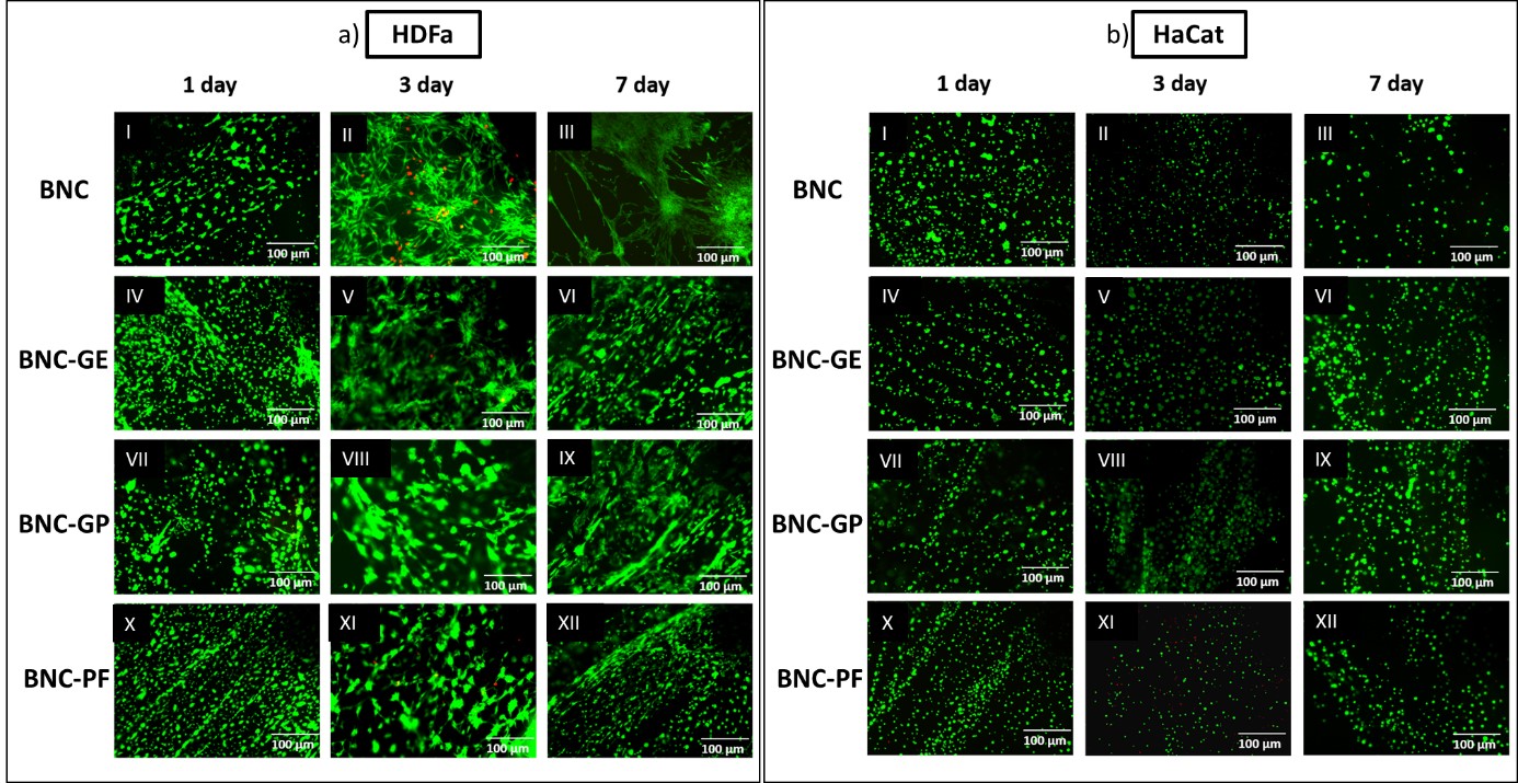
A complementary qualitative assay was performed to observe the presence of live/dead cells cultured on BNC and BNC-Aloe membranes. Cell viability of epithelial cells cultured on BNC and BNC-Aloe membranes was evaluated over 7 days of culture, as shown on Figure 5. According to the Live/Dead® stain, the green stained dots were related to living cells and the red dots were related to dead cells. Figure 4a shows the behavior of HDFa cells cultured on BNC and BNC-Aloe membranes. Dead cells were identified after 3 days of HDFa cells cultured on BNC (Figure 5 a-II), BNC-GE (Figure 5 a-V) and BNC-GP (Figure 5a-XI). HDFa cells remained viable when they were cultured on BNC and BNC-Aloe membranes over the entire cultivation period (7 days). Red dots were not observed in Figure 5b, thus viable cells were observed in all experimental times.

Viability of epithelial cells by fluorescence microscopy using Live/Dead® assay. Live cells are stained in green (calcein) and dead cells are in red (ethidium homodimer). Epithelial cells were cultured on BNC and BNC-Aloe biomembranes over 7 days. (a) HDFa cells and (b) HaCat cells.
The incorporation of A. vera fractions into BNC does not cause any cytotoxic effect to HaCat and HDFa cells. This evidence is consistent with other in vitro cytotoxic study involving A. vera/Gellan Gum sponges[4141 Silva, S. S., Oliveira, M. B., Mano, J. F., & Reis, R. L. (2014). Bio-inspired Aloe vera sponges for biomedical applications. Carbohydrate Polymers, 112, 264-270. http://dx.doi.org/10.1016/j.carbpol.2014.05.042. PMid:25129743.
http://dx.doi.org/10.1016/j.carbpol.2014...
].
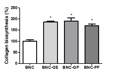
The percentage of collagen biosynthesis by HDFa cells cultured on BNC-Aloe membranes for 7 days is shown in Figure 6. As noticed, the concentration of collagen is significantly higher in all BNC-Aloe membranes compared to the control (BNC). The collagen concentration reached 90% of increase on the BNC-GP, 86% on BNC-GE and 70% on BNC-PF membranes, suggesting that BNC-Aloe membranes stimulated the collagen biosynthesis over the entire culture time.

. Quantification of collagen biosynthesis by fibroblasts cultured on BNC and BNC-Aloe membranes. Data normalized to the BNC control. Bars represent the mean and standard deviation and (* ) represents significant differences at p<0.05.
The biosynthesis of collagen was stimulated in the presence of fractions of A. vera and the concentration reached 90% of increase in the BNC-GP, 86% in BNC-GE and 70% in BNC-PF membranes. It was reported in the literature that solutions containing acemannan, one of the major components present in A. vera fractions, stimulated the synthesis of collagen[3838 Atiba, A., Nishimura, M., Kakinuma, S., Hiraoka, T., Goryo, M., Shimada, Y., Ueno, H., & Uzuka, Y. (2011). Aloe vera oral administration accelerates acute radiation-delayed wound healing by stimulating transforming growth factor-β and fibroblast growth factor production. American Journal of Surgery, 201(6), 809-818. http://dx.doi.org/10.1016/j.amjsurg.2010.06.017. PMid:21396624.
http://dx.doi.org/10.1016/j.amjsurg.2010...
,4242 Jettanacheawchankit, S., Sasithanasate, S., Sangvanich, P., Banlunara, W., & Thunyakitpisal, P. (2009). Acemannan stimulates gingival fibroblast proliferation; expressions of keratinocyte growth factor-1, vascular endothelial growth factor, and type I collagen; and wound healing. Journal of Pharmacological Sciences, 109(4), 525-531. http://dx.doi.org/10.1254/jphs.08204FP. PMid:19372635.
http://dx.doi.org/10.1254/jphs.08204FP...
]. The healing of skin lesions in rats using collagen-based membranes containing A. vera gel was also reported by in vivo studies[2222 Chithra, P., Sajithlal, G. B., & Chandrakasan, G. (1998). Influence of Aloe vera on the glycosaminoglycans in the matrix of healing dermal wounds in rats. Journal of Ethnopharmacology, 59(3), 179-186. http://dx.doi.org/10.1016/S0378-8741(97)00112-8. PMid:9507902.
http://dx.doi.org/10.1016/S0378-8741(97)...
]. The histological analysis of the lesions demonstrated the increased synthesis of collagen type I and III, as well as the proliferation of fibroblasts and macrophages in the injured areas after the membrane’s application. These results suggest the potential of BNC-Aloe membranes in the healing process, which can stimulate the components present in the extracellular matrix[4343 Boonyagul, S., Banlunara, W., Sangvanich, P., & Thunyakitpisal, P. (2014). Effect of acemannan, an extracted polysaccharide from Aloe vera, on BMSCs proliferation, differentiation, extracellular matrix synthesis, mineralization, and bone formation in a tooth extraction model. Odontology, 102(2), 310-317. http://dx.doi.org/10.1007/s10266-012-0101-2. PMid:23315202.
http://dx.doi.org/10.1007/s10266-012-010...
]. Therefore, the data indicate that the BNC-Aloe membranes present a favorable environment for cell growth, adhesion and proliferation, as well as stimulating collagen production.
4. Conclusions
In this work, we developed membranes with distinct fractions of A. vera polysaccharide extract to investigate the cellular behavior on the porous side of BNC membranes. The presence of A. vera extracts on membranes improved the adaptation of epithelial cells when compared to pure BNC. The use of human dermal and epidermal cells identified a great potential to be applied for wound repair, especially because they may facilitate the healing process by promoting keratinocyte proliferation and collagen synthesis by fibroblasts in vitro. Future in vivo assays will be further conducted in order to confirm our findings.
5. Acknowledgements
The authors thank the Brazilian agencies CAPES, CNPq and FINEP for financial support. The authors also thank André Tosello Foundation for the kind donation of Gluconacetobacter hansenii strain; and Cellular and Molecular Immunology Laboratory of Biological Sciences Institute (UFMG) and Antitumor Substances Laboratory - UFMG for providing the cells. Electron Microscopy Central Laboratory (LCME, UFSC) is also acknowledged.
-
How to cite: Piaia, L., Pittella, C. Q. P., Souza, S. S., Berti, F. V., & Porto, L. M. (2022). Incorporation of Aloe vera extract in bacterial nanocellulose membranes. Polímeros: Ciência e Tecnologia, 32(1), e2022002. https://doi.org/10.1590/0104-1428.210062
6. References
-
1Lin, W., Lien, C., Yeh, H., Yu, C., & Hsu, S. (2013). Bacterial cellulose and bacterial cellulose–chitosan membranes for wound dressing applications. Carbohydrate Polymers, 94(1), 603-611. http://dx.doi.org/10.1016/j.carbpol.2013.01.076 PMid:23544580.
» http://dx.doi.org/10.1016/j.carbpol.2013.01.076 -
2Pang, M., Huang, Y., Meng, F., Zhuang, Y., Liu, H., Du, M., Ma, Q., Wang, Q., Chen, Z., Chen, L., Cai, T., & Cai, Y. (2020). Application of bacterial cellulose in skin and bone tissue engineering. European Polymer Journal, 122, 109365. http://dx.doi.org/10.1016/j.eurpolymj.2019.109365
» http://dx.doi.org/10.1016/j.eurpolymj.2019.109365 -
3Souto, L. R. M., Rehder, J., Vassallo, J., Cintra, M. L., Kraemer, M. H. S., & Puzzi, M. B. (2006). Model for human skin reconstructed in vitro composed of associated dermis and epidermis. Sao Paulo Medical Journal, 124(2), 71-76. http://dx.doi.org/10.1590/S1516-31802006000200005 PMid:16878189.
» http://dx.doi.org/10.1590/S1516-31802006000200005 -
4Souto, L. R. M., Vassallo, J., Rehder, J., Pinto, G. A., & Puzzi, M. B. (2009). Immunoarchitectural characterization of a human skin model reconstructed in vitro. Sao Paulo Medical Journal, 127(1), 28-33. http://dx.doi.org/10.1590/S1516-31802009000100007 PMid:19466292.
» http://dx.doi.org/10.1590/S1516-31802009000100007 -
5Bell, E., Sher, S., Hull, B., Merrill, C., Rosen, S., Chamson, A., Asselineau, D., Dubertret, L., Coulomb, B., Lapiere, C., Nusgens, B., & Neveux, Y. (1983). The reconstitution of living skin. The Journal of Investigative Dermatology, 81(Suppl. 1), 2S-10S. http://dx.doi.org/10.1111/1523-1747.ep12539993 PMid:6306115.
» http://dx.doi.org/10.1111/1523-1747.ep12539993 -
6Grøn, B., Stoltze, K., Andersson, A., & Dabelsteen, E. (2002). Oral fibroblasts produce more HGF and KGF than skin fibroblasts in response to co-culture with keratinocytes. Acta Pathologica, Microbiologica, et Immunologica Scandinavica, 110(12), 892-898. http://dx.doi.org/10.1034/j.1600-0463.2002.1101208.x PMid:12645668.
» http://dx.doi.org/10.1034/j.1600-0463.2002.1101208.x -
7Sayag, J., Lieaume, S., & Bohbot, S. (1996). Healing properties of calcium alginate dressings. Journal of Wound Care, 5(8), 357-362. http://dx.doi.org/10.12968/jowc.1996.5.8.357 PMid:27935753.
» http://dx.doi.org/10.12968/jowc.1996.5.8.357 -
8Koide, M., Osaki, K., Konishi, J., Oyamada, K., Katakura, T., Takahashi, A., & Yoshizato, K. (1993). A new type of biomaterial for artificial skin: dehydrothermally cross-linked composites of fibrillar and denatured collagens. Journal of Biomedical Materials Research, 27(1), 79-87. http://dx.doi.org/10.1002/jbm.820270111 PMid:8421002.
» http://dx.doi.org/10.1002/jbm.820270111 -
9Badylak, S. F. (2007). The extracellular matrix as a biologic scaffold material. Biomaterials, 28(25), 3587-3593. http://dx.doi.org/10.1016/j.biomaterials.2007.04.043 PMid:17524477.
» http://dx.doi.org/10.1016/j.biomaterials.2007.04.043 -
10Brown, R. M., Jr., & Montezinos, D. (1976). Cellulose microfibrils: visualization of biosynthetic and orienting complexes in association with the plasma membrane. Proceedings of the National Academy of Sciences of the United States of America, 73(1), 143-147. http://dx.doi.org/10.1073/pnas.73.1.143 PMid:1061108.
» http://dx.doi.org/10.1073/pnas.73.1.143 -
11Rambo, C. R., Recouvreux, D. O. S., Carminatti, C. A., Pitlovanciv, A. K., Antônio, R. V., & Porto, L. M. (2008). Template assisted synthesis of porous nanofibrous cellulose membranes for tissue engineering. Materials Science and Engineering C, 28(4), 549-554. http://dx.doi.org/10.1016/j.msec.2007.11.011
» http://dx.doi.org/10.1016/j.msec.2007.11.011 -
12Recouvreux, D. O. S., Carminatti, C. A., Pitlovanciv, A. K., Rambo, C. R., Porto, L. M., & Antônio, R. V. (2008). Cellulose biosynthesis by the beta-proteobacterium, Chromobacterium violaceum Current Microbiology, 57(5), 469-476. http://dx.doi.org/10.1007/s00284-008-9271-0 PMid:18820969.
» http://dx.doi.org/10.1007/s00284-008-9271-0 -
13Souza, S. S., Berti, F. V., Oliveira, K. P. V., Pittella, C. Q. P., Castro, J. V., Pelissari, C., Rambo, C. R., & Porto, L. M. (2019). Nanocellulose biosynthesis by Komagataeibacter hansenii in a defined minimal culture medium. Cellulose, 26(3), 1641-1655. http://dx.doi.org/10.1007/s10570-018-2178-4
» http://dx.doi.org/10.1007/s10570-018-2178-4 -
14Sperotto, G., Stasiak, L. G., Godoi, J. P. M. G., Gabiatti, N. C., & Souza, S. S. (2021). A review of culture media for bacterial cellulose production: complex, chemically defined and minimal media modulations. Cellulose, 28(5), 2649-2673. http://dx.doi.org/10.1007/s10570-021-03754-5
» http://dx.doi.org/10.1007/s10570-021-03754-5 -
15Anton-Sales, I., Beekmann, U., Laromaine, A., Roig, A., & Kralisch, D. (2019). Opportunities of bacterial cellulose to treat epithelial tissues. Current Drug Targets, 20(8), 808-822. http://dx.doi.org/10.2174/1389450120666181129092144 PMid:30488795.
» http://dx.doi.org/10.2174/1389450120666181129092144 -
16Rahman, S., Carter, P., & Bhattarai, N. (2017). Aloe vera for tissue engineering applications. Journal of Functional Biomaterials, 8(1), 6. http://dx.doi.org/10.3390/jfb8010006 PMid:28216559.
» http://dx.doi.org/10.3390/jfb8010006 -
17Tanaka, M., Yamada, M., Toida, T., & Iwatsuki, K. (2012). Safety evaluation of supercritical carbon dioxide extract of Aloe vera gel. Journal of Food Science, 77(1), T2-T9. http://dx.doi.org/10.1111/j.1750-3841.2011.02452.x PMid:22260137.
» http://dx.doi.org/10.1111/j.1750-3841.2011.02452.x -
18Godinho, J. F., Berti, F. V., Müller, D., Rambo, C. R., & Porto, L. M. (2016). Incorporation of Aloe vera extracts into nanocellulose during biosynthesis. Cellulose(1), 23, 545-555. http://dx.doi.org/10.1007/s10570-015-0844-3
» http://dx.doi.org/10.1007/s10570-015-0844-3 -
19Davis, R. H., Donato, J. J., Hartman, G. M., & Haas, R. C. (1994). Anti-inflammatory and wound healing activity of a growth substance in Aloe vera. Journal of the American Podiatric Medical Association, 84(2), 77-81. http://dx.doi.org/10.7547/87507315-84-2-77 PMid:8169808.
» http://dx.doi.org/10.7547/87507315-84-2-77 -
20Kang, M., Kim, S. Y., Kim, Y. T., Kim, E., Lee, S., Ko, S., Wijesinghe, W. A. J. P., Samarakoon, K. W., Kim, Y., Cho, J. H., Jang, H., & Jeon, Y. (2014). In vitro and in vivo antioxidant activities of polysaccharide purified from aloe vera (Aloe barbadensis) gel. Carbohydrate Polymers, 99, 365-371. http://dx.doi.org/10.1016/j.carbpol.2013.07.091 PMid:24274519.
» http://dx.doi.org/10.1016/j.carbpol.2013.07.091 -
21Sierra-García, G. D., Castro-Ríos, R., González-Horta, A., Lara-Arias, J., & Chávez-Montes, A. (2014). Acemannan, an extracted polysaccharide from Aloe vera: a literature review. Natural Product Communications, 9(8), 1217-1221. http://dx.doi.org/10.1177/1934578X1400900836 PMid:25233608.
» http://dx.doi.org/10.1177/1934578X1400900836 -
22Chithra, P., Sajithlal, G. B., & Chandrakasan, G. (1998). Influence of Aloe vera on the glycosaminoglycans in the matrix of healing dermal wounds in rats. Journal of Ethnopharmacology, 59(3), 179-186. http://dx.doi.org/10.1016/S0378-8741(97)00112-8 PMid:9507902.
» http://dx.doi.org/10.1016/S0378-8741(97)00112-8 -
23Boudreau, M. D., & Beland, F. A. (2006). An evaluation of the biological and toxicological properties of Aloe barbadensis (miller), Aloe vera Journal of Environmental Science and Health. Part C, Environmental Carcinogenesis & Ecotoxicology Reviews, 24(1), 103-154. http://dx.doi.org/10.1080/10590500600614303 PMid:16690538.
» http://dx.doi.org/10.1080/10590500600614303 -
24Saibuatong, O., & Phisalaphong, M. (2010). Novo Aloe vera-bacterial cellulose composite film from biosynthesis. Carbohydrate Polymers, 79(2), 455-460. http://dx.doi.org/10.1016/j.carbpol.2009.08.039
» http://dx.doi.org/10.1016/j.carbpol.2009.08.039 -
25Stumpf, T. R., Pértile, R. A. N., Rambo, C. R., & Porto, L. M. (2013). Enriched glucose and dextrin mannitol-based media modulates fibroblast behavior on bacterial cellulose membranes. Materials Science and Engineering C, 33(8), 4739-4745. http://dx.doi.org/10.1016/j.msec.2013.07.035 PMid:24094182.
» http://dx.doi.org/10.1016/j.msec.2013.07.035 -
26Moniri, M., Moghaddam, A. B., Azizi, S., Rahim, R. A., Ariff, A. B., Saad, W. Z., Navaderi, M., & Mohamad, R. (2017). Production and status of bacterial cellulose in biomedical engineering. Nanomaterials, 7(9), 257. http://dx.doi.org/10.3390/nano7090257 PMid:32962322.
» http://dx.doi.org/10.3390/nano7090257 -
27Berti, F. V., Rambo, C. R., Dias, P. F., & Porto, L. M. (2013). Nanofiber density determines endothelial cell behavior on hydrogel matrix. Materials Science and Engineering C, 33(8), 4684-4691. http://dx.doi.org/10.1016/j.msec.2013.07.029 PMid:24094176.
» http://dx.doi.org/10.1016/j.msec.2013.07.029 -
28Silva, A. R. P., Paula, A. C. C., Martins, T. M. M., Goes, A. M., & Pereria, M. M. (2014). Synergistic effect between bioactive glass foam and a perfusion bioreactor on osteogenic differentiation of human adipose stem cells. Journal of Biomedical Materials Research, Part A, 102(3), 818-827. http://dx.doi.org/10.1002/jbm.a.34758 PMid:23625853.
» http://dx.doi.org/10.1002/jbm.a.34758 -
29Godinho, J. (2014). Hidrogéis de celulose bacteriana incorporados com frações de Aloe vera (Dissertação de Mestrado). Universidade Federal de Santa Catarina, Santa Catarina, Brasil.
-
30Piaia, L., Paes, C. Q., & Porto, L. M. (2014). Viability of human dermal fibroblasts cultured on bacterial cellulose and Aloe vera composites. BMC Proceedings, 8(4), 61. http://dx.doi.org/10.1186/1753-6561-8-S4-P61
» http://dx.doi.org/10.1186/1753-6561-8-S4-P61 -
31Dayal, M. S., & Catchmark, J. M. (2016). Mechanical and structural property analysis of bacterial cellulose composites. Carbohydrate Polymers, 144, 447-453. http://dx.doi.org/10.1016/j.carbpol.2016.02.055 PMid:27083837.
» http://dx.doi.org/10.1016/j.carbpol.2016.02.055 -
32Murphy, C. M., & O’Brien, F. J. (2010). Understanding the effect of mean pore size on cell activity in collagen-glycosaminoglycan scaffolds. Cell Adhesion & Migration, 4(3), 377-381. http://dx.doi.org/10.4161/cam.4.3.11747 PMid:20421733.
» http://dx.doi.org/10.4161/cam.4.3.11747 -
33Fu, L., Zhang, J., & Yang, G. (2013). Present status and applications of bacterial cellulose-based materials for skin tissue repair. Carbohydrate Polymers, 92(2), 1432-1442. http://dx.doi.org/10.1016/j.carbpol.2012.10.071 PMid:23399174.
» http://dx.doi.org/10.1016/j.carbpol.2012.10.071 -
34Tokoh, C., Takabe, K. J., & Fujita, M. (2002). Cellulose synthesized by Acetobacter xylinum in the presence of plant cell wall polysaccharides. Cellulose, 9(1), 65-74. http://dx.doi.org/10.1023/A:1015827121927
» http://dx.doi.org/10.1023/A:1015827121927 -
35Petersen, A., Princ, A., Korus, G., Ellinghaus, A., Leemhuis, H., Herrera, A., Klaumünzer, A., Schreivogel, S., Woloszyk, A., Schmidt-Bleek, K., Geissler, S., Heschel, I., & Duda, G. N. (2018). A biomaterial with a channel-like pore architecture induces endochondral healing of bone defects. Nature Communications, 9(1), 4430. http://dx.doi.org/10.1038/s41467-018-06504-7 PMid:30361486.
» http://dx.doi.org/10.1038/s41467-018-06504-7 -
36Carter, P., Rahman, S. M., & Bhattarai, N. (2016). Facile fabrication of Aloe vera containing PCL nanofibers for barrier membrane application. Journal of Biomaterials Science. Polymer Edition, 27(7), 692-708. http://dx.doi.org/10.1080/09205063.2016.1152857 PMid:26878323.
» http://dx.doi.org/10.1080/09205063.2016.1152857 -
37McAnalley, B. H., Carpenter, R. H., & McDaniel, H. R. (1995). US 5468737A. USA. Retrieved in 2021, December 20, from http://www.google.com/patents/US5468737
» http://www.google.com/patents/US5468737 -
38Atiba, A., Nishimura, M., Kakinuma, S., Hiraoka, T., Goryo, M., Shimada, Y., Ueno, H., & Uzuka, Y. (2011). Aloe vera oral administration accelerates acute radiation-delayed wound healing by stimulating transforming growth factor-β and fibroblast growth factor production. American Journal of Surgery, 201(6), 809-818. http://dx.doi.org/10.1016/j.amjsurg.2010.06.017 PMid:21396624.
» http://dx.doi.org/10.1016/j.amjsurg.2010.06.017 -
39Deters, A., Dauer, A., Schnetz, E., Fartasch, M., & Hensel, A. (2001). High molecular compounds (polysaccharides and proanthocyanidins) from Hamamelis virginiana bark: influence on human skin keratinocyte proliferation and differentiation and influence on irritated skin. Phytochemistry, 58(6), 949-958. http://dx.doi.org/10.1016/S0031-9422(01)00361-2 PMid:11684194.
» http://dx.doi.org/10.1016/S0031-9422(01)00361-2 -
40Bell, E., Parenteau, N., Gay, R., Nolte, C., Kemp, P., Bilbo, P., Ekstein, B., & Johnson, E. (1991). The living skin equivalent: its manufacture, its organotypic properties and its responses to irritants. Toxicology In Vitro, 5(5-6), 591-596. http://dx.doi.org/10.1016/0887-2333(91)90099-Y PMid:20732083.
» http://dx.doi.org/10.1016/0887-2333(91)90099-Y -
41Silva, S. S., Oliveira, M. B., Mano, J. F., & Reis, R. L. (2014). Bio-inspired Aloe vera sponges for biomedical applications. Carbohydrate Polymers, 112, 264-270. http://dx.doi.org/10.1016/j.carbpol.2014.05.042 PMid:25129743.
» http://dx.doi.org/10.1016/j.carbpol.2014.05.042 -
42Jettanacheawchankit, S., Sasithanasate, S., Sangvanich, P., Banlunara, W., & Thunyakitpisal, P. (2009). Acemannan stimulates gingival fibroblast proliferation; expressions of keratinocyte growth factor-1, vascular endothelial growth factor, and type I collagen; and wound healing. Journal of Pharmacological Sciences, 109(4), 525-531. http://dx.doi.org/10.1254/jphs.08204FP PMid:19372635.
» http://dx.doi.org/10.1254/jphs.08204FP -
43Boonyagul, S., Banlunara, W., Sangvanich, P., & Thunyakitpisal, P. (2014). Effect of acemannan, an extracted polysaccharide from Aloe vera, on BMSCs proliferation, differentiation, extracellular matrix synthesis, mineralization, and bone formation in a tooth extraction model. Odontology, 102(2), 310-317. http://dx.doi.org/10.1007/s10266-012-0101-2 PMid:23315202.
» http://dx.doi.org/10.1007/s10266-012-0101-2
Publication Dates
-
Publication in this collection
24 Jan 2022 -
Date of issue
2022
History
-
Received
10 Sept 2021 -
Reviewed
23 Nov 2021 -
Accepted
20 Dec 2021