ABSTRACT:
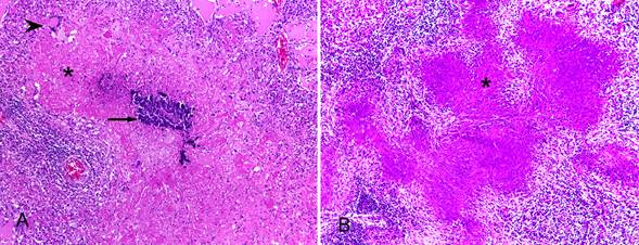
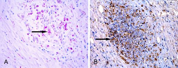
Tuberculosis is a debilitating infecto-contagious disease, caused by an acid-fast bacillus (AFB) that belong to different species of the Mycobacterium tuberculosis complex (MTC). Mycobacteriosis are important diseases in veterinary medicine because of their zoonotic potential and worldwide distribution, affecting all classes of vertebrates. In wild animals the mycobacteriosis have been reported mainly as a problem in captivity. There are also reports in free-ranging wildlife, endangering and hampering tuberculosis erradication programs in animal production. The diagnosis of the disease in wildlife is usually postmortem, because the tuberculin test is not standardized for wildlife species, and also it is not reliable for screening. The postmortem diagnosis is based on macroscopic findings, microscopic detection of AFB at Ziehl-Neelsen staining (ZN), and mainly isolation and identification of the agent. However, only gross and microscopic exams do not allow to distinguish the species of Mycobacterium involved. The immunostaining with polyclonal anti-Mycobacterium tuberculosis confirms tuberculosis infection, but is not specific; there may be marking of other mycobacteria. The aim of this study was to describe the histologic findings, of ZN staining and immunohistochemistry technique (IHC) of 13 cases of wildlife herbivores diagnosed with tuberculosis in the Setor de Patologia Veterinária of the Universidade Federal do Rio Grande do Sul (SPV-UFRGS) during the 2003- 2015 period. Formalin fixed paraffin embedded tissues were recut, stained with hematoxylin and eosin and ZN, and samples were submitted to the IHC (anti-M. tuberculosis marking, streptavidin-biotin peroxidase method). All animals included were adults living in captivity and belonged to the following species: llama (5/13), sambar deer (4/13), camel (1/13), red deer (1/13), Brazilian tapir (1/13) and Nilgai antelope (1/13). The IHC revealed immunostaining of accentuated (4/13), moderate (4/13) and discrete (4/13) intensity, except in a case with insufficient material. The histological features, findings in ZN staining and IHC in the wild herbivores with tuberculosis lesions allowed the diagnosis of infection with Mycobacterium sp., turning into fast and efficient methods of diagnosis, which can help to prevent the spread of this disease in animals from the same herd.
INDEX TERMS:
Mycobacterium sp.; tuberculosis; herbivores; camelids; deer; histopathology; immunohistochemistry; diagnosis
 Thumbnail
 Thumbnail
 Thumbnail
 Thumbnail
 Thumbnail