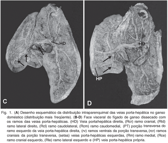
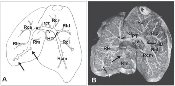
The intraparenchymal distribution of the hepatic portal veins in 30 domestic geese were studied. Stained Neoprene latex was injected into the isquiatic vessels, and the animals were fixed in 10% formaldehyde by immersion and intramuscular injection. The liver of geese was composed of a large right and a smaller left hepatic lobe, connected by a parenchyma bridge. The right hepatic lobe had vessels exclusively from the hepatic portal system composed by intraparenchymal distribution of the right hepatic portal vein, while the vessels of the left hepatic lobe came from the right hepatic portal vein and from small left hepatic portal veins. The right hepatic portal vein emitted the right caudal branch, which emitted a small right caudolateral branch and a large right caudomedial branch. Cranially this vein emitted right cranial and right lateral branches. The tranverse portion of the right hepatic portal vein crossed to the left hepatic lobe, emitting 1 to 6 small cranial and caudal branches to the medial area of the liver. In the left hepatic lobe, the left branch from the right hepatic vein emitted the left cranial, left lateral and left median branches. One to six left hepatic portal veins were identified arising from the left branch or from the transverse portion of the right hepatic portal vein. These vessels arose from the gizzard and pro-ventricle. In 40% of geese one proper hepatic portal vein originated from venous vessels of the gizzard and was distributed into the caudal extremity of the left hepatic isolated lobe.
Morphology; hepatic portal vein; liver; geese


