Abstract:
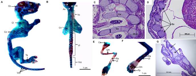
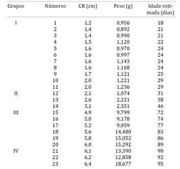
The aim of this study was to describe the skeletogenesis in punaré (Thrichomys laurentinus). We used 11 embryos and 12 fetuses in different stages of development, allocated into 4 groups. Samples were obtained from the Multiplication Center of Wild Animals, at Federal Rural University of the Semi-Arid, Mossoro/RN, Brazil. After fixed in formalin (10%) or glutaraldehyde (2.5%) the morphological analysis was performed with a magnifying glass, and the macroscopic characteristics were photographed. Analysis of X-rays and alizarin red staining was made to better understand the development of bone structures. In x-ray analysis, it was possible to verify that the embryos showed no radiopacity, unlike fetuses that had gradual radiopacity along of the groups. In group II, there was an increase in radiopacity in the spine, mandibular and maxillary regions. In group III, the radiopacity was increased in the hind limbs, ribs and in the frontal region, and group IV showed higher radiopacity in the thoracic limbs and occipital, temporal and frontal skull. These characteristics were confirmed by histological and alizarin red analysis. We concluded that the knowledge of normal skeletal embryology is critical for understanding of adverse effects caused by nutrition and use of drugs during the development.
Index Terms:
Skeletogenesis; punare; forelimbs; hind limb
 Thumbnail
 Thumbnail
 Thumbnail
 Thumbnail
 Thumbnail