 Histiocytic sarcoma in dogs: epidemiology, anatomopathology, and immunohistochemistry
ABSTRACT:
Histiocytic sarcomas have been described in veterinary medicine since 1980, but studies on the subject are still scarce. Based on this, the objective of this article is to describe the epidemiological, anatomopathological and immunohistochemical aspects of histiocytic sarcoma in dogs submitted to necropsy in a diagnostic service covering the midwestern region of Rio Grande do Sul State, Brazil. From 2007 to 2021, 4,310 dogs were necropsied, of which 598 died or were euthanized due to some type of cancer. At least 18 cases of histiocytic sarcoma were diagnosed, i.e., 3% of cancer deaths and 0.4% of total deaths. The criterion used to establish the definitive diagnosis and inclusion in the study was an interaction between characteristic histopathology and positive immunostaining for CD204. Almost all (17/18, 94.4%) of these patients were of a defined breed and were large, with the vast majority (14/18, 77.8%) being Rottweiler. There was a predominance of disseminated histiocytic sarcoma (15/18, 83.3%) affecting several organs, while 10 (66.7%) affecting the lungs, liver, spleen and lymph nodes were affected concomitantly. Of the few cases (3/18, 16.7%) diagnosed as localized histiocytic sarcoma, where lungs were affected. Five different presentation patterns were observed macroscopically, not mutually exclusive: multinodular, massive, diffuse, peribronchiolar, and placoid. The most affected organs were the lungs (17/18, 94.4%), lymph nodes (15/18, 83.3%), liver (13/18, 72.2%), spleen (12/18, 66.7%), kidneys (6/15, 60%) and heart (6/15, 40%). Other less affected organs included adrenals (4/15, 26.7%), skeletal muscle (diaphragm) (4/15, 26.7%), bones (2/15, 13.3%), pancreas (2/15, 13.3%), pericardial sac (2/15, 13.3%), joint (1/15, 6.7%), omentum (1/15, 6.7%) and parietal pleura (1/15, 6.7%). Histologically, histiocytic sarcoma was characterized by a non-delimited, mantle-shaped proliferation with a scant stroma of round cells, many markedly anaplastic, often giving the tumor a rather pleomorphic appearance. A hallmark was the occurrence of a variable, but often high, number of mono, bi and multinucleated giant cells (30-100 µm in diameter), which always had large nuclei (karyomegaly) formed by loose chromatin and with nucleoli almost always multiple and conspicuous. Although there are peculiarities in the neoplastic involvement in each affected organ, in general, this proliferation tends to obscure the affected parenchyma and often invades and obliterates lymphatic and blood vessels. About 90% of neoplastic cells, including the most anaplastic and many of the multinucleated ones, immunostained strongly for CD204 and MHC-II but not for CD11d, confirming that they were histiocytes, other than splenic/bone marrow macrophages. It is hoped that this information will contribute to a better characterization of histiocytic sarcoma in the canine species and may help veterinary pathologists in their diagnostic routines.
INDEX TERMS:
Malignant histiocytosis; histiocytic tumors; pathology; oncology; diseases of dogs
RESUMO:
Os sarcomas histiocíticos têm sido descritos na medicina veterinária desde meados de 1980, mas os estudos sobre o tema ainda são escassos. Com base nisso, o objetivo deste artigo foi determinar os aspectos epidemiológicos, anatomopatológicos e imuno-histoquímicos do sarcoma histiocítico em cães submetidos à necropsia em um serviço de diagnóstico que abrange a região centro-oeste do Rio Grande do Sul, Brasil. Entre os anos de 2007 e 2021 foram necropsiados 4.310 cães, dos quais 598 morreram ou foram submetidos à eutanásia devido a algum tipo de câncer. Pelo menos 18 casos de sarcoma histiocítico foram diagnosticados, ou seja, 3% das mortes por câncer e 0,4% das mortes totais. O critério utilizado para estabelecer o diagnóstico definitivo e inclusão no estudo foi uma interação entre a histopatologia característica e a imunomarcação positiva para CD204. Quase a totalidade (17/18; 94,4%) desses pacientes tinha raça definida e era de porte grande, sendo a grande maioria (14/18; 77,8%) da raça Rottweiler. A maior parte dos casos (15/18; 83,3%) eram sarcomas histiocíticos disseminados, sendo que em 10 (66,7%), os pulmões, o fígado, o baço e os linfonodos foram acometidos concomitantemente. Dos poucos casos (3/18; 16,7%) diagnosticados como sarcoma histiocítico localizado, os pulmões foram sempre afetados. Macroscopicamente foram observados cinco padrões de apresentação, não mutualmente excludentes, a saber: multinodular, massivo, difuso, peribronquiolar e placoide. Os órgãos mais afetados foram: pulmões (17/18; 94,4%), linfonodos (15/18; 83,3%), fígado (13/18; 72,2%), baço (12/18; 66,7%), rins (6/15; 60%) e coração (6/15; 40%). Outros órgãos menos afetados incluíram: adrenais (4/15; 26,7%), músculo esquelético (diafragma) (4/15; 26,7%), ossos (2/15; 13,3%), pâncreas (2/15; 13,3%), saco pericárdico (2/15; 13,3%), articulação (1/15; 6,7%), omento (1/15; 6,7%) e pleura parietal (1/15; 6,7%). Histologicamente, o diagnóstico do sarcoma histiocítico sempre foi suspeitado pela presença de um tumor de células redondas ou fusiformes, marcadamente anaplásico e pleomórfico, rico em células gigantes mononucleadas e frequentemente associado à presença de variável quantidade de células gigantes (com 30-100 µm de diâmetro) mono, bi e multinucleadas, as quais sempre possuíam grandes núcleos (cariomegalia) formados por cromatina frouxa e com nucléolos quase sempre múltiplos e conspícuos. Apesar de em cada órgão afetado haver peculiaridades no acometimento neoplásico, no geral essa proliferação tendia a obscurecer o parênquima afetado e frequentemente invadir e obliterar vasos linfáticos e sanguíneos. A maior parte das células neoplásicas (cerca de 90%), incluindo as mais anaplásicas e muitas das multinucleadas, imunomarcaram fortemente para CD204 e MHC-II, mas não para CD11d, confirmando tratar-se de histiócitos outros que não macrófagos esplênicos/medulares ósseos. Espera-se que essas informações contribuam para uma melhor caracterização do sarcoma histiocítico na espécie canina e que possam auxiliar patologistas veterinários em suas rotinas diagnósticas.
TERMOS DE INDEXAÇÃO:
Histiocitose maligna; tumores histiocíticos; patologia; oncologia; doenças de cães
Introduction
Histiocytic sarcomas were first described in veterinary medicine in the 1980s (Moore & Rosin 1986), when Peter Moore demonstrated that anaplastic large cell carcinomas (giant cell type) previously reported in the lungs of dogs (Stünzi 1973, Stünzi et al. 1974) were, in fact, histiocytic tumors. This form of cancer had only been recognized by the World Health Organization (WHO) for humans two decades earlier, as until then, it was mistakenly included as lymphoma (histiocytic lymphoma) (Weiss et al. 2017). In the original description in dogs (Moore & Rosin 1986), and for some time, the term malignant histiocytosis was widely used until it was gradually abandoned and replaced by histiocytic sarcoma (Affolter & Moore 2002). Since then, many scientific articles have been published and given notoriety to this aggressive form of cancer, described mainly in dogs, less commonly in cats and only rarely in other animal species. Currently, histiocytic sarcoma has been used as an animal model for the study of molecular cytogenetics, as it occurs with a certain frequency in dogs, unlike the rarity with which it affects humans (Davis & Ostrander 2014, Schiffman & Breen 2015).
Although canine histiocytic sarcoma has been studied more in recent years, the number of scientific articles available in the international literature is still small compared to other important forms of canine cancer, such as lymphomas, osteosarcomas and mammary carcinomas. Likewise, the main pathology books still describe much less about histiocytic sarcoma than these other tumors mentioned, meaning that the pathologist knows little about the subject and recognize these cases less in their routine. Furthermore, the macroscopic and histopathological presentation of histiocytic sarcoma makes it a great imitator of many other forms of cancer. In this sense, this article aims to: 1) determine the main patterns of anatomopathological presentation (macroscopic, histological and immunohistochemical) of histiocytic sarcoma and establish the frequency with which they occur; 2) establish its prevalence among canine cancers; and 3) characterize the epidemiological profile of affected dogs.
Materials and Methods
Ethical approval. No approval of research ethics committees was required to accomplish the goals of this study since it is retrospective and no animal experiments were performed.
Initially, in the Report Archive of the “Laboratório de Patologia Veterinária” (Veterinary Pathology Laboratory) of the “Universidade Federal de Santa Maria” (LPV/UFSM), reports of canine histiocytic sarcoma, diagnosed from 2007 to 2021 (15 years), were accessed. Only necropsy cases were considered. Subsequently, the respective paraffin blocks were tracked in the LPV/UFSM Block Archive. An individual analysis of the reports referring to all cases included in the study was carried out, collecting epidemiological, macroscopic and histopathological information. All cases were reevaluated histologically through slides from tissue sections embedded in paraffin and stained with hematoxylin and eosin (HE). The LPV/UFSM Image Archive was reviewed for a detailed and systematized macroscopic characterization. The definitive diagnosis of the cases was made based on the histological criteria used in the classification of hematopoietic tumors in domestic animals published by the Armed Forces Institute of Pathology (AFIP) (Valli et al. 2002), including its most recent updates for domestic animals in leading veterinary pathology (Valli et al. 2016), tumor pathology (Valli et al. 2017), and hematopathology (Valli 2007) textbooks.
At least one tissue sample from each case under study was subjected to the immunohistochemistry (IHC) technique, carried out at the “Laboratório de Patologia Morfológica e Molecular” (Laboratory of Morphological and Molecular Pathology - LAPMOL), “Faculdade de Medicina Veterinária e Zootecnia” (FMVZ) of “Universidade de São Paulo” (USP) to confirm the origin of the neoplastic cells. For canine histiocytic, the primary antibodies were used: CD204 (Clone SRA-C6, TransGenic Inc., Kobe, Japan, produced in mice; dilution 1:1,000), MHC-II (Clone TAL. 1B5, Dako Cytomation, Glostrup, Germany, produced in mice; dilution 1:2,000) and CD11d (Clone CA18.3C6, a non-commercial antibody produced by Peter Moore, Davis, California, United States, in rabbit; dilution 1:1,000). The technique was performed following the following protocol: After deparaffinization and rehydration of the paraffin-embedded tissues, antigen retrieval was performed under heating with a citrate solution (pH 6.0) in a high-power pressure cooker for 10 minutes. To block the endogenous peroxidase, hydrogen peroxide (10 volumes) was used in an oven at 37 °C for 30 minutes in a reaction protected from light. Blocking of non-specific reactions was carried out with Protein Block (NovolinkTM Kit) for 10 minutes, at room temperature, in a humid chamber, and with a solution of powdered milk diluted to 5% at 37 °C for 30 minutes. The primary antibodies were incubated in a humid chamber in a refrigerator at 4 °C for 18 hours (overnight). The secondary antibody (NovolinkTM post Primary) and the Polymer (NovolinkTM Polymer) were used consecutively and incubated at room temperature in a humid chamber for 30 minutes each. The development was carried out by adding the chromogen 3,3’-diaminobenzidine-tetrahydrochloride-dihydrate (DAB) in a humid chamber for five minutes. Washing between technique steps was carried out with TTBS (pH 7.6). Counterstaining was performed with Harris hematoxylin. Tissues were dehydrated, and slides were mounted using Permount dilution with xylene. Dog mesenteric lymph nodes were used as a positive control, while the negative control was represented by the same sections, replacing the primary antibody with TTBS. For the CD11d antibody, the following modifications were used: 1) antigen retrieval was performed with Tris-EDTA (pH 9.0) in a water bath at 90 °C for 30 minutes; 2) the development was done through the addition of ImmPACT NovaRED (peroxidase), at room temperature, for three minutes; 3) After counterstaining, assembly proceeded directly, without dehydration; and 4) the assembly was carried out only by Permount, without the use of xylene. All cells immunostained for MHC-II were considered leukocytes, all cells immunostained for CD204 as histiocytes, and all cells immunostained for CD11d as macrophages of splenic origin.
Results
For the study, 24 cases previously diagnosed as histiocytic sarcoma were gathered by several pathologists of LPV/UFSM, two of whom (R.A.F and G.D.K.) are authors of this study. From 2007 to 2021, 4,310 dogs were necropsied at LPV/UFSM. Of these, 598 (13.9%) died or were euthanized due to some type of cancer. Of the total dogs with cancer included in this study, 18 met the criteria to be diagnosed as having histiocytic sarcoma. It represented 3% of cancer cases and 0.4% of total cases. Of the total number of dogs with histiocytic sarcoma, nine (50%) died spontaneously, and nine (50%) were euthanized.
Epidemiological findings
Of the total number of dogs with histiocytic sarcoma, 10 were females (55.6%), while eight were males (44.4%). The affected dogs’ ages ranged from two to 14 years, with a higher frequency between five and 12 years (14/18, 77.8%). The age average was 8.5 years. Almost all dogs were purebred (17/18, 94.4%), mainly Rottweiler (14/18, 77.8%). Only one dog (5.6%) did not have a defined breed, and it was classified as large. Other pure breeds included Dogo Argentino (1/18, 5.6%), Labrador Retriever (1/18, 5.6%), and Shar-Pei (1/18, 5.6%). Based on these breeds, 17 (94.4%) dogs were included as large (Rottweiler, Labrador Retriever, Dogo Argentino and mixed breed) and only one (5.6%) as medium size (Shar-Pei). A detailed distribution of each case regarding epidemiological aspects can be found in Table 1.
Macroscopic findings
Of the total number of dogs with histiocytic sarcoma, 15 (83.3%) were classified as disseminated and three (16.7%) as localized. From the 15 cases diagnosed as disseminated histiocytic sarcoma, 10 (66.7%) had the lungs, liver, spleen and lymph nodes concomitantly affected. At least three of these four organs were affected in the other five cases (33.3%). All three cases (100%) were diagnosed as localized histiocytic sarcoma, and the lungs were affected. The retropharyngeal lymph nodes were also involved in one (33.3%) of these three cases.
In cases with pulmonary involvement (17/18, 94.4%), four different patterns of macroscopic presentation were observed. The first (6/17, 35.3%) was characterized by multiple yellowish-white nodules (multinodular pattern), of variable dimensions and distributed multifocally throughout the lung lobes (Fig.1). These nodules were soft to the touch and cut, with a homogeneously white or yellowish cut surface. In the second pattern (6/17, 35.3%), one or two lung lobes were completely replaced by a yellowish-white and irregular mass (massive pattern) (Fig.2), not very firm and with a cut surface similar to the one described for the first pattern. In the third pattern (4/17, 23.5%), the lungs did not collapse when the thoracic cavity was accessed (diffuse pattern), being reddish or painted in different shades of red and white (Fig.3), including one or more lobes that were diffusely firm to touch and cut. Some of these cases (3/17, 17.6%), when cut, presented as equally firm, with a cut surface characterized by marked peribronchial thickening (diffuse peribronchial pattern) accompanied or not by nodules or masses obliterating the parenchyma (Fig.4). In the fourth pattern, seen only in two localized cases (2/17, 11.7%), white plaques (placoid pattern), more or less circumferential, could be visualized in one of the lung lobes (Fig.5). In one case (1/17, 5.9%) there was an overlap between the multinodular and massive patterns (mixed pattern), which means, multifocal nodules and a lobar mass were seen together in different lung lobes. In another case (1/17, 5.9%), one lobe had a diffuse pattern, while another had a mass. There was also one case (1/17, 5.9%) with small nodules around a plaque.

Histiocytic sarcoma, lungs, dog. Multiple multifocal and coalescent white nodules were randomly distributed throughout the parenchyma of all lung lobes (multinodular pattern).

Histiocytic sarcoma, lungs, dog. A large, irregular, multilobulated mass completely obliterated the right cranial lobe (massive pattern).

Histiocytic sarcoma, lungs, dog. The lungs were diffusely red and did not collapse after the chest opening cavity (diffuse pattern). Near the apex of the left caudal, middle, and right cranial lobes, there were circumferential, slightly raised areas that varied from white to red.

Histiocytic sarcoma, lung (cut surface), dog. Multiple white and prominent annular lesions. Microscopically, these lesions corresponded to marked bronchial thickening.

Histiocytic sarcoma, left lung, dog. Yellowish-white plaque that protruded from the natural surface of the middle lobe.
In 13 of 18 cases (72.2%) of histiocytic sarcoma, 13 of 15 (86.7%) cases of disseminated histiocytic sarcoma, the liver was affected, and three distinct macroscopic presentations were observed. In most cases (8/15, 53.3%), the liver was diffuse and moderately enlarged, brownish, containing several yellowish-white nodules (multinodular pattern) (Fig.6), sometimes umbilicated (Fig.7), distributed multifocally and randomly throughout the hepatic lobes. These nodules were soft on touch and cut, with a yellowish-white cut surface. In other cases (4/15, 26.7%), a large mass (massive pattern) completely surrounded and replaced one of the hepatic lobes. In some cases (3/15, 20%), the liver was diffuse (diffuse pattern) and markedly increased in volume, with a marked accentuation of the lobular pattern, better seen on the cut surface. In at least two cases (2/15, 13.3%), there were overlapping patterns (mixed pattern) in which a diffuse liver markedly increased in volume and demonstrated some nodules protruding from the capsule and on cutting. In one case (1/15, 6.7%), there was an overlap between the multinodular and massive patterns; multifocal nodules and a lobular mass were seen together in different hepatic lobes.

Histiocytic sarcoma, liver, dog. Multifocal and coalescent nodules ranging from 1-2 millimeters to 2-3 centimeters in diameter, distributed randomly throughout all liver lobes (multinodular pattern).

Histiocytic sarcoma, liver, dog. Close-up view of one of the liver nodules seen in Figure 6. Note the depressed central area. Nodules of this type are known as umbilicated and reflect the collapse of the outermost portion of the lesion due to central necrosis.
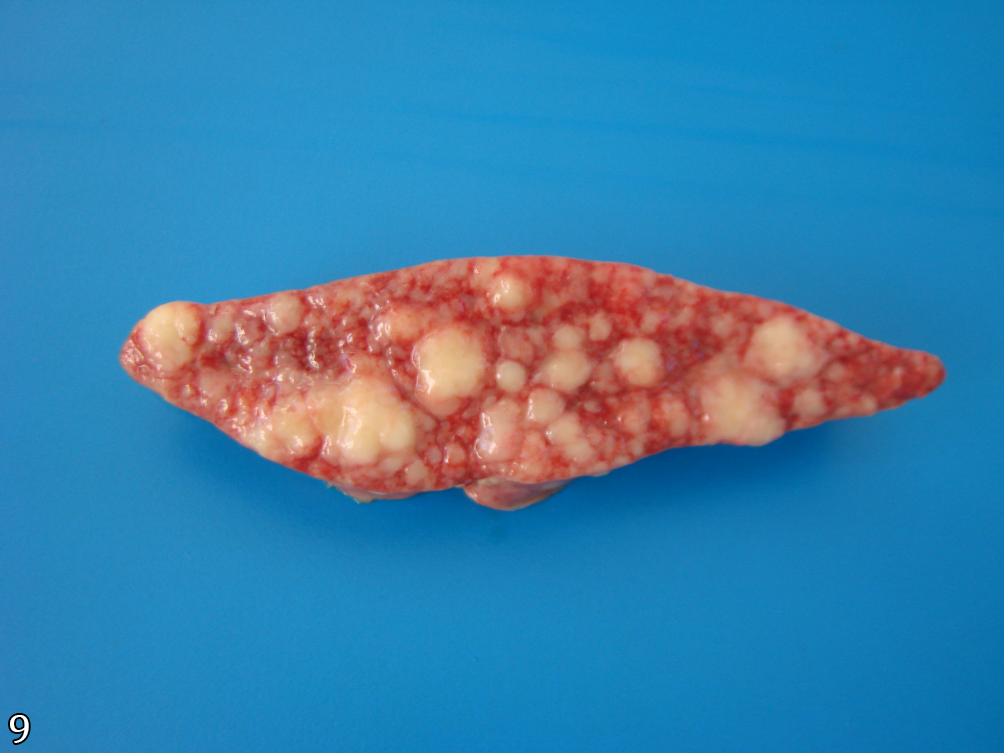
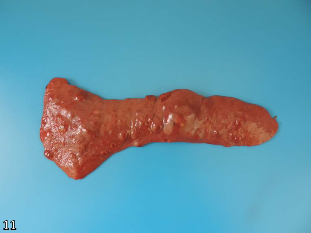
In 12 of 18 cases (66.7%) of histiocytic sarcoma, 12 of 15 (80%) cases of disseminated histiocytic sarcoma, the spleen was affected, and three distinct macroscopic presentations were observed. In the most frequent presentation (8/15, 53.3%), nodules (multinodular pattern) yellowish-white and soft on touch were seen multifocally on the splenic surface (Fig.8). When cut, they were soft with a homogeneously yellowish-white cut surface (Fig.9). In the other cases (4/15, 26.7%), a large mass (massive pattern) protruded from the splenic parenchyma, mostly (3/4) at anterior or posterior end (Fig.10), but occasionally (1 /4) of the central portion of it. In some cases (3/15, 20%), the spleen was diffuse and slightly enlarged (diffuse pattern). In at least two of these three cases, there were also multifocal hemispherical nodules, similar in color to the parenchyma, which protruded from the surface of the organ (Fig.11) and/or yellowish dots and small, multifocal or coalescent white nodules, varying from 0.1 to 1 cm in diameter (Fig.12).

Histiocytic sarcoma, spleen, dog. Multiple multifocal and coalescent white nodules distributed throughout the parenchyma (multinodular pattern).

Histiocytic sarcoma, spleen (cut surface of Figure 8), dog. Note the millimetric white nodules that coalesce to form larger nodules, a pattern reminiscent of that is seen in follicular lymphomas.

Histiocytic sarcoma, spleen, dog. Large spherical, dark red mass emerging from one end of the spleen (massive pattern). Splenic masses like this usually lead to the diagnosis of vascular tumors.

Histiocytic sarcoma, spleen, dog. Spleen markedly increased in volume, with bulging edges and multiple nodules projecting hemispherically from the parenchyma.

Histiocytic sarcoma, spleen, dog. Spleen markedly enlarged with multiple yellowish-white nodules on the edges and white dots dotted across the capsular surface.
Macroscopically, in 15 of 18 cases (83.3%) of histiocytic sarcoma, 14 of 15 cases (93.3%) of disseminated histiocytic sarcoma and in one of the three cases (33.3%) of localized histiocytic sarcoma, the lymph nodes were moderately or markedly enlarged (diffuse pattern), reddish or yellowish-white, soft on touch and cut. The cut surface was homogeneously white or mottled (white and red), with partial or complete loss of the corticomedullary delimitation (Fig.13). Additionally, in some of these cases (6/15, 40%), the enlarged lymph nodes formed large multilobed, yellowish-white or reddish masses (massive pattern) in the subcutaneous tissue of the axillary region (2/15, 13.3%) or in the cranial mediastinum (4/15, 26.7%), extraluminally compressing the trachea, large bronchi and/or esophagus (Fig.14).

Histiocytic sarcoma, lymph node (cut surface), dog. Complete replacement of normal nodal morphology by a white, multilobulated, moist and shiny tissue with multifocal red areas (necrosis and hemorrhage).

Histiocytic sarcoma, thoracic cavity, mediastinal lymph nodes, dog. Large yellowish-white mass, irregular and markedly vascularized. Many dogs with this form of cancer have swelling, completely occupying the cranial mediastinum, a pattern similar to that seen in cases of mediastinal lymphoma and thymoma.
Other organs affected in cases of histiocytic sarcoma included kidneys (6/15, 60%) (Fig.15 and 16), heart (6/15, 40%) (Fig.17), adrenals (4/15, 26.7%), skeletal muscle (4/15, 26.7%), bones (2/15, 13.3%), pancreas (2/15, 13.3%), pericardial sac (2/15, 13.3%), bone joint (1/15, 6.7%), omentum (1/15, 6.7%) and parietal pleura (1/15, 6.7%). All cases in which skeletal muscle was affected occurred in the diaphragm. Cases in which bones were affected included mainly long bones (2/15, 13.3%), such as femur, humerus, radius, tibia, and, less frequently, ribs and sternum (1/15, 6.7%). A single case (1/15, 6.7%) in which articular and periarticular tissues were affected occurred in the patellofemoral joint (“knee joint”). A detailed distribution of each case, regarding the distribution of lesions by organ and their macroscopic pattern of presentation, can be seen in Table 1 and 2, respectively. The affected lymph nodes in each case are described in Table 3.

Histiocytic sarcoma, abdominal cavity, kidneys, dog. Multiple white and multifocal nodules protruded from the subcapsular surface of both kidneys.

Histiocytic sarcoma, kidney (cut surface), dog. Multifocal, coalescent, beige to light brown nodules almost completely obliterated the cortex and corticomedullary transition.

Histiocytic sarcoma, heart (cut surface - longitudinal), dog. Multilobulated white mass that infiltrated the cardiac apex. The apical portion of the left ventricle, the interventricular septum and the lower portion of the left papillary muscle were replaced by this mass.
Histopathological findings
Regardless of the affected organ, there was a densely cellular proliferation, poorly demarcated, non-encapsulated and infiltrative, which partially or completely obliterated and replaced the affected tissue parenchyma. In cases where proliferation occurred as a round cell tumor (15/18, 83.3%), which were the majority, the arrangement was typically in the form of a mantle. A pattern of intersecting bundles was observed in cases where proliferation occurred as a spindle cell tumor (3/18, 16.7%). The stroma was always sparse and consisted of thin septa of fibrovascular tissue.
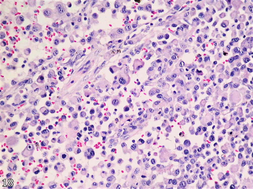
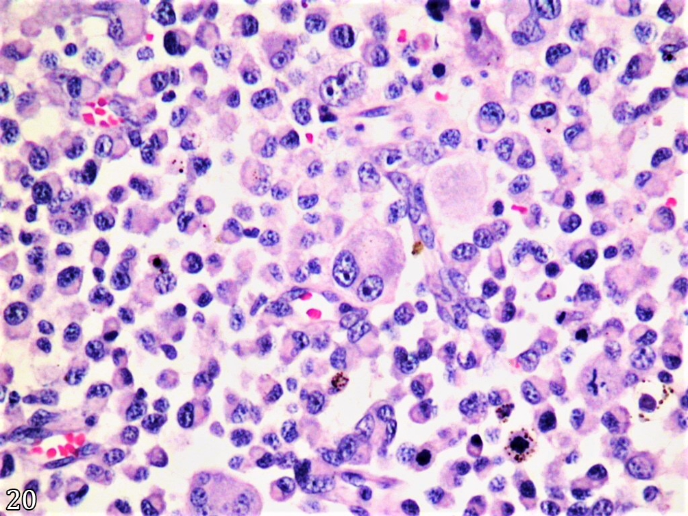
The cells that composed this proliferation were predominantly (15/18, 83.3%) round, oval or polyhedral and, in some cases (3/18, 16.7%), slightly elongated to markedly fusiform. These cells varied greatly in volume, but the majority (60 to 80%) was the size of a histiocyte seen in an inflammatory lesion, i.e., measuring from 20 to 30 µm in diameter (Fig.18). Among this population, there were variable amounts (20 to 40%) of cells with similar morphology, however much larger, measuring around 30 to 100 µm (mononuclear giant cells) (Fig.19). These cells varied in shape, sometimes being round or oval, but not uncommonly also somewhat elongated. Many of these giant cells were binucleated or multinucleated. The nuclei of bi- or trinucleate cells mostly varied in size but not much in their shape. When the nuclei were identical in size and shape, a mirror appearance, which mimicked Reed-Sternberg cells (Fig.20), could be perceived. Cells with multiple nuclei (more than three), the multinucleated giant cells, identical to those seen in granulomatous inflammation, were seen in many cases.

Histiocytic sarcoma, lymph node, dog. Neoplastic infiltrate formed by round, oval and polyhedral cells, ranging from 20 to 30 µm in diameter. These cells had morphological characteristics closely resembling histiocytes, with abundant cytoplasm, round, oval or kidney-shaped nuclei and heterogeneous chromatin. HE, obj.20x.

Histiocytic sarcoma, lymph node, dog. Among the neoplastic cells with histiocytoid morphology, there were cells similar in morphology but much larger, ranging from 30 to 60 µm in diameter. These cells were called mononucleated giant cells. HE, obj.20x.

Histiocytic sarcoma, lymph node, dog. Cells with two nuclei within the histiocytoid infiltrate were common. Generally, these cells were large, up to 80 µm in diameter, and appeared as a “mirror image”, reminiscent of the Reed-Sternberg cells typical of Hodgkin’s lymphoma. These cells were called binucleate giant cells. HE, obj.20x.
The neoplastic cells had distinct cell boundaries and abundant and variably eosinophilic cytoplasm. In some of them, multiple small cytoplasmic vacuoles could be seen. The nuclei were round, oval or kidney-shaped, eccentric and formed by heterogeneous and loosely arranged chromatin. The nucleoli were almost always multiple, evident, intensely basophilic and of varying sizes. Only rare cells had a single nucleolus. Commonly, one of the multiple nucleoli in the same cell was much larger than the others.
The majority of multinucleated giant cells had abundant, eosinophilic and homogeneous cytoplasm. The nuclei of the cells were: 1) pushed back towards the periphery in a circumferential manner, similar to a garland (Fig.21), or like a horseshoe; or 2) randomly distributed throughout the cytoplasm, in a completely disorganized pattern. Such neoplastic cells were similar to those described in granulomatous inflammatory infiltrates as Langhans-like cells and foreign body-like cells, respectively. A few giant cells had more centralized nuclei, in the shape of a pearl necklace, and foamy cytoplasm, mainly at the periphery, a pattern similar to Touton cells seen in xanthomas and xanthogranulomas.

Histiocytic sarcoma, lymph node, dog. In all the cases, there were neoplastic cells with multiple nuclei. Such cells were easily 80 to 100 µm in diameter and called multinucleated giant cells. The nuclear arrangement often resembled a garland, similar to that described for Langhans cells in granulomatous inflammation. HE, obj.20x.
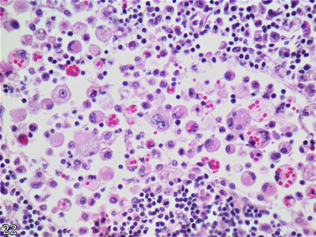
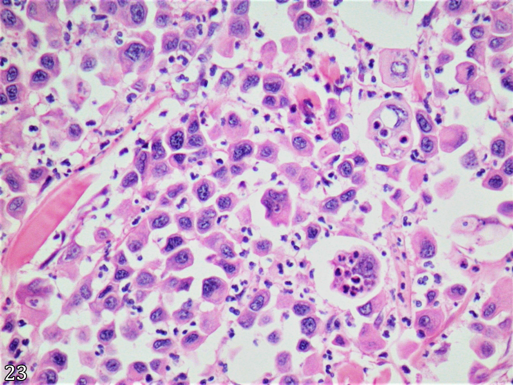
The hemophagocytic activity was seen in all cases, always occurring in a few randomly distributed cells, sometimes predominantly erythrophagocytic (Fig.22) or leukophagocytic (Fig.23). The presence of intracytoplasmic neoplastic cells in another neoplastic cell, which was interpreted as phagocytosis (“cellular cannibalism”), was an unusual finding.

Histiocytic sarcoma, lymph node, dog. Multiple histiocytoid cells and multinucleated giant cells with vacuolated cytoplasm filled with erythrocytes (erythrophagia) in a medullary sinus. A hemophagocytic (erythrophagocytic) pattern occasionally occurred. HE, obj.20x.

Histiocytic sarcoma, subcutaneous tissue, dog. Two binucleated giant cells with cytoplasm full of neutrophils (leukophagy). A hemophagocytic (leukophagocytic) pattern occasionally occurred. HE, obj.20x.
Findings indicative of malignancy were seen in all cases and included varying degrees but generally present in all microscopic fields of anisocytosis, anisokaryosis, karyomegaly, pleomorphism, and cellular atypia. The presence of a large number of mitotic figures, including bizarre mitoses, was common. On average, regardless of the organ affected, there were five mitoses per high-power field (hpf), varying from 1 to 8/hpf.
Areas formed by a grossly floccular and homogeneously eosinophilic material and cellular debris (foci of necrosis) were randomly seen among the neoplastic cells, especially in the central areas of larger tumors. Occasionally, in these areas, there was a large amount of empty space in the form of cholesterol clefts. Around these clefts was an infiltration of non-neoplastic macrophages and multinucleated giant cells in a typical formation of cholesterol granuloma. Neutrophils could also be randomly identified in these areas.
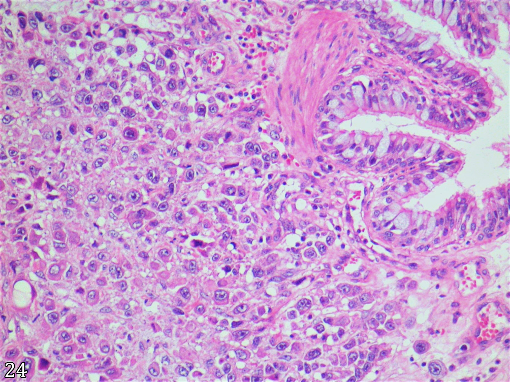
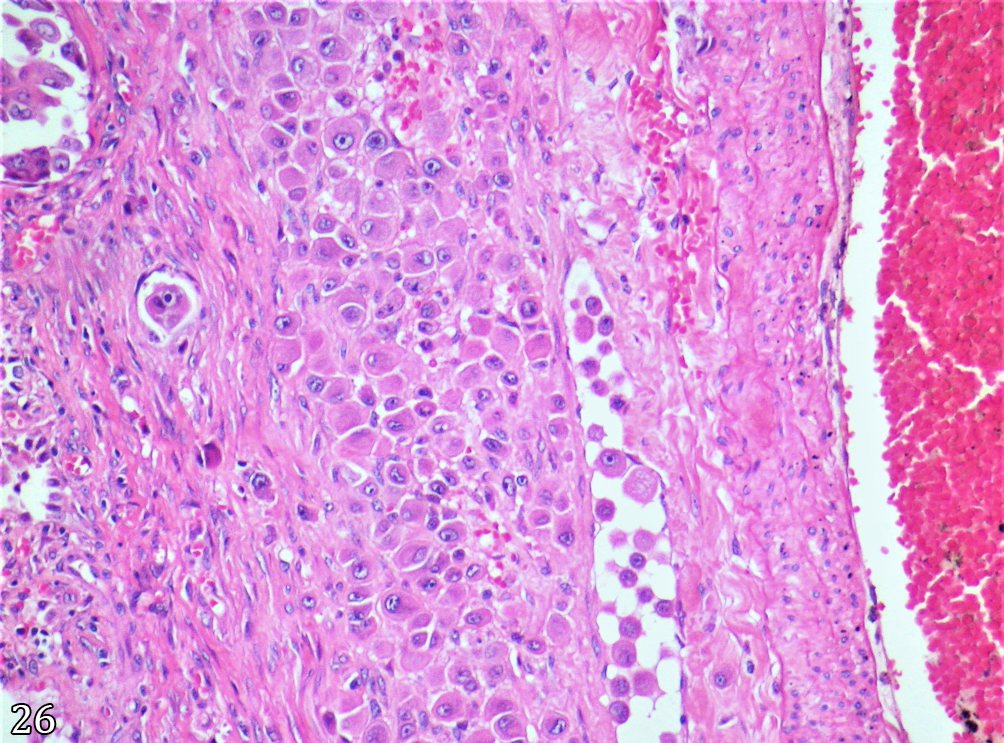
Additionally, the neoplastic infiltrate in the lungs was generally organized around small bronchi and bronchioles. This microscopic lung pattern was seen in all cases where the lung was affected. The large bronchi were affected in some cases (8/17, 47.1%). In both large and small bronchi, the neoplastic infiltrate gradually invaded and replaced the mucosa (Fig.24), making it bare in some areas. In others, it was only possible to determine that it was the bronchial tree by the persistence of the cartilage amid neoplastic proliferation. Obliterated bronchioles could be identified only by the persistence of smooth muscle fragments. The peribronchial and peribronchiolar lymphatic vessels were often filled (Fig.25) and, in some areas, obliterated with neoplastic cells. In some cases (6/17, 35.3%), the tunica media of the pulmonary artery branches were infiltrated and variably obscured by the presence of neoplastic infiltrate. In these cases, neoplastic cells were visualized in the lumen of the small blood vessels that irrigated the pulmonary arterial wall (vasa vasorum) (Fig.26).

Histiocytic sarcoma, lung, dog. Histiocytoid neoplastic infiltrate surrounded the large subsegmental bronchi (histological section referring to the annular bronchial thickening seen macroscopically in Figure 4), obliterating the submucosa and compressing the bronchial mucosa. HE, obj.20x.

Histiocytic sarcoma, lung, dog. Lymphatic vessel adjacent to a large subsegmental bronchus filled with histiocytoid cells and mononucleated giant cells. HE, obj.20x.

Histiocytic sarcoma, lung, pulmonary artery, dog. Middle layer of one of the branches of the pulmonary artery infiltrated and partially obscured by neoplastic histiocytoid cells. Note that the small blood vessel that supplies the arterial wall (vasa vasorum) has the same neoplastic cells in its lumen. HE, obj.20x.
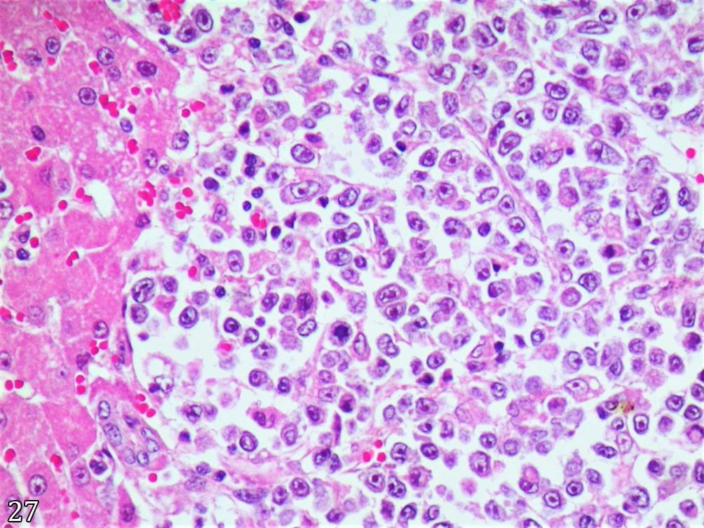
In the liver, at least three microscopic presentation patterns could be visualized. In the most common of them (12/15, 80%), there were clusters of neoplastic cells forming nodules randomly distributed throughout the parenchyma (Fig.27). These nodules were sometimes closer to the portal spaces or closer to the centrilobular vein, without a more apparent distribution. In other cases (8/15, 53.3%), an infiltrate filled the sinusoids, similar to the infiltrative pattern of leukemias and leukemoid reactions. In these cases, no nodule formation was observed, only occasional cellular aggregates in areas of hepatocyte loss, similar to Kupffer cell granulomas. In some cases (5/15, 33.3%), a mixed pattern could be observed, in which both presentations were seen, even in the same histological section. In those nodules with a depressed center area, previously referred to in the macroscopic description as having an umbilicated center, there was necrosis and formation of cholesterol granulomas.

Histiocytic sarcoma, liver, dog. Histiocytoid neoplastic infiltrate, demonstrating mild pleomorphism and distributed as a mantle with scarce fibrovascular stroma into the liver parenchyma. HE, obj.20x.
At least two histological patterns were observed in the affected lymph nodes. In the main pattern, seen in all cases (15/15, 100%) in which macroscopically the lymph nodes were interpreted as affected, there was complete nodal replacement by a mass of neoplastic cells with a pattern identical to that previously described. In eight of 15 cases (53.3%), in the additional lymph nodes, which were not altered at necropsy but were collected and processed, there were varying degrees of involvement of the drainage spaces, from just the presence of cells neoplastic lesions in the subcapsular, peritrabecular and medullary sinuses until their complete obliteration (Fig.28), with parenchymal compression and atrophy. Commonly, the medullary cords were very thin, frequently ruptured and infiltrated by neoplastic histiocytes. Paracortical infiltration could be noted in these cases as a mixture of neoplastic histiocytes and residual lymphocytes.

Histiocytic sarcoma, lymph node, dog. Presence of a large number of histiocytoid neoplastic cells in the medullary sinuses. Note that some of these cells have already invaded the medullary cords and are mixed with the resident plasma cells. HE, obj.20x.
Immunohistochemical findings
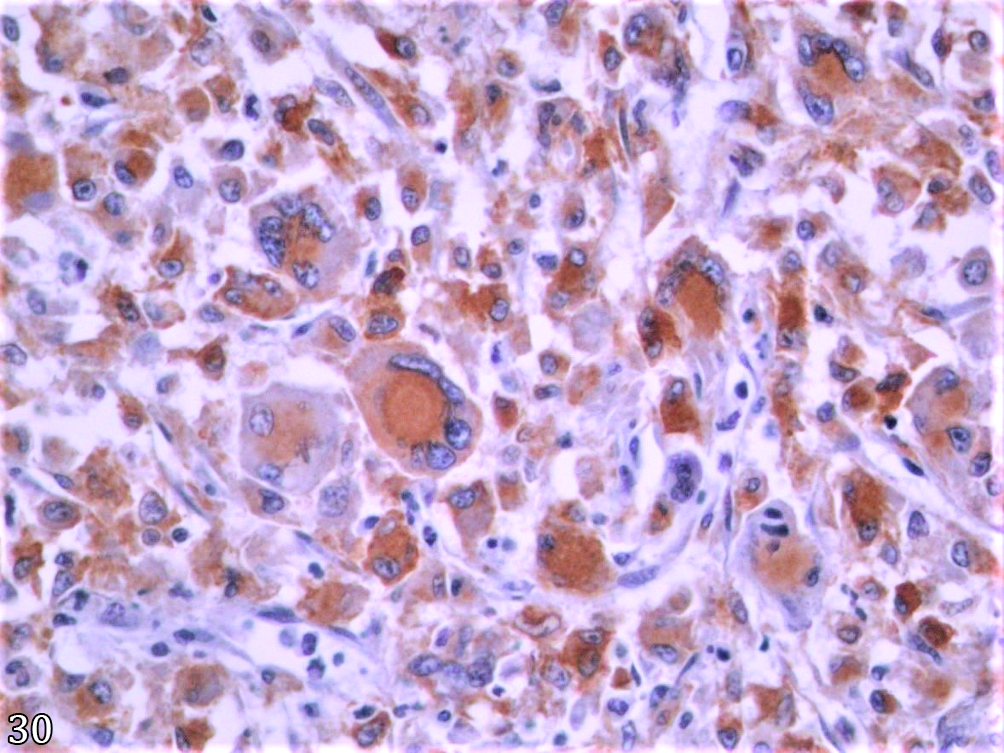
Immunostaining for CD204, the criterion here used for definitive validation of cases, demonstrated an intense dark brown stain in the cytoplasm of neoplastic cells. Although there was variation in the number of immunostained cells, in all cases, more than 90% of these cells were considered positive (Fig.29). The intensity of cytoplasmic immunostaining did not vary between mononucleate, binucleate, trinucleate and multinucleate cells (Fig.30). Identical cells to those immunostained and randomly distributed throughout the tissue proliferation did not demonstrate any staining. Similar immunostaining was observed for the MHC-II antibody - none of the cases selected for this study were immunostained for CD11d. In three cases where immunohistochemistry was performed on the spleen, cells from the peritumoral splenic tissue demonstrated marked immunostaining for this antibody. These cells were interpreted as normal splenic macrophages. Nonimmunostained cells for any of these antibodies were considered as other and, therefore, did not belong to the object of this study.

Histiocytic sarcoma, lung, dog. Neoplastic cells with intense cytoplasmic immunostaining confirming they are histiocytes. IHC (CD204), obj.20x.

Histiocytic sarcoma, lung, dog. Most neoplastic cells, including giant mono-, bi- and multinucleated cells, demonstrated an intense cytoplasmic staining pattern. IHC (MHC-II), obj.40x.
Discussion
Although histiocytic sarcoma was recognized relatively recently (Moore & Rosin 1986), previous studies carried out with samples from this same laboratory and representing a regional canine population (Fighera 2008), histiocytic sarcoma was not even mentioned as one of the thousands of tumor diagnoses (n = 4,844) carried out from 1965 to 2004. In the last significant retrospective study (Flores 2016), carried out specifically on cancer epidemiology, by using case series (n = 7,780) of identical origin but with different time intervals (1964-2013), the prevalence of cancer as a cause of death or reason for euthanasia was 11.1%. The most common types of neoplasia were: 1st) mammary carcinomas (24.5% of total cancer deaths - TCD, and 2.7% of total deaths in general - TDG), 2nd) lymphomas (8.8% of TCD and 1% of TDG), 3rd) skeletal osteosarcomas (7.8% of TCD and 0.9% of TDG), 4th), intrahepatic cholangiocarcinomas (5.5% of TCD and 0.6% of TDG), 5th) cutaneous mast cell tumors (5.4% of TCD and 0.6% of TDG) and 6th) multicentric hemangiosarcomas (3.3% of TCD and 0.4% of TDG). Therefore, if these previous results are compared to the current prevalence of histiocytic sarcoma in our routine (3% of TCD and 0.4% of TDG), histiocytic sarcoma would be classified in the seventh place among the main neoplasms diagnosed as a cause of death or reason for euthanasia of dogs in our region. Furthermore, despite this study only considering cases from 2007 onwards, it is plausible to state that the diagnoses increased significantly, as histiocytic sarcoma was not even mentioned in the two previous major studies (Fighera 2008, Flores 2016).
Based on the findings described here, histiocytic sarcoma has been epidemiologically observed in our necropsy routine as a tumor that occurs more frequently in large dogs, mainly in Rottweiler breed, with no evidence of sexual predisposition, affecting individuals of any age, but with its highest age concentration between five and 12 years (average of 8.5 years). When it was first described (Moore & Rosin 1986) and for some years ahead, histiocytic sarcoma, as well as other histiocytic diseases, such as systemic histiocytosis (Moore 1984), were closely associated with the Bernese Moutain Dog breed. Over the years, the disease has been described in other breeds, initially in the Golden Retriever (Hayden et al. 1993) and later in the Flatcoated Retriever (Dobson et al. 2009, Boerkamp et al. 2013) and the Labrador Retriever (Moore 2014). Despite this, histiocytic sarcoma as an epidemiological important disease has always been highlighted in the literature only for the Bernese Mountain Dog (Voegeli et al. 2006, Abadie et al. 2009, Nielsen et al. 2010) and for Retriever dogs (Dervisis et al. 2017). However, Rottweiler dogs were much more affected in our study region (77.8% of cases). Rottweiler is recognized as one of the five most predisposed breeds to developing cancer, including the Bernese Mountain Dog and the Flatcoated Retriever, with an average death age of 8.9 years (Dobson 2013).
In our pathology routine, Rottweilers makeup only 2.8% of dogs necropsied (Flores et al. 2018), which strongly contrasts with the prevalence of this form of cancer in this breed (77.8%). Making up a simple comparison, mixed breed dogs comprised 40.2% of our routine (Flores et al. 2018), representing only 5.6% of cases of histiocytic sarcoma. Thus, it is possible to observe that a Rottweiler dog is more likely to develop histiocytic sarcoma when compared to mixed-breed dogs. This same epidemiological observation was described by other authors (Toyoda et al. 2020) in a recent study evaluating histiocytic sarcomas of the central nervous system. In this study, Toyoda et al. (2020) demonstrated that Rottweiler contributed to only 2.6% of a hospital routine but made up 15.4% of dogs affected by the tumor. Therefore, we understand that the introduction and increase in the Rottweiler breed in our region strongly modulated the occurrence of histiocytic sarcoma since between the second (1984-1993) and fourth (2004-2013) decades of operation of this diagnostic service, necropsy of this breed twelvefold increased, from 0.6% to 7.2% pure breed dogs (Flores et al. 2018). Results lead to this same reasoning thinking described for Bernese Mountain and Flatcoated Retriever dogs (Erich et al. 2013).
The anatomopathological findings described here demonstrate that histiocytic sarcoma almost always (around 85% of cases) affects several organs at the same time in a disseminated presentation, that is, a disseminated histiocytic sarcoma (previously malignant histiocytosis), which in two thirds (66.7%) of cases include lungs, liver, spleen and lymph nodes. Less commonly (about 15% of cases), this neoplasm can occur as an isolated tumor mainly affecting the lungs, described as a localized histiocytic sarcoma, as also observed by other authors (Marlowe et al. 2018). Regarding the affected organs, in cases of disseminated histiocytic sarcoma, the veterinary literature commonly includes as the most frequently affected sites: lungs, lymph nodes, liver, spleen, pancreas, mediastinum, skin/subcutaneous tissue, central nervous system, bones, including bone marrow, joints/periarticular tissues and eyes (Valli 2007, Valli et al. 2016, Moore 2014, 2017, Mullin & Clifford 2019). For humans, according to the WHO (Weiss et al. 2017), about a third of the cases occur in the lymph nodes, a third of them affect the skin, while the remaining third part occurs in the viscera, mainly in the intestines. A systemic presentation similar to that described in dogs is rare in humans.
In cases of lung localization of histiocytic sarcoma, which corresponds to 100% of localized histiocytic sarcomas and approximately 95% of disseminated histiocytic sarcomas, five patterns of presentation have been described (multinodular, massive, diffuse, peribronchiolar and placoid). For some authors, pulmonary histiocytic sarcomas can occur primarily or as metastases. In the case of metastases, it has been associated with primary splenic origin and concomitant hepatic dissemination (Caswell & Williams 2016, Moore 2017). When cases are evaluated only through necropsy, as in our study, it is not possible to set the organ of origin for these tumors, nor even whether they occur sequentially or synchronously. Therefore, it is plausible to assume that perhaps many of these histiocytic sarcomas started in the spleen, metastasizing later to the liver, lymph nodes and lungs. However, this sequence did not occur in at least three localized cases this sequence did not occur, and the neoplasms emerged in the lungs. At least some of these disseminated histiocytic sarcomas may even have started in the lung, perhaps as localized tumors, as has already been suggested for other organs. In any case, lungs and histiocytic sarcomas have a close relation (McPhetridge et al. 2021), which seems to be important enough to some of the most classic books on oncology (Withrow 2007) and veterinary pathology (López 2007) dedicate some text room in their chapters on lung tumors to this important form of lung cancer.
Two other commonly affected organs were the kidneys (60% of disseminated histiocytic sarcoma cases) and the heart (40% of disseminated histiocytic sarcoma cases). Classic veterinary pathology books (Valli 2007, Valli et al. 2016, Moore 2017) do not mention the occurrence of histiocytic sarcoma in these organs, possibly because they do not observe its involvement with a certain frequency. In our routine, histiocytic sarcomas are the most common hematopoietic tumors in the kidney and heart, even surpassing the involvement seen in cases of multicentric lymphoma, in which they occur with low frequency (21% and 14%, respectively) (Fighera et al. 2006).
Although several authors (Valli 2007, Valli et al. 2016, Moore 2017) state that the skin and subcutaneous tissue are frequently affected in cases of histiocytic sarcoma, none of the patients described here presented involvement of the integument. Histiocytic sarcoma in dogs’ skin generally occurs as a localized tumor, which may later metastasize, mainly to local lymph nodes (Gross et al. 2005). However, it is common for some authors (Moore 2014, Mullin & Clifford 2019) to include the skin as one of the organs most frequently affected in cases of disseminated histiocytic sarcoma, which was not observed in the cases studied here.
From the results presented here, it is possible to assume, at least in our routine, that the diagnosis of histiocytic sarcoma is generally suspected through anatomopathological findings, mainly due to the presence of a markedly anaplastic and pleomorphic round or spindle cell tumor, always rich in mononucleated giant cells and often associated with the presence of variable amounts of bi-, tri- and multinucleated giant cells. Although other systemic tumors constituted of round cells, such as lymphocytes (lymphomas), plasma cells (plasmacytomas and myelomas) and mast cells (mast cells and mastocytosis), occasionally occur as anaplastic and pleomorphic proliferations, rich in karyomegalic cells and, sometimes, with a large number of multinucleated giant cells, it is possible to state, based on the results described here, that it is a hallmark of histiocytic sarcomas. The differential diagnosis for these tumors will be described below.
High-grade mast cell tumors, according to the classification proposed by Kiupel et al. (2011), are diagnosed when there are: 1) seven mitoses in 10 high power fields (hpf); 2) 10% of neoplastic cells demonstrating karyomegaly (with at least twice the volume of other cells); or 3) three bizarre cores/10 hpf. In these cases (Kiupel et al. 2011), and in those previously described as Grade III mast cell tumors (Patnaik et al. 1984) and anaplastic mast cell tumors (Bostock 1973), multinucleated cells (with three or more nuclei) are frequent. However, even in these cases, other morphological findings may suggest that it is a mast cell tumor, ranging from the morphology of other non-anaplastic cells, which may demonstrate varying degrees of cytoplasmic granulation, to the presence of eosinophils as part of the infiltrate (Bostock 1973, Patnaik et al. 1984, Kiupel et al. 2011). It is worth noting here that mast cell tumors generally occur as primary cutaneous or subcutaneous tumors, which often demonstrate a high risk for the development of local metastases (Kiupel et al. 2010). None of the histiocytic sarcoma described in this study showed involvement of the integument. Therefore, the differential diagnosis for mast cell tumors must take into account visceral mastocytosis (Welle et al. 2008), a much less common form of canine cancer. Furthermore, based on the fact that the skin is not always affected in cases of systemic mastocytosis (O’Keefe et al. 1987), this is also a difference to be considered. In most cases of visceral or systemic mastocytosis described in the veterinary literature, the neoplastic mast cells infiltrating these organs are well differentiated and easily recognized and generally do not demonstrate cellular anaplasia, pleomorphism and atypia (Moirano et al. 2018, Aceino et al. 2021) as observed in cases of histiocytic sarcoma.
Plasma cell tumors have been described in dogs originating mainly from the skin (Mauldin & Peters-Kennedy 2016, Hendrick 2017) and oral mucosa (Smithson et al. 2012, Pargass et al. 2017). Furthermore, cases of plasmacytomas arising from the viscera (Aoki et al. 2004, Johnson et al. 2021) and the central nervous system (Sheppard et al. 1997) have also been described. However, visceral plasmacytomas generally behave as localized tumors (Mikiewicz et al. 2016), unlike the histiocytic sarcomas described here, which present a multicentric behavior. Despite this benign behavior (Miller et al. 2013), metastatic (Trevor et al. 1993) and disseminated (Dagher et al. 2019, Brown et al. 2021), plasmacytomas have already been described in dogs. Plasma cell tumors are classified as well-differentiated (indolent) or anaplastic. (Valli et al. 2002). The neoplastic cells observed in anaplastic cases generally demonstrate a certain degree of atypia; some are giant and can even be bi- or multinucleated (Baer et al. 1989). In cases of multiple myeloma, the neoplastic plasma cells are often very large, similar to the mononucleated giant cells described for histiocytic sarcoma (Palgrave et al. 2010). However, in most cases, these cells are confined to the bone marrow and rarely metastasize. When it occurs, only the spleen and liver are affected (Valli 2007). More recently, canine cutaneous and visceral plasmacytomas cases have been described, evolving into plasma cell leukemia, which can secondarily spread to other organs (Rout et al. 2017, Dagher et al. 2019).
There is no question that the most important differential diagnoses for histiocytic sarcoma are some types of lymphoma, especially because for years, this form of cancer was mistakenly considered a lymphoproliferative disorder and generically called “histiocytic lymphoma”. Currently, the formerly called “histiocytic lymphomas” include diffuse large B-cell lymphoma (DLBCL), anaplastic large cell lymphoma (ALCL) and histiocytic sarcoma (Weiss et al. 2017). Based on cell size, lymphomas that arise from immature and large cells (larger than the volume of two erythrocytes), mainly immunoblasts (e.g., immunoblastic lymphoma - IBL, a DLBCL), are the main differentials. Furthermore, despite its rarity in dogs, ALCL, mainly T but also null cell, should be placed as a differential diagnosis. Due to the high frequency of lung involvement, lymphomatoid granulomatosis was also considered in the differential diagnosis for our cases. These lymphoid differential diagnoses will be described below.
Immunoblastic lymphoma is a common histopathological presentation of canine lymphoma (7.7%, Ponce et al. 2010; 8.5%, Arespacochaga et al. 2007; 10.5%, Vezzali et al. 2010; or 24.9% of cases, Carter et al. 1986). Generally, it presents itself as multicentric (91.5% of cases, Ponce et al. 2010), affecting lymph nodes, spleen and liver, that is, as a multicentric lymphoma in Stage 3 (Vail & Young 2006). This is a macroscopic presentation that is very close to the one described for histiocytic sarcoma in this study. However, pulmonary involvement in cases of multicentric lymphoma is very rare (Fry & McGavin 2007, Fighera & Graça 2016, Valli et al. 2016, 2017), and this should be a criterion to differentiate these cases initially. However, as it has been shown, cases of histiocytic sarcoma may occasionally not affect the lungs, and, therefore, it is not a definitive differentiation criterion. Immunoblastic lymphoma occurs as diffuse and intermediate to high-grade, characterized by cellular pleomorphism. Neoplastic cells are large, with markedly indented nuclei and are made up of heterogeneous chromatin, with mixed areas, from a finely granular pattern to a very loose one (parachromatin) (Valli et al. 2002, 2016, 2017, Valli 2007), which closely resembles the typical cellular pattern seen in histiocytic sarcoma. The main differences for histiocytic sarcoma include the presence of mononucleated, binucleated and multinucleated giant cells, multiple nucleoli (versus the single nucleolus of immunoblasts), cytoplasmic vacuolation and the occurrence of hematophagy. Because histiocytic sarcomas do not often present such differentiating criteria, immunohistochemistry for B lymphocytes can definitively exclude this differential diagnosis. The most used markers for this are CD79, CD20 and PAX-5 (Valli et al. 2002, 2016, 2017).
A DLBCL, closely similar microscopically to that described in the previous paragraph but which occurs in a typical mediastinal presentation, is described in young dogs, originating in the thymus, and it is called thymic B-cell lymphoma or mediastinal B-cell lymphoma (Valli et al. 2002, 2016, 2017). Although similar to the previously described immunoblastic lymphoma, the neoplastic cells are much more anaplastic, and mono-, bi-, tri-, and multinucleated giant cells are often present (Valli et al. 2002, 2016, 2017). This rare form of canine lymphoma (0.2%, Ponce et al. 2010 to 0.8% of cases, Vezzali et al. 2010) was considered a differential diagnosis in the four cases in which mediastinal masses were observed in dogs in this study. In this more anaplastic and localized type of immunoblastic lymphoma, other organs are generally not affected; on the other hand, in these four cases of histiocytic sarcoma, the lungs, liver and spleen were always involved.
Anaplastic large T-cell lymphoma is a rare histopathological presentation of canine lymphoma (0.8%, Ponce et al. 2010 to 1% of cases, Mazaro et al. 2020), and it usually manifests itself as an extranodal condition that initially affects isolated organs (Valli et al. 2002), may mimetizing the localized histiocytic sarcoma. However, despite the occurrence of this form of primary lung lymphoma in humans (Han et al. 2014, Carvalho et al. 2019), similar cases in dogs have not yet been described. It is known that, unlike our results, canine histiocytic sarcoma can occur primarily and isolated in: skin (Mastrorilli et al. 2012), spleen (Latifi et al. 2020), brain (Chandra & Ginn 1999, Thio et al. 2006, Mariani et a. 2015, Barrot et al. 2017, Toyoda et al. 2020, Takahashi et al. 2021), spinal cord (Mariani et al. 2015, Taylor et al. 2015), ocular globe (Naranjo et al. 2007), stomach (Fant et al. 2004, Elliott 2016), heart, including aortic valve (Kovacevic et al. 2019), bones (Schultz et al. 2007), periarticular tissues (Harasen & Simko 2008, Klahn et al. 2011, Van Kuijk et al. 2013, Manor et al. 2018) and gums (Carioto 1997). Some of these locations, such as the skin (Azuma et al. 2022), and others, such as the small intestine (Stranahan et al. 2019, Kojima et al. 2021), the urethrovaginal junction (Kaneguchi et al. 2021) and skeletal muscle (Thuilliez et al. 2008) have already been described as the origin of canine ALCL.
Anaplastic large T-cell lymphomas occur as diffuse, high-grade tumors characterized by their cellular pleomorphism (Valli et al. 2002). The neoplastic cells are large, with nuclei of various shapes (anisokaryosis), chromatin pattern very similar to that previously described for immunoblastic lymphoma, and, therefore, similar to the neoplastic histiocytes of histiocytic sarcoma. Due to the frequent occurrence of cells with irregular nuclei, sometimes similar to a horse’s shoe or a kidney (Kaneguchi et al. 2021), such “hallmark cells” (Mazaro et al. 2020), this type of lymphoma may resemble the neoplastic histiocytes of histiocytic sarcoma. The presence of giant cells is frequent, many of which may be bi- or multinucleated (Gross et al. 2005, Cerroni 2014). This is certainly the most important histopathological differential diagnosis of histiocytic sarcoma. Although some small details may suggest one diagnosis or another, immunohistochemistry must be performed to definitively clarify any doubts regarding this diagnosis. The most used marker for this procedure is CD3 (Kaneguchi et al. 2021). More recently (Pittaway et al. 2018), an anaplastic large cell lymphoma, negative for T (CD3) and B (CD79α) lymphocyte markers, was confirmed to originate itself from null cells through immunostaining with CD30, similar to what has been described in humans (Feldman et al. 2017). Therefore, this last information is important to ensure that lymphoma must not be completely discarded in cases without immunostaining for B and T lymphocytes.
Lymphomatoid granulomatosis is an uncommon (2% of cases, Mazaro et al. 2020) and peculiar presentation of canine lymphoma in which the infiltrate mixes neoplastic cells in smaller amounts and non-neoplastic cells in greater amounts (Valli et al. 2002). It was initially described in dogs as a primary and specifically pulmonary lesion (Lucke et al. 1979), and it has been studied in the species as an animal model since then (Postorino et al. 1989, Berry et al. 1990, Leblanc et al. 1990, Fitzgerald et al. 1991). For humans, it has been proven for some years that neoplastic cells are immature B lymphocytes, while reactive cells are mainly mature T lymphocytes, as well as plasma cells and other inflammatory cells, such as eosinophils. This same hypothesis took a long time to be confirmed for dogs (Magi et al. 2009), and it was even suspected that this neoplasm was an atypical form of T-cell lymphoma and primary pulmonary Hodgkin’s lymphoma (Park et al. 2007). Thus, lymphomatoid granulomatosis is a T-cell-rich B-cell lymphoma that usually emerges from the lungs (Feldman et al. 2017). Although the infiltrate is distinct from that described here, for histiocytic sarcoma, the fact that the lungs are frequently affected, and often in conjunction with the liver, spleen and lymph nodes, and less frequently kidneys and heart (Fitzgerald et al. 1991), define that this form of lymphoma has to be included in the differential diagnosis. Furthermore, the fact that this infiltrate is distributed in the lungs as an angiocentric pattern, sometimes angioinvasive and angiodestructive (Magi et al. 2009, Hatoya et al. 2011), makes it very similar to the histological description of histiocytic sarcoma.
Based on the previously discussed, immunophenotypic confirmation is needed to differentiate histiocytic sarcoma from other mesenchymal tumors, especially the round cell tumors described above. In this way, the immunohistochemical method on tissue sections previously processed for histology and embedded in paraffin proved to be a very promising tool when it used an anti-CD204 antibody called class A macrophage scavenger receptor (MSR-A), as described by other authors (Kato et al. 2013). Anti-CD204 is a monoclonal antibody produced in mice, and it was initially employed to detect histiocytes in human atheromas in atherogenesis studies (Tomokiyo et al. 2002). Binding occurs with low-density lipoproteins (LDL) on CD204, a surface receptor present on the plasma membrane of histiocytes (Fukuhara-Takaki et al. 2005). Previous studies have confirmed the effectiveness of anti-CD204 in diagnosing histiocytic sarcoma in dogs (Kato et al. 2013, Thongtharb et al. 2016). More recently, this same marker was used in characterizing cases of canine histiocytic ulcerative colitis (Nolte et al. 2017) and as another tool inferring prognostic value in canine mammary tumors (Seung et al. 2018), similar to what had already been performed for human lower urinary tract carcinomas (Ichimura et al. 2014) and lung adenocarcinomas (Ohtaki et al. 2010).
Conclusion
Based on the anatomopathological findings obtained in this study, it is possible to infer that histiocytic sarcoma is currently one of the ten most important forms of cancer that affects dogs, almost exclusively the large ones and especially for the Rottweiler breed. Most of the time, sarcoma histiocytic is seen as a generalized disease with a presentation very close to lymphomas; however, unlike lymphomas, it affects the lungs in most cases. It should be suspected whenever an architectural pattern in the mantle, composed of anaplastic round or fusiform cells, presents giant cells, mono-, bi- and multinucleated. Confirmation of this diagnosis can be carried out using the anti-CD204 antibody for the immunohistochemistry technique in paraffin-embedded tissues. It is expected that this information will contribute to providing a better characterization of histiocytic sarcoma in dogs, assisting veterinary pathologists in their diagnostic routines.
Acknowledgments
The authors thank the “Conselho Nacional de Desenvolvimento Científico e Tecnológico” (CNPq) and the “Coordenação de Aperfeiçoamento de Pessoal de Nível Superior” (CAPES) for granting postgraduate scholarships.
References
-
Abadie J, Hédan B, Cadieu E, De Brito C, Devauchelle P, Bourgain C, Parker HG, Vaysse A, Margaritte-Jeannin P, Galibert F, Ostrander EA, André C. Epidemiology, pathology, and genetics of histiocytic sarcoma in the Bernese mountain dog breed. J Hered 2009; https://doi.org/10.1093/jhered/esp039, PMid:19531730
» https://doi.org/10.1093/jhered/esp039 -
Aceino A, Jeffery U, Piccione J, Hodo CL. Systemic mastocytosis with subcutaneous hemorrhage and edema in a Greyhound dog: case report and review of diagnostic criteria. J Vet Diagn Invest 2021; https://doi.org/10.1177/1040638720972500, PMid:33164718
» https://doi.org/10.1177/1040638720972500 -
Affolter VK, Moore PF. Localized and disseminated histiocytic sarcoma of dendritic cell origin in dogs. Vet Pathol 2002; https://doi.org/10.1354/vp.39-1-74, PMid:12102221
» https://doi.org/10.1354/vp.39-1-74 -
Aoki M, Kim T, Shimada T, Kuwamura M, Yamate J, Ohashi F. A primary hepatic plasma cell tumor in a dog. J Vet Med Sci 2004; https://doi.org/10.1292/jvms.66.445, PMid:15133277
» https://doi.org/10.1292/jvms.66.445 -
Arespacochaga AG, Schwendenwein I, Weissenböck H. Retrospective study of 82 cases of canine lymphoma in Austria based on the working formulation and immunophenotyping. J Comp Pathol 2007; https://doi.org/10.1016/j.jcpa.2007.01.009, PMid:17416237
» https://doi.org/10.1016/j.jcpa.2007.01.009 -
Azuma K, Ohmi A, Goto-Koshino Y, Tomiyasu H, Ohno K, Chambers J.K., Uchida K, Namba H, Nagata M, Nagamine E, Nibe K, Irie M, Tsujimoto H. Outcomes and prognostic factors in canine epitheliotropic and nonepitheliotropic cutaneous T lymphoma. Vet Comp Oncol 2022; https://doi.org/10.1111/vco.12752, PMid:34250731
» https://doi.org/10.1111/vco.12752 -
Baer KE, Patnaik AK, Gilbertson SR, Hurvitz AI. Cutaneous plasmacytomas in dogs: a morphologic and immunohistochemical study. Vet Pathol 1989; https://doi.org/10.1177/030098588902600305, PMid:2669312
» https://doi.org/10.1177/030098588902600305 - Barrot A-C, Bédard A, Dunn M. Syndrome of inappropriate antidiuretic hormone secretion in a dog with a histiocytic sarcoma. Can Vet J 2017;58(7):713-715. PMid:28698689.
-
Berry CR, Moore PF, Thomas WP, Sisson D, Koblik PD. Pulmonary lymphomatoid granulomatosis in seven dogs (1976-1987). J Vet Intern Med 1990; https://doi.org/10.1111/j.1939-1676.1990.tb00890.x, PMid:2366226
» https://doi.org/10.1111/j.1939-1676.1990.tb00890.x -
Boerkamp KM, Van der Kooij M, Van Steenbeek FG, Van Wolferen ME, Groot Koerkamp MJA, Van Leenen D, Grinwis GCM, Penning LC, Wiemer EAC, Rutteman GR. Gene expression profiling of histiocytic sarcomas in a canine model: the predisposed flatcoated retriever dog. PLoS One 2013; https://doi.org/10.1371/journal.pone.0071094, PMid:23936488
» https://doi.org/10.1371/journal.pone.0071094 -
Bostock DE. The prognosis following surgical removal of mastocytomas in dogs. J Small Anim Pract 1973; https://doi.org/10.1111/j.1748-5827.1973.tb06891.x, PMid:4199921
» https://doi.org/10.1111/j.1748-5827.1973.tb06891.x -
Brown JE, Russell EB, Moore AR, Oscos-Snowball A, Stent A, Courtman NF. Hypoglobulinemia in a dog with disseminated plasma cell neoplasia: Case report and review of the diagnostic. Vet Clin Pathol 2021; https://doi.org/10.1111/vcp.12948, PMid:33913178
» https://doi.org/10.1111/vcp.12948 - Carioto L. Malignant histiocytosis in a Bernese mountain dog presenting as a mandibular mass. Can Vet J 1997;38(2):105-107. PMid:9028594.
- Carter RF, Valli VEO, Lumsden JH. The cytology, histology and prevalence of cell types in canine lymphoma classified according to the National Cancer Institute working formulation. Can J Vet Res 1986;50(2):154-164. PMid:3756669.
-
Carvalho J, Marques DP, Oliveira I, Claudino C. A rare case of primary pulmonary anaplastic large cell lymphoma. Eur J Case Rep Intern Med 2019; https://doi.org/10.12890/2019_001249, PMid:31583219
» https://doi.org/10.12890/2019_001249 - Caswell JL, Williams KJ. Respiratory system, p.465-591. In: Maxie MG. Jubb, Kennedy, and Palmer’s Pathology of Domestic Animals. Vol.2. 6th ed. St. Louis: Elsevier; 2016.
- Cerroni L. Cutaneous anaplastic large cell lymphoma, p.99-108. In: Cerroni L. Skin lymphoma: the illustrated guide. 4th ed. Oxford: Wiley Blackwell; 2014.
-
Chandra AMS, Ginn PE. Primary malignant histiocytosis of the brain in a dog. J Comp Pathol 1999; https://doi.org/10.1053/jcpa.1998.0296, PMid:10373296
» https://doi.org/10.1053/jcpa.1998.0296 -
Dagher E, Soetart N, Chocteau F, Dequéant B, Piccirillo E, Ibisch C, Abadie J, Jaillardon L. Plasma cell leukemia with plasmablastic morphology in a dog. J Vet Diagn Invest 2019; https://doi.org/10.1177/1040638719882045, PMid:31609183
» https://doi.org/10.1177/1040638719882045 -
Davis BW, Ostrander EA. Domestic dogs and cancer research: a breed-based genomics approach. ILAR J 2014; https://doi.org/10.1093/ilar/ilu017, PMid:24936030
» https://doi.org/10.1093/ilar/ilu017 -
Dervisis NG, Kiupel M, Qin Q, Cesario L. Clinical prognostic factors in canine histiocytic sarcoma. Vet Comp Oncol 2017; https://doi.org/10.1111/vco.12252, PMid:27334037
» https://doi.org/10.1111/vco.12252 -
Dobson J, Hoather T, McKinley TJ, Wood JLN. Mortality in a cohort of flat-coated retrievers in the UK. Vet Comp Oncol 2009; https://doi.org/10.1111/j.1476-5829.2009.00181.x, PMid:19453365
» https://doi.org/10.1111/j.1476-5829.2009.00181.x -
Dobson JM. Breed-predispositions to cancer in pedigree dogs. ISRN Vet Sci 2013; https://doi.org/10.1155/2013/941275, PMid:23738139
» https://doi.org/10.1155/2013/941275 -
Elliott J. Gastric histiocytic sarcoma in a dog. J Small Anim Pract 2016; https://doi.org/10.1111/jsap.12602, PMid:27925661
» https://doi.org/10.1111/jsap.12602 -
Erich SA, Rutteman GR, Teske E. Causes of death and the impact of histiocytic sarcoma on the life expectancy of the Dutch population of Bernese mountain dogs and Flat-coated retrievers. Vet J 2013; https://doi.org/10.1016/j.tvjl.2013.09.062, PMid:24231621
» https://doi.org/10.1016/j.tvjl.2013.09.062 -
Fant P, Caldin M, Furlanello T, De Lorenzi D, Bertolini G, Bettini G, Morini M, Masserdotti C. Primary gastric histiocytic sarcoma in a dog - a case report. J Vet Med A 2004; https://doi.org/10.1111/j.1439-0442.2004.00645.x, PMid:15533119
» https://doi.org/10.1111/j.1439-0442.2004.00645.x - Feldman AL, Harris NL, Stein H, Campo E, Kinney MC, Jaffe ES, Falini B, Inghirami GG, Pileri SA. Anaplastic large cell lymphoma, ALK negative, p.418-421. In: Swerdlow SH, Campo E, Harris NL, Jaffe ES, Pileri SA, Stein H, Thiele J, Vardiman JW. WHO Classification of tumours of haematopoietic and lymphoid tissues. Revised 4th ed. Lyon: World Health Organization; 2017.
- Fighera RA, Graça DL. Sistema hematopoético, p.311-405. In: Santos RL, Alessi AC. Patologia veterinária. 2ª ed. Rio de Janeiro: Roca; 2016.
- Fighera RA, Souza TM, Rodrigues A, Barros CSL. Aspectos clinicopatológicos de 43 casos de linfoma em cães. MEDVEP, Rev Científ Med Vet, Pequenos Anim Estim 2006;4(12):139-146.
- Fighera RA. Causas de morte e razões para eutanásia de cães. Tese de Doutorado, Universidade Federal de Santa Maria, Santa Maria, 2008, 171p.
-
Fitzgerald SD, Wolf DC, Carlton WW. Eight cases of canine lymphomatoid granulomatosis. Vet Pathol 1991; https://doi.org/10.1177/030098589102800308, PMid:1858255
» https://doi.org/10.1177/030098589102800308 -
Flores MM, Mazaro RD, Poeta APS, Kommers GD, Fighera RA. Caracterização do gênero, da raça e da idade de uma população de 7.780 cães da Região Central do Rio Grande do Sul submetidos à necropsia ao longo de cinco décadas (1964-2013). Pesq Vet Bras 2018; https://doi.org/10.1590/1678-5150-PVB-5587
» https://doi.org/10.1590/1678-5150-PVB-5587 - Flores MM. Aspectos epidemiológicos do câncer em cães da Região Central do Rio Grande do Sul: 50 anos (1964-2013). Tese de Doutorado, Universidade Federal de Santa Maria, Santa Maria, 2016, 91p.
- Fry MM, McGavin MD. Bone marrow, blood cells, and lymphatic system, p.743-832. In: McGavin MD, Zachary JF. Pathologic basis of veterinary disease. 4th ed. St. Louis: Mosby Elsevier; 2007.
-
Fukuhara-Takaki K, Sakai M, Sakamoto Y-I, Takeya M, Horiuchi S. Expression of class A scavenger receptor is enhanced by high glucose in vitro and under diabetic conditions in vivo. J Biol Chem 2005; https://doi.org/10.1074/jbc.M408715200, PMid:15556945
» https://doi.org/10.1074/jbc.M408715200 -
Gross TL, Ihrke PJ, Walder EJ, Affolter VK. Lymphocytic tumors, p.866-893. In: Gross TL, Ihrke PJ, Walder EJ, Affolter VK. Skin diseases of the dog and cat: clinical and histopathologic diagnosis. 2005; https://doi.org/10.1002/9780470752487.ch37
» https://doi.org/10.1002/9780470752487.ch37 -
Han SH, Maeng YH, Kim YS, Jo JM, Kwon JM, Kim WK, Kim MO. Primary anaplastic large cell lymphoma of the lung presenting with acute atelectasis. Thorac Cancer 2014; https://doi.org/10.1111/1759-7714.12027, PMid:26766977
» https://doi.org/10.1111/1759-7714.12027 -
Harasen GLG, Simko E. Histiocytic sarcoma of the stifle in a dog with cranial cruciate ligament failure and TPLO treatment. Vet Comp Orthop Traumatol 2008; https://doi.org/10.3415/vcot-07-05-0054, PMid:18704246
» https://doi.org/10.3415/vcot-07-05-0054 -
Hatoya S, Kumagai D, Takeda S, Yamamoto E, Nakanishi M, Kuwamura M, Sugiura K, Sasai H, Yamate J, Inaba T. Successful management with CHOP for pulmonary lymphomatoid granulomatosis in a dog. J Vet Med Sci 2011; https://doi.org/10.1292/jvms.10-0301, PMid:21139353
» https://doi.org/10.1292/jvms.10-0301 -
Hayden DW, Waters DJ, Burke BA, Manivel JC. Disseminated malignant histiocytosis in a Golden Retriever: clinicopathologic, ultrastructural, and immunohistochemical findings. Vet Pathol 1993; https://doi.org/10.1177/030098589303000306, PMid:8333107
» https://doi.org/10.1177/030098589303000306 -
Hendrick MJ. Mesenchymal tumors of the skin and soft tissues, p.142-175. In: Meuten DJ. Tumors in domestic animals. 2017; https://doi.org/10.1002/9781119181200.ch5
» https://doi.org/10.1002/9781119181200.ch5 -
Ichimura T, Morikawa T, Kawai T, Nakagawa T, Matsushita H, Kakimi K, Kume H, Ishikawa S, Homma Y, Fukayama M. Prognostic significance of CD204-positive macrophages in upper urinary tract cancer. Ann Surg Oncol 2014; https://doi.org/10.1245/s10434-014-3503-2, PMid:24492923
» https://doi.org/10.1245/s10434-014-3503-2 - Johnson J, Hung G, Larson V, Manasse J, Spotswood T. Renal extramedullary plasmacytoma in a dog. Can Vet J 2021;62(10):1077-1082. PMid:34602635
-
Kaneguchi A, Izawa T, Tanaka M, Suzuki H, Tani H, Kuwamura M, Yamate J. Systemic anaplastic large t-cell lymphoma with initial presentation of dysuria in a dog. J Comp Pathol 2021; https://doi.org/10.1016/j.jcpa.2021.09.006, PMid:34886983
» https://doi.org/10.1016/j.jcpa.2021.09.006 -
Kato Y, Murakami M, Hoshino Y, Mori T, Maruo K, Hirata A, Nakagava TLDR, Yanai T, Sakai H. The class A macrophage scavenger receptor CD204 is a useful immunohistochemical marker of canine histiocytic sarcoma. J Comp Pathol 2013; https://doi.org/10.1016/j.jcpa.2012.06.009, PMid:22901707
» https://doi.org/10.1016/j.jcpa.2012.06.009 -
Kiupel M, Webster JD, Bailey KL, Best S, DeLay J, Detrisac CJ, Fitzgerald SD, Gamble D, Ginn PE, Goldschmidt MH, Hendrick MJ, Howerth EW, Janovitz EB, Langohr I, Lenz SD, Lipscomb TP, Miller MA, Misdorp W, Moroff S, Mullaney TP, Neyens I, O’Toole D, Ramos-Vara J, Scase TJ, Schulman FY, Sledge D, Smedley RC, Smith K, Snyder PW, Southorn E, Stedman NL, Steficek BA, Stromberg PC, Valli VE, Weisbrode SE, Yager J, Heller J, Miller R. Proposal of a 2-tier histologic grading system for canine cutaneous mast cell tumors to more accurately predict biological behavior. Vet Pathol 2011; https://doi.org/10.1177/0300985810386469, PMid:21062911
» https://doi.org/10.1177/0300985810386469 - Kiupel M, Webster JD, Yuzbasiyan-Gurkan V. Prognostic indicators for canine mast cell tumors: margin evaluation and histologic grading. J Small Anim Dermatol Pract 2010;3(2):8-14.
-
Klahn SL, Kitchell BE, Dervisis NG. Evaluation and comparison of outcomes in dogs with periarticular and nonperiarticular histiocytic sarcoma. J Am Vet Med Assoc 2011; https://doi.org/10.2460/javma.239.1.90, PMid:21718201
» https://doi.org/10.2460/javma.239.1.90 -
Kojima K, Chambers JK, Li T, Nibe K, Mizuno T, Uchida K. Histopathological features and immunophenotyping of canine transmural gastrointestinal lymphoma using full-thickness biopsy samples. Vet Pathol 2021; https://doi.org/10.1177/03009858211030523, PMid:34282671
» https://doi.org/10.1177/03009858211030523 -
Kovacevic A, Rossier C, Gurtner C. Primäres histiozytäres Sarkom der Aortenklappe als Auslöser von Herzversagen bei einem Mischlingshund. Schweiz Arch Tierheilkd 2019; https://doi.org/10.17236/sat00199, PMid:30843523
» https://doi.org/10.17236/sat00199 -
Latifi M, Tuohy JL, Coutermarsh-Ott SL, Klahn SL, Leeper H, Dervisis N. Clinical outcomes in dogs with localized splenic histiocytic sarcoma treated with splenectomy with or without adjuvant chemotherapy. J Vet Intern Med 2020; https://doi.org/10.1111/jvim.15910, PMid:32986268
» https://doi.org/10.1111/jvim.15910 -
Leblanc B, Masson MT, Andreu M, Bonnet MC, Paulus G. Lymphomatoid granulomatosis in a beagle dog. Vet Pathol 1990; https://doi.org/10.1177/030098589002700412, PMid:2402856
» https://doi.org/10.1177/030098589002700412 - López A. Respiratory system, p.463-558. In: McGavin MD, Zachary JF. Pathologic basis of veterinary disease. 4th ed. St. Louis: Mosby Elsevier; 2007.
-
Lucke VM, Kelly DF, Harrington GA, Gibbs C, Gaskeli CJ. A lymphomatoid granulomatosis of the lungs in young dogs. Vet Pathol 1979; https://doi.org/10.1177/030098587901600402, PMid:452314
» https://doi.org/10.1177/030098587901600402 -
Magi GE, Di Pasquale S, Renzoni G. Cutaneous lymphomatoid granulomatosis (angiotropic lymphoma) in a dog: immunophenotyping analysis. Vet Res Commun 2009; https://doi.org/10.1007/s11259-009-9264-x, PMid:19578958
» https://doi.org/10.1007/s11259-009-9264-x -
Manor EK, Craig LE, Sun X, Cannon CM. Prior joint disease is associated with increased risk of periarticular histiocytic sarcoma in dogs. Vet Comp Oncol 2018; https://doi.org/10.1111/vco.12338, PMid:28836371
» https://doi.org/10.1111/vco.12338 -
Mariani CL, Jennings MK, Olby NJ, Borst LB, Brown Jr. JC, Robertson ID, Seiler GS, MacKillop E. Histiocytic sarcoma with central nervous system involvement in dogs: 19 cases (2006-2012). J Vet Intern Med 2015; https://doi.org/10.1111/jvim.12554, PMid:25711602
» https://doi.org/10.1111/jvim.12554 -
Marlowe KW, Robat CS, Clarke DM, Taylor A, Touret M, Husbands BD, Vail DM. Primary pulmonary histiocytic sarcoma in dogs: A retrospective analysis of 37 cases (2000-2015). Vet Comp Oncol 2018; https://doi.org/10.1111/vco.12437, PMid:30246460
» https://doi.org/10.1111/vco.12437 -
Mastrorilli C, Spangler EA, Christopherson PW, Aubry OA, Newton JC, Smith AN, Kennis RA, Weismann JL, Moore PF. Multifocal cutaneous histiocytic sarcoma in a young dog and review of histiocytic cell immunophenotyping. Vet Clin Pathol 2012; https://doi.org/10.1111/j.1939-165X.2012.00449.x, PMid:22747712
» https://doi.org/10.1111/j.1939-165X.2012.00449.x - Mauldin EA, Peters-Kennedy J. Integumentary system, p.509-736. Maxie MG. Jubb, Kennedy, and Palmer’s Pathology of Domestic Animals. Vol.1. 6th ed. St. Louis: Elsevier; 2016.
-
Mazaro RD, Luz FS, Herbichi AP, Lorenzetti DM, Cogliati B, Fighera RA. Uncommon anatomical sites and anatomopathological presentations of lymphomas in dogs. Pesq Vet Bras 2020; https://doi.org/10.1590/1678-5150-PVB-6488
» https://doi.org/10.1590/1678-5150-PVB-6488 -
McPhetridge JB, Scharf VF, Regier PJ, Toth D, Lorange M, Tremolada G, Dornbusch JA, Selmic LE, Bae S, Townsend KL, McAdoo JC, Thieman KM, Solari F, Walton RA, Romeiser J, Tuohy JL, Oblak ML. Distribution of histopathologic types of primary pulmonary neoplasia in dogs and outcome of affected dogs: 340 cases (2010-2019). J Am Vet Med Assoc 2021; https://doi.org/10.2460/javma.20.12.0698, PMid:34851850
» https://doi.org/10.2460/javma.20.12.0698 -
Mikiewicz M, Otrocka-Domagala I, Paździor-Czapula K, Gesek M. Morphology and immunoreactivity of canine and feline extramedullary plasmacytomas. Pol J Vet Sci 2016; https://doi.org/10.1515/pjvs-2016-0042, PMid:27487508
» https://doi.org/10.1515/pjvs-2016-0042 - Miller WH, Griffin CE, Kirk KC. Neoplastic and non-neoplastic tumors, p.774-843. In: Miller WH, Griffin CE, Campbell KL. Muller & Kirk’s Small Animal Dermatology. 7th ed. St. Louis: Elsevier; 2013.
-
Moirano SJ, Lima SF, Hume KR, Brodsky EM. Association of prognostic features and treatment on survival time of dogs with systemic mastocytosis: A retrospective analysis of 40 dogs. Vet Comp Oncol 2018; https://doi.org/10.1111/vco.12373, PMid:29239110
» https://doi.org/10.1111/vco.12373 -
Moore PF, Rosin A. Malignant histiocytosis of Bernese Mountain dogs. Vet Pathol 1986; https://doi.org/10.1177/030098588602300101, PMid:3946051
» https://doi.org/10.1177/030098588602300101 -
Moore PF. A review of histiocytic diseases of dogs and cats. Vet Pathol 2014; https://doi.org/10.1177/0300985813510413, PMid:24395976
» https://doi.org/10.1177/0300985813510413 -
Moore PF. Canine and feline histiocytic diseases, p.322-336. In: Meuten DJ. Tumors in Domestic Animals. 2017; https://doi.org/10.1002/9781119181200.ch8
» https://doi.org/10.1002/9781119181200.ch8 -
Moore PF. Systemic histiocytosis of Bernese mountain dogs. Vet Pathol 1984; https://doi.org/10.1177/030098588402100602, PMid:6334931
» https://doi.org/10.1177/030098588402100602 -
Mullin C, Clifford CA. Histiocytic sarcoma and hemangiosarcoma update. Vet Clin Small Anim 2019; https://doi.org/10.1016/j.cvsm.2019.04.009, PMid:31186126
» https://doi.org/10.1016/j.cvsm.2019.04.009 -
Naranjo C, Dubielzig RR, Friedrichs KR. Canine ocular histiocytic sarcoma. Vet Ophthalmol 2007; https://doi.org/10.1111/j.1463-5224.2007.00534.x, PMid:17445080
» https://doi.org/10.1111/j.1463-5224.2007.00534.x -
Nielsen L, Andreasen SN, Andersen SD, Kristensen AT. Malignant histiocytosis and other causes of death in Bernese mountain dogs in Denmark. Vet Rec 2010; https://doi.org/10.1136/vr.b4756, PMid:20154310
» https://doi.org/10.1136/vr.b4756 -
Nolte A, Junginger J, Baum B, Hewicker-Trautwein M. Heterogeneity of macrophages in canine histiocytic ulcerative colitis. Innate Immunity 2017; https://doi.org/10.1177/1753425916686170, PMid:28100085
» https://doi.org/10.1177/1753425916686170 -
Ohtaki Y, Ishii G, Nagai K, Ashimine S, Kuwata T, Hishida T, Nishimura M, Yoshida J, Takeyoshi I, Ochiai A. Stromal macrophage expressing CD204 is associated with tumor aggressiveness in lung adenocarcinoma. J Thorac Oncol 2010; https://doi.org/10.1097/JTO.0b013e3181eba692, PMid:20802348
» https://doi.org/10.1097/JTO.0b013e3181eba692 -
O’Keefe DA, Couto CG, Burke-Schwartz C, Jacobs RM. Systemic mastocytosis in 16 dogs. J Vet Intern Med 1987; https://doi.org/10.1111/j.1939-1676.1987.tb01990.x, PMid:3506091
» https://doi.org/10.1111/j.1939-1676.1987.tb01990.x -
Palgrave CJ, Hunter SA, Clarke DM, Hess PR. Pathology in practice. Diagnosis: Multicentric plasma cell tumor (multiple myeloma [MM]). J Am Vet Med Assoc 2010; https://doi.org/10.2460/javma.237.8.911, PMid:20946077
» https://doi.org/10.2460/javma.237.8.911 -
Pargass I, Bally A, Suepaul R. Oral plasmacytoma in a dog. Vet Sci 2017; https://doi.org/10.3390/vetsci4040068, PMid:29240688
» https://doi.org/10.3390/vetsci4040068 -
Park H-M, Hwang D-N, Kang B-T, Jung D-I, Song G-S, Lee S-J, Yhee J-Y, Yu C-H, Doster AR, Sur J-H. Pulmonary lymphomatoid granulomatosis in a dog: evidence of immunophenotypic diversity and relationship to human pulmonary lymphomatoid granulomatosis and pulmonary Hodgkin’s disease. Vet Pathol 2007; https://doi.org/10.1354/vp.44-6-921, PMid:18039906
» https://doi.org/10.1354/vp.44-6-921 -
Patnaik AK, Ehler WJ, MacEwen EG. Canine cutaneous mast cell tumor: morphologic grading and survival time in 83 dogs. Vet Pathol 1984; https://doi.org/10.1177/030098588402100503, PMid:6435301
» https://doi.org/10.1177/030098588402100503 -
Pittaway R, Wu Y, Szladovits B, Suárez-Bonnet A, Scurrell EJ, Garden OA, Polton G, Priestnall SL. Diagnosis of anaplastic large-cell lymphoma in a dog using CD30 immunohistochemistry. J Vet Diagn Invest 2018; https://doi.org/10.1177/1040638718760965, PMid:29455626
» https://doi.org/10.1177/1040638718760965 -
Ponce F, Marchal T, Magnol JP, Turinelli V, Ledieu D, Bonnefont C, Pastor M, Delignette ML, Fournel-Fleury C. A Morphological study of 608 cases of canine malignant lymphoma in France with a focus on comparative similarities between canine and human lymphoma morphology. Vet Pathol 2010; https://doi.org/10.1177/0300985810363902, PMid:20472804
» https://doi.org/10.1177/0300985810363902 -
Postorino NC, Wheeler SL, Park RD, Powers BE, Withrow SJ. A syndrome resembling lymphomatoid granulomatosis in the dog. J Vet Intern Med 1989; https://doi.org/10.1111/j.1939-1676.1989.tb00323.x, PMid:2926717
» https://doi.org/10.1111/j.1939-1676.1989.tb00323.x -
Rout ED, Shank AMM, Waite AHK, Siegel A, Avery AC, Avery PR. Progression of cutaneous plasmacytoma to plasma cell leukemia in a dog. Vet Clin Pathol 2017; https://doi.org/10.1111/vcp.12463, PMid:28186653
» https://doi.org/10.1111/vcp.12463 -
Schiffman JD, Breen M. Comparative oncology: what dogs and other species can teach us about humans with cancer. Phil Trans R Soc B 2015; https://doi.org/10.1098/rstb.2014.0231, PMid:26056372
» https://doi.org/10.1098/rstb.2014.0231 -
Schultz RM, Puchalski SM, Kent M, Moore PF. Skeletal lesions of histiocytic sarcoma in nineteen dogs. Vet Radiol Ultrasound 2007; https://doi.org/10.1111/j.1740-8261.2007.00292.x, PMid:18018725
» https://doi.org/10.1111/j.1740-8261.2007.00292.x -
Seung B-J, Lim H-Y, Shin J-I, Kim H-W, Cho S, Kim S-H, Sur J-H. CD204-Expressing tumor-associated macrophages are associated with malignant, high-grade, and hormone receptor-negative canine mammary gland tumors. Vet Pathol 2018; https://doi.org/10.1177/0300985817750457, PMid:29343199
» https://doi.org/10.1177/0300985817750457 -
Sheppard BJ, Chrisman CL, Newell SM, Raskin RE, Homer BL. Primary encephalic plasma cell tumor in a dog. Vet Pathol 1997; https://doi.org/10.1177/030098589703400612, PMid:9396145
» https://doi.org/10.1177/030098589703400612 -
Smithson CW, Smith MM, Tappe J, Beaudin A, Bradley M. Multicentric oral plasmacytoma in 3 dogs. J Vet Dent 2012; https://doi.org/10.1177/089875641202900205, PMid:23008858
» https://doi.org/10.1177/089875641202900205 -
Stranahan LW, Whitley D, Thaiwong T, Kiupel M, Oliveira F. Anaplastic large t-cell lymphoma in the intestine of dogs. Vet Pathol 2019; https://doi.org/10.1177/0300985819852132, PMid:31170900.
» https://doi.org/10.1177/0300985819852132 - Stünzi H, Head KW, Nielsen SW. Tumours of the lung. Bull Org Mond Sante (Bull Wid Hlth Org) 1974;50(1/2):9-19. PMid:4371738.
-
Stünzi H. Das anaplastische lungenkarzinom des hundes. Vet Pathol 1973; https://doi.org/10.1177/030098587301000203, PMid:4358528
» https://doi.org/10.1177/030098587301000203 -
Takahashi T, Kagawa Y, Ito D. Successful use of prednisolone and radiation therapy in a dog with intracranial histiocytic sarcoma. J Vet Med Sci 2021; https://doi.org/10.1292/jvms.21-0206, PMid:34556593
» https://doi.org/10.1292/jvms.21-0206 -
Taylor A, Eichelberger B, Hodo C, Cooper J, Porter B. Imaging diagnosis - spinal cord histiocytic sarcoma in a dog. Vet Radiol Ultrasound 2015; https://doi.org/10.1111/vru.12135, PMid:24382300
» https://doi.org/10.1111/vru.12135 -
Thio T, Hilbe M, Grest P, Pospischil A. Malignant histiocytosis of the brain in three dogs. J Comp Pathol 2006; https://doi.org/10.1016/j.jcpa.2005.09.005, PMid:16537081
» https://doi.org/10.1016/j.jcpa.2005.09.005 -
Thongtharb A, Uchida K, Chambers JK, Kagawa Y, Nakayama H. Histological and immunohistochemical studies on primary intracranial canine histiocytic sarcomas. J Vet Med Sci 2016; https://doi.org/10.1292/jvms.15-0627, PMid:26668164
» https://doi.org/10.1292/jvms.15-0627 -
Thuilliez C, Watrelot-Virieux D, Chanut F, Fournel-Fleury C, Ponce F, Marchal T. Presumed primary muscular lymphoma in a dog. J Vet Diagn Invest 2008; https://doi.org/10.1177/104063870802000621, PMid:18987239
» https://doi.org/10.1177/104063870802000621 -
Tomokiyo RI, Jinnouchi K, Honda M, Wada Y, Hanada N, Hiraoka T, Suzuki H, Kodama T, Takahashi K, Takeya M. Production, characterization, and interspecies reactivities of monoclonal antibodies against human class A macrophage scavenger receptors. Atherosc 2002; https://doi.org/10.1016/s0021-9150(01)00624-4, PMid:11882324
» https://doi.org/10.1016/s0021-9150(01)00624-4 -
Toyoda I, Vernau W, Sturges BK, Vernau KM, Rossmeisl J, Zimmerman K, Crowe CM, Woolard K, Giuffrida M, Higgins RJ, Dickinson PJ. Clinicopathological characteristics of histiocytic sarcoma affecting the central nervous system in dogs. J Vet Intern Med 2020; https://doi.org/10.1111/jvim.15673, PMid:31919895
» https://doi.org/10.1111/jvim.15673 -
Trevor PB, Saunders GK, Waldron DR, Leib MS. Metastatic extramedullary plasmacytoma of the colon and rectum in a dog. J Am Vet Med Assoc 1993; https://doi.org/10.2460/javma.1993.203.03.406, PMid:7693631
» https://doi.org/10.2460/javma.1993.203.03.406 - Vail DM, Young KM. Canine lymphoma and lymphoid leukemia, p.699-733. In: Withrow SJ, Vail DM. Withrow & MacEwen’s Small Animal Clinical Oncology. 4th ed. St. Louis: Saunders Elsevier; 2006.
-
Valli VE, Bienzle D, Meuten DJ. Tumors of the hemolymphatic system, p.203-321. In: Meuten DJ. Tumors in domestic animals. 2017; https://doi.org/10.1002/9781119181200.ch7
» https://doi.org/10.1002/9781119181200.ch7 - Valli VE, Jacobs RM, Parodi AL, Vernau W, Moore PF. WHO Histological classification of hematopoietic tumors of domestic animals. Vol. VIII. Second Series. Washington: Armed Forces Institute of Pathology and American Registry of Pathology; 2002.
- Valli VE, Kiupel M, Bienzle D, Wood RD. Hematopoietic system, p.102-268. In: Maxie MG. Jubb, Kennedy, and Palmer’s Pathology of Domestic Animals. Vol.3. 6th ed. St. Louis: Elsevier; 2016.
-
Valli VE. Veterinary comparative hematopathology. 2007; https://doi.org/10.1002/9780470344545
» https://doi.org/10.1002/9780470344545 -
Van Kuijk L, Van Ginkel K, Vos JP, Brearley MJ, Butinar J, Gielen I, Van Garderen E, Chiers K, Verhoeven PS. Peri-articular histiocytic sarcoma and previous joint disease in Bernese Mountain dogs. J Vet Intern Med 2013; https://doi.org/10.1111/jvim.12059, PMid:23458888
» https://doi.org/10.1111/jvim.12059 -
Vezzali E, Parodi AL, Marcato PS, Bettini G. Histopathologic classification of 171 cases of canine and feline non-Hodgkin lymphoma according to the WHO. Vet Comp Oncol 2010; https://doi.org/10.1111/j.1476-5829.2009.00201.x, PMid:20230580
» https://doi.org/10.1111/j.1476-5829.2009.00201.x -
Voegeli E, Welle M, Hauser B, Dolf G, Flückiger M. Das histiozytäre Sarkom beim Berner Sennenhund in der Schweiz:Eine retrospektive Studie über seine genetische Prädisposition. Schweiz Arch Tierheilkd 2006; https://doi.org/10.1024/0036-7281.148.6.281, PMid:16826705
» https://doi.org/10.1024/0036-7281.148.6.281 - Weiss LM, Pileri SA, Chan JKC, Fletcher CDM. Histiocytic sarcoma, p.468-470. In: Swerdlow SH, Campo E, Harris NL, Jaffe ES, Pileri SA, Stein H, Thiele J, Vardiman JW. WHO classification of tumours of haematopoietic and lymphoid tissues. Revised 4th ed. Lyon: World Health Organization; 2017.
-
Welle MM, Bley CR, Howard J, Rüfenacht S. Canine mast cell tumours: A review of the pathogenesis, clinical features, pathology and treatment. Vet Dermatol 2008; https://doi.org/10.1111/j.1365-3164.2008.00694.x, PMid:18980632
» https://doi.org/10.1111/j.1365-3164.2008.00694.x - Withrow SJ. Lung cancer, p.517-525. In: Withrow SJ, Vail DM. Withrow & MacEwen’s small animal clinical oncology. 4th ed. St. Louis: Saunders Elsevier; 2007.
-
Data Availability Statement
The authors confirm that the data supporting the findings of this study are available within the article. Raw data were generated from LPV-UFSM files. Derived data supporting the findings of this study are available from the corresponding author (R.A.F.) on request.
The authors confirm that the data supporting the findings of this study are available within the article. Raw data were generated from LPV-UFSM files. Derived data supporting the findings of this study are available from the corresponding author (R.A.F.) on request.