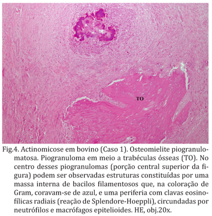
Cases of actinomycosis with atypical presentation are described in two oxen. In both cases there was a hard irregular and extensive lesions in the maxilla. The maxilla of both cattle became enlarged and honeycombed as a result of destructive rarefaction and regenerative bone proliferation. The cut surface of the lesions consisted of white glistening fibrous tissue within which numerous yellow caseous granules could be seen. Sinus tracts could be demonstrated within the lesions. In hematoxylin-eosin stained sections the lesions consisted of island of pyogranulomatous inflammation within an extensive fibrous stroma. In the center of the granuloma there was a basophilic irregular shaped mass surrounded by a zone radially arranged eosinophilic projections (Splendore-Hoeppli material). Around the radiating mass there was a zone of neutrophils, surrounded by a layer of epithelioid macrophages and occasional multinucleated giant cells. An outer layer of lymphocytes and plasma cells was present that limit the granuloma from the abundant fibrous tissue surrounding it. Up on Gram stain the central part of the colony revealed a tangled mass of rod shaped organisms morphologically consistent with Actinomyces bovis. Since the unusual presentation of this lesions misled the initial diagnosis the detailed description of these cases are reported here in the hopes it can help in the differential diagnosis by veterinary practitioners and met inspectors.
Actinomycosis; Actinomyces bovis; osteomyelitis; atypical presentation; diseases of cattle