Abstracts
The Galea spixii inhabits semiarid vegetation of Caatinga in the Brazilian Northeast. They are bred in captivity for the development of researches on the biology of reproduction. Therefore, the aim of this study is characterize the estrous cycle of G. spixii, in order to provide information to a better knowledge of captive breeding of the species. The estrous cycle was monitored by vaginal exfoliative cytology in 12 adult females. After the detection of two complete cycles in each animal, the same were euthanized. Then, histological study of the vaginal epithelium, with three females in each phase of the estrous cycle was performed; five were paired with males for performing the control group for estrous cycle phases, and three other were used to monitor the formation and rupture of vaginal closure membrane. By vaginal exfoliative cytology, predominance of superficial cells in estrus, large intermediate cells in proestrus, intermediate and parabasal cells, with neutrophils, in diestrus and metestrus respectively was found. Estrus was detected by the presence of spermatozoa in the control group. By histology, greater proliferation of the vaginal epithelium in proestrus was observed. We conclude that the estrous cycle of G. spixii lasts 15.8 ± 1.4 days and that the vaginal closure membrane develops until complete occlusion of the vaginal ostium, breaking after few days. Future studies may reveal the importance of this fact for the reproductive success of this animal.
Galea spixii; cavies; female; preservation; reproduction; rodents.
Os Galea spixii habitam a vegetação semiárida da Caatinga, no Nordeste brasileiro. Eles são criados em cativeiro para realização de pesquisas relacionadas a biologia da reprodução. Sendo assim, o objetivo deste trabalho foi caracterizar o ciclo estral de G. spixii para obtenção de informações que melhorem o conhecimento do manejo reprodutivo da espécie em cativeiro. O ciclo estral foi monitorado por citologia esfoliativa vaginal em doze fêmeas adultas. Após a detecção de dois ciclos completos em cada animal, os mesmos foram eutanasiados. Em seguida foi realizado estudo histológico do epitélio vaginal com três fêmeas em cada fase do ciclo estral; cinco foram pareadas com machos para realização do grupo controle e outras três fêmeas foram utilizadas para monitorar a formação e ruptura da membrana de oclusão vaginal. Através de citologia esfoliativa vaginal, constatou-se predomínio de células superficiais em estro, células intermediárias grandes em proestro, células intermediárias pequenas e células parabasais com presença de neutrófilos em diestro e metaestro, respectivamente. O estro foi detectado pela presença de espermatozoides no grupo controle. Através de histologia, observou-se uma maior proliferação no epitélio vaginal no proestro. Concluiu-se que o ciclo estral de G. spixii dura em média 15.8 ± 1.4 dias e a membrana de oclusão vaginal se desenvolve até completa oclusão do óstio vaginal externo, rompendo-se em poucos dias. Futuros estudos podem revelar a importância deste último fato para o sucesso reprodutivo deste animal.
Galea spixii; fêmeas; preás; preservação; reprodução; roedores.
Introduction
The Spix' yellow-toothed cavies (Galea spixii) are rodents that belong to the Caviinae subfamily and Caviidae family. They live in semiarid vegetation of Caatinga at Brazilian Northeast (Oliveira et al. 2008Oliveira M.F., Mess A., Ambrósio C.E., Dantas C.A.G., Favaron P.O. & Miglino M.A. 2008. Chorioallantoic placentation in Galea Spixii (Rodentia, Caviomorpha, Caviidae). Reprod. Biol. Endocrinol. 6(39):1-8.), where they are constantly used as alternative source of protein for inhabitants of this region (Santos et al. 2014aSantos A.C., Olio R.L., Viana D.C., Oliveira M.F., Miglino M.A.& Assis-Neto A.C. 2014b. Morphological description of unusual urinary tract in the female of a rodent, Galea spixii (Wagler, 1831). Pakistan J. Zool. 46(6):1617-1623.).
In Brazil, they are bred in captivity for conservation of the species, which is found as
vulnerable in the "red list of threatened species" (IUCN
2014IUCN 2014. Red List of Threatened Species, version 2013.1. International
Union of Conservation of Nature. Available at <www.iucnredlist.org> Access on
February 2014.
http://www.iucnredlist.org...
). Moreover, they are used for development of a new experimental model for
research about reproductive biology (Rodrigues et al.
2013Rodrigues M.N., Oliveira G.B., Albuquerque J.F.B., Menezes D.J.A.,
Assis-Neto A.C., Miglino M.A.& Oliveira M.F. 2013. Aspectos anatômicos do
aparelho genital masculino de preás adultos (Galea spixii Wagler, 1831). Revta
Biotemas 26(1):181-188., Santos et al. 2014bSantos A.C., Olio R.L., Viana D.C., Oliveira M.F., Miglino M.A.&
Assis-Neto A.C. 2014b. Morphological description of unusual urinary tract in the
female of a rodent, Galea spixii (Wagler, 1831). Pakistan J. Zool.
46(6):1617-1623.).
Researches related with reproductive biology have shown that G. spixii has continuous poliestral cycle, developing a type of inverted choriovitellinic placenta during pregnancy (Oliveira et al. 2008Oliveira M.F., Mess A., Ambrósio C.E., Dantas C.A.G., Favaron P.O. & Miglino M.A. 2008. Chorioallantoic placentation in Galea Spixii (Rodentia, Caviomorpha, Caviidae). Reprod. Biol. Endocrinol. 6(39):1-8., 2012Oliveira M.F., Vale A.M., Favaron P.O., Vasconcelos B.G., Oliveira G.B., Miglino M.A.& Mess A. 2012. Development of yolk sac inversion in Galea spixii and Cavia porcellus (Rodentia, Caviidae). Placenta 33(10):878-881.); gestation period lasts about 48 days (Oliveira et al. 2008Oliveira M.F., Mess A., Ambrósio C.E., Dantas C.A.G., Favaron P.O. & Miglino M.A. 2008. Chorioallantoic placentation in Galea Spixii (Rodentia, Caviomorpha, Caviidae). Reprod. Biol. Endocrinol. 6(39):1-8.); the onset of puberty in males occurs after 45th postnatal day (Santos et al. 2012Santos P.R., Oliveira M.F., Silva A.R. & Assis-Neto A.C. 2012. Development of spermatogenesis in captive-bred Spix's yellow-toothed cavy (Galea spixii). Reprod. Fertil. Develop. 24(6):877-885.); and females have masculinized external genitalia (Santos et al. 2014bSantos A.C., Olio R.L., Viana D.C., Oliveira M.F., Miglino M.A.& Assis-Neto A.C. 2014b. Morphological description of unusual urinary tract in the female of a rodent, Galea spixii (Wagler, 1831). Pakistan J. Zool. 46(6):1617-1623.).
It is known that the majority of female rodents can breed throughout the year and the reproductive cycle is systematically repeated, alternating periods of sexual activity and preparation of the reproductive tract for possible pregnancy (Selle 1922Selle R.M. 1922. Changes in the vaginal epithelium of the Guinea-pig during the oestrous cycle. Am. J. Anat.30(4):429-449., Lilley et al. 1997Lilley K.G., Epping R.J. & Hafner L.M. 1997. The guinea pig estrous cycle: correlation of vaginal impedance measurements with vaginal cytologic findings. Lab. Anim. Sci. 47(6):632-637., Touma et al. 2001Touma C., Palme R. & Sachser N. 2001. Different types of oestrous cycle in two closely related South American rodents (Cavia aperea and Galea musteloides) with different social and mating systems. Reproduction 121(5):791-801., Mahoney et al. 2011Mahoney M.M., Brooke V., Rossi B.V., Megan H., Hagenauer M.H. & Lee T.M. 2011. Characterization of the estrous cycle in Octodon degus. Biol. Reprod. 84(4):664-671.). In this sense, tools to detect features related to receptivity of the female for copulation in order to assist captive breeding of threatened species is very important (Binelli et al. 2014Binelli M., Pugliesi G., Hoeck V.V., Sponchiado M., Ramos R.S., Oliveira M.L., França M.R., D'Alexandri F.L., Mesquita F.S. & Membrive C.M.B. 2014. The role of proestrus on fertility and postovulatory uterine function in the cow. Anim. Reprod. 11(3):246-253.).
Regarding Caviidae as Cavia porcellus (Stockard & Papanicolaou 1917Stockard C.R. & Papanicolaou G.N. 1917. The existence of a typical oestrous cycle in the guinea-pig with a study of its histological and physiological changes. Am. J. Anat.22(2):225-283.; Kelly & Papanicolaou 1927Kelly G.L. & Papanicolaou G.N. 1927. The mechanism of the periodical opening and closing of the vaginal orifice in the Guinea-pig. Am. J. Anat. 40(2):387-411.), C. aperea and G. musteloides (Touma et al. 2001Touma C., Palme R. & Sachser N. 2001. Different types of oestrous cycle in two closely related South American rodents (Cavia aperea and Galea musteloides) with different social and mating systems. Reproduction 121(5):791-801.), the authors report that females show development of a vaginal closure membrane during the estrous cycle. This fact may be related to intraspecific sexual selection and formation of various mating systems among Caviidae (Adrian & Sachser 2011Adrian O. & Sachser N. 2011. Diversity of social and mating systems in cavies: a review. J. Mammal. 92(1):39-53.).
Due to the lack of studies on the reproductive cycle of G. spixii females, characterize the estrous cycle by vaginal exfoliative cytology and corresponding vaginal epithelium variations and check the presence of vaginal closure membrane, providing more data for captive breeding of this species were the aim of this study.
Materials And Methods
Animals. Twenty adults, non-pregnant, primiparous females and two adult males, prevenient of the Center for Wild Animals Multiplication of Federal Rural University of the Semi-Arid, Mossoró, RN, Brazil were used. This research was approved by Brazilian Institute of Environment and Renewable Resources (IBAMA, 2028236/2008) and Bioethics Committee of the School of Veterinary Medicine and Animal Science of the University of São Paulo, Brazil (protocol 2400/2011).
Detection of estrous cycle phases by vaginal cytology. Twelve adults, non-pregnant females in three separate boxes, each of which contained four properly identified females were used. During the experiment the females were fed with fruits, grasses, corn, rabbit feed and water.
In another box, five females were paired with adult males for comparison of cell types along the estrous cycle, especially in the estrous phase, with possible detection of spermatozoa in vaginal exfoliative cytology.
The vaginal exfoliative cytology was performed daily by the same collector. The collection of vaginal smears was performed using swabs of sterile cotton and subsequent deposit of biological material in histological slides, which were stained with fast Panoptic, according to the manufacturer (Laborclin(r), Vargem Grande/Pinhais, PR, Brazil). The females were independently monitored and there was no synchronization.
Subsequently, the samples were analyzed and photo documented by light microscopy. Different cell types: superficial cells; large and small intermediate cells; parabasal cells and neutrophils were identified (Stockard & Papanicolaou 1917Stockard C.R. & Papanicolaou G.N. 1917. The existence of a typical oestrous cycle in the guinea-pig with a study of its histological and physiological changes. Am. J. Anat.22(2):225-283., Selle 1922Selle R.M. 1922. Changes in the vaginal epithelium of the Guinea-pig during the oestrous cycle. Am. J. Anat.30(4):429-449., Lilley et al. 1997Lilley K.G., Epping R.J. & Hafner L.M. 1997. The guinea pig estrous cycle: correlation of vaginal impedance measurements with vaginal cytologic findings. Lab. Anim. Sci. 47(6):632-637., Touma et al. 2001Touma C., Palme R. & Sachser N. 2001. Different types of oestrous cycle in two closely related South American rodents (Cavia aperea and Galea musteloides) with different social and mating systems. Reproduction 121(5):791-801., Allison et al. 2008Allison R.W., Thrall M.A. & Olson P.N. 2008. Vaginal cytology, p.378-389. In: Conwelle R.L., Tyler R.D., Meincoth J.H. & Denicola D.B (Eds), Diagnostic Cytology and Hematology of the Dog and Cat. 3rd ed. Elsevier, Toronto.).
For a better monitoring of the estrous cycle phases, cells were counted in each sample of vaginal exfoliative cytology into fields containing from 0 to 100 cells in the cytological slides (Guimarães et al. 1997Guimarães D.A., Moreira D. & Vale W.G. 1997. Determinação do ciclo reprodutivo da cutia (Dasyprocta prymnolopha) através do diagnóstico calpocitológico. Acta Amazonica 27(1):55-64.). During the counting, cell types were grouped according to the criteria of morphology of each cell as above described.
After counting of two complete estrous cycles, average of the estrous cycle length of each female; of the females that showed estrous cycle of the same duration; and, finally, the average of all females were established. Statistical analysis was applied to the Student-t test for paired and unpaired values, with p < 0.05 of significance.
Day 1 was established by absence of neutrophils and predominance of superficial cells (Allison et al., 2008Allison R.W., Thrall M.A. & Olson P.N. 2008. Vaginal cytology, p.378-389. In: Conwelle R.L., Tyler R.D., Meincoth J.H. & Denicola D.B (Eds), Diagnostic Cytology and Hematology of the Dog and Cat. 3rd ed. Elsevier, Toronto.); and by observation of copulation in the control group.
Monitoring of the development and rupture of the vaginal closure membrane. In this phase of the experiment, another box containing three females, isolated from males were used. We started the analysis after observation of rupture of vaginal closure membrane. Development of vaginal closure membrane in all females was monitored until complete occlusion of the external vaginal ostium, ending at the moment of rupture. In this group, vaginal cytology was not used to avoid mechanical rupture of the vaginal closure membrane. Photo documentation was performed by camera Olympus SP 810UZ 14 mp.
Histological analysis of the vaginal epithelium during the estrous cycle phases. After analysis of the estrous cycle by vaginal cytology, the 12 females separated from males were anesthetized with xylazine (4mg/kg/IM) and ketamine (60mg/kg/IM), and then euthanized with sodium thiopental (2.5% 60mg/kg) by intracardiac cannulation. Three females were in estrus; three in proestrus; three in diestrus; and three in metestrus.
Vaginal tissue samples from all females were fixed in 10% formalin solution, processed for light microscopy and stained with H/E (hematoxylin/eosin). Microscopic photodocumentation was performed using BX61VS Olympus photomicroscope.
Results
Detection of the phases of the estrous cycle by vaginal exfoliative cytology
The cell types found by vaginal exfoliative cytology were: (1) parabasal cells, which were small and round, with a central nucleus occupying an area greater than the cytoplasm; (2) small intermediate cells, which were ovoid, with a central nucleus smaller than the cytoplasm; (3) large intermediate cells, which were polygonal cells, with a nucleus smaller than the cytoplasm, when compared to small intermediate cell; and (4) superficial cells with three different morphologies: nucleated, enucleated, and with picnotic nucleus (Fig.1). In addition, polymorphonuclear neutrophils were also found.
Cell types by vaginal exfoliative cytology in Galea spixii. (A) Superficial nucleated cells (arrows), proestrus; (B) enucleated superficial cell (arrow), estrus; (C) superficial cells with picnotic nuclei (arrow) and spermatozoa (arrowhead), estrus; (D) parabasal cell (arrow) and neutrophils (arrowhead), metestrus; (E) large intermediate cells (filled arrow), small intermediate cells (white arrowhead), parabasal cells (empty arrow), and neutrophils (black arrowhead), diestrus. Bars = 100µm.
We observed that the amount of these different cell types varied in each phase of the estrous cycle. During estrus, there was a predominance of superficial cells, which went from nucleated to enucleated. In metestrus, there was predominance of parabasal cells with presence of large numbers of neutrophils. Superficial and intermediate cells were also present in this phase. In diestrus there was predominance of small and large intermediate cells, with presence of neutrophils and parabasal cells. In proestrus, there was predominance of large intermediate cells and superficial cells, with little or no presence of neutrophils. We found six different estrous cycle lengths (Fig.1 and 2).
(A)Percentage of parabasal cells, (B) small intermediate cells (C) large intermediate cells, and (D) superficial cells by vaginal exfoliative cytology, reflecting different estrous cycle durations in Galea spixii. Day 1 includes the onset of estrus, while days 14, 15, 16, 17, 18 and 19 indicate the last day of proestrus.
In the control group, the cell types were the same as those found in the experimental group, but the duration of the estrous cycle could not be found, because of the copula, which interrupted the experiment. Copulation was confirmed by presence of spermatozoa in vaginal smears. After copulation, females followed pregnancy. The vaginal exfoliative cytology in this group showed that, in estrus phase, enucleated superficial cells and superficial cell with picnotic nuclei were predominant, and neutrophils were not found. Prior estrus, we observed predominance of large intermediate cells, which gradually were replaced by superficial cells (Fig.1).
Development and rupture of the vaginal closure membrane
The development of vaginal closure membrane was observed in those three females separated from males. In these females, the membrane developed gradually, blocking the external vaginal ostium, and then, naturally, this membrane ruptured within a few days. During a first moment, vaginal closure was broken. In few days, this membrane begins to grow and occlude the external vaginal ostium. Then, this membrane completely occludes the external vaginal ostium, giving an aspect of absent external vaginal ostium during this period. Then in few days, vaginal closure membrane breaks again. These events occurred cyclically (Fig. 3).
Modifications in vulva and clitoris of Galea spixii. (A) Vulva (black arrow) with broken vaginal closure membrane (arrow) and clitoris (empty arrow); (B) vulva (black arrow) with partially developed vaginal closure membrane (arrow) and clitoris (empty arrow); (C) vulva (black arrow) with fully developed vaginal closure membrane (arrow) and clitoris (empty arrow). Bars = 1cm.
Histological analysis of the vaginal epithelium throughout the estrous cycle
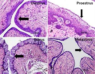
By histological studies, data performed by vaginal exfoliative cytology were confirmed. We found that the vaginal epithelium underwent proliferation, cornification and cell desquamation along the estrous cycle. In estrus phase, vaginal epithelium contained one or a few layer of cornified superficial cells. After the estrus phase (metestrus), the epithelium lost the superficial cells. Then, the epithelium presented one or a few parabasal layers. In diestrus, the epithelium initiated the cell proliferation, consisting on evident stratified epithelium. In proestrus, epithelium underwent massive proliferation. Then, the outer cells began the cornification process, with the approximation of next estrus (Fig. 4).
Vaginal epithelium of Galea spixii. (A) Stratified epithelium in diestrus (arrow); (B) stratified epithelium with cornification in proestrus (arrow); (C) superficial epithelial cells (cornified) in estrus (arrow); (D) epithelium with less stratified epithelium in early metaestrus (arrow). HE, Bars = 200μm.
Estrous cycle length by vaginal exfoliative cytology
We found females with estrous cycle ranging between 14 and 18 days with a mean of 15.6 ± 1.3 days in the first estrous cycle. In the second estrous cycle, we found females with cycles ranging between 14 and 19 days, with mean of 16.1 ± 1.5 days. The average of two complete cycles was 15.8 ± 1.4 days. The Student-t test for paired samples showed no difference between cycles (p<0.05).
Discussion
Understanding the reproductive behavior of animals can contribute to the development of techniques and tools for the biotechnology of reproduction, including artificial insemination, in vitro fertilization, embryo transfer and cryopreservation in several species bred in captivity, in order to ensure better reproductive performance and preservation of genetic variability (Domingues & Caldas-Bussiere 2007Domingues S.F.S. & Caldas-Bussiere M.C. 2007. Fisiologia e biotécnicas da reprodução desenvolvidas em fêmeas de primatas neotropicais importantes para a pesquisa biomédica. Revta Bras. Reprod. Anim. 30(1):57-71., Guimarães et al. 2011Guimarães D.A., Ramos L.R., Ohashi O.M., Garcia G.W. & Vale W.G. 2011. Plasma concentration of progesterone and 17-beta-estradiol of black-rumped agouti (Dasyprocta prymnolopha) during the estrous cycle. RevtaBiol. Trop. 59(1):29-35., Binelli et al. 2014Binelli M., Pugliesi G., Hoeck V.V., Sponchiado M., Ramos R.S., Oliveira M.L., França M.R., D'Alexandri F.L., Mesquita F.S. & Membrive C.M.B. 2014. The role of proestrus on fertility and postovulatory uterine function in the cow. Anim. Reprod. 11(3):246-253.).
Thus, in the present study, we found satisfactory results for detection of estrous cycles by vaginal exfoliative cytology, and there was no statistical difference (p<0.05) between the duration of the first and second estrous cycles. In addition, histological tests confirmed the results found in vaginal exfoliative cytology. The histological analysis showed cell proliferation and cornification more pronounced in proestrus and estrus, as found in Cavia porcellus (Stockard & Papanicolaou 1917Stockard C.R. & Papanicolaou G.N. 1917. The existence of a typical oestrous cycle in the guinea-pig with a study of its histological and physiological changes. Am. J. Anat.22(2):225-283.) and Lagostomus maximus (Weir 1971Weir B.J. 1971. The reproductive organs of the female plains viscacha, Lagostomus maximus. J. Reprod. Fertil. 25(3):365-373.). In this sense Deanesly (1966)Deanesly R. 1966. Pro-oestrus in the guinea-pig: hormonal stimulation of the vaginal epithelium. Reproduction 12(1):205-212. showed that estradiol and sometimes small amount of androstenedione combined with estradiol stimulated cell proliferation. In other hand, progesterone combined with estradiol showed negative results for epithelial proliferation in vagina of Cavia porcellus.
In G. spixii, the predominant cell types in the different phases of the estrous cycle were easier to detect during the proestrus and estrus by vaginal exfoliative cytology, similar to that found in other rodents such as Cavia porcellus (Selle 1922Selle R.M. 1922. Changes in the vaginal epithelium of the Guinea-pig during the oestrous cycle. Am. J. Anat.30(4):429-449., Lilley et al. 1997Lilley K.G., Epping R.J. & Hafner L.M. 1997. The guinea pig estrous cycle: correlation of vaginal impedance measurements with vaginal cytologic findings. Lab. Anim. Sci. 47(6):632-637.) Dasyprocta prymnolopha (Guimarães et al. 1997Guimarães D.A., Moreira D. & Vale W.G. 1997. Determinação do ciclo reprodutivo da cutia (Dasyprocta prymnolopha) através do diagnóstico calpocitológico. Acta Amazonica 27(1):55-64.), Agouti paca (Guimarães et al. 2008Guimarães D.A., Bastos L.V., Ferreira A.C.S., Luz-Ramos R.S., Ohashi O.M. & Ribeiro H.L. 2008. Características reprodutivas da paca fêmea (Agouti paca) criada em cativeiro. Acta Amazonica 38(3):531-538.) and Myocastor coypus (Felipe et al. 2001Felipe A.E., Cabodevila J. & Callejas S. 2001. Characterization of the estrous cycle of the Myocastor coypus (coypu) by means of exfoliative colpocytology. Mastozool. J. Neotrop. Mammal. 8(2):129-137.). Our results also agree with those described in domestic mammals (Allison et al. 2008Allison R.W., Thrall M.A. & Olson P.N. 2008. Vaginal cytology, p.378-389. In: Conwelle R.L., Tyler R.D., Meincoth J.H. & Denicola D.B (Eds), Diagnostic Cytology and Hematology of the Dog and Cat. 3rd ed. Elsevier, Toronto.). Lilley et al. (1997)Lilley K.G., Epping R.J. & Hafner L.M. 1997. The guinea pig estrous cycle: correlation of vaginal impedance measurements with vaginal cytologic findings. Lab. Anim. Sci. 47(6):632-637. showed positive correlation between vaginal impedance measurements and vaginal exfoliative cytology used for monitoring of estrous cycle in Cavia porcellus.
We believe that the satisfactory detection of estrous cycle phases was possible due to the fact of G. spixii presents continuous poliestral cycle (Oliveira et al. 2008Oliveira M.F., Mess A., Ambrósio C.E., Dantas C.A.G., Favaron P.O. & Miglino M.A. 2008. Chorioallantoic placentation in Galea Spixii (Rodentia, Caviomorpha, Caviidae). Reprod. Biol. Endocrinol. 6(39):1-8.) similar that found in other rodents, as Myocastor coypus (Felipe et al. 2001Felipe A.E., Cabodevila J. & Callejas S. 2001. Characterization of the estrous cycle of the Myocastor coypus (coypu) by means of exfoliative colpocytology. Mastozool. J. Neotrop. Mammal. 8(2):129-137.), Dasyprocta prymnolopha (Guimarães et al. 2011Guimarães D.A., Ramos L.R., Ohashi O.M., Garcia G.W. & Vale W.G. 2011. Plasma concentration of progesterone and 17-beta-estradiol of black-rumped agouti (Dasyprocta prymnolopha) during the estrous cycle. RevtaBiol. Trop. 59(1):29-35.), Agouti paca (Guimarães et al. 2008Guimarães D.A., Bastos L.V., Ferreira A.C.S., Luz-Ramos R.S., Ohashi O.M. & Ribeiro H.L. 2008. Características reprodutivas da paca fêmea (Agouti paca) criada em cativeiro. Acta Amazonica 38(3):531-538.), Paca cuniculus (Reis et al. 2011Reis A.C.G., Gerbasi S.H.B., Martins C., Machado M.R.F. & Oliveira C.A. 2011. Morfologia do sistema genital feminino da paca (Cuniculus paca Linnaeus, 1766). Braz. J. Vet. Res. Anim. Sci. 48(3):183-191.) and Rattus norvegicus (Marcondes et al. 2002Marcondes F.K., Bianchi F.J. & Tanno A.P. 2002. Determination of the estrous cycle phases of rats: some helpful considerations. Braz. J. Biol. 62(4A):609-614.).
Galea spixii showed formation of the vaginal closure membrane, which can also be found in other rodents, as Thryonomys swinderianus (Addo et al. 2007Addo P.G., Awumbila B., Awotwi E. & Ankrah N.-A. 2007. Reproductive characteristics of the female grasscutter (Thryonomys swinderianus) and formulation of colony breeding strategies. Livest. Res. Rural Develop. 19(1):28-37.), Lagostomus maximus (Weir 1971Weir B.J. 1971. The reproductive organs of the female plains viscacha, Lagostomus maximus. J. Reprod. Fertil. 25(3):365-373.), Galea musteloides and Cavia aperea (Touma et al. 2001Touma C., Palme R. & Sachser N. 2001. Different types of oestrous cycle in two closely related South American rodents (Cavia aperea and Galea musteloides) with different social and mating systems. Reproduction 121(5):791-801.), Octodon degus (Mahoney et al. 2011Mahoney M.M., Brooke V., Rossi B.V., Megan H., Hagenauer M.H. & Lee T.M. 2011. Characterization of the estrous cycle in Octodon degus. Biol. Reprod. 84(4):664-671.), Cavia porcellus (Selle 1922Selle R.M. 1922. Changes in the vaginal epithelium of the Guinea-pig during the oestrous cycle. Am. J. Anat.30(4):429-449., Lilley et al. 1997Lilley K.G., Epping R.J. & Hafner L.M. 1997. The guinea pig estrous cycle: correlation of vaginal impedance measurements with vaginal cytologic findings. Lab. Anim. Sci. 47(6):632-637.) and Rattus norvegicus (Lephart et al. 1989Lephart E.D., Mathews D., Noble J.L. & Ojeda S.R. 1989. The vaginal epithelium of immature rats metabolizes androgens trough an aromatase-like reaction: changes during the time of puberty. Biol. Reprod. 40(2):259-267.).
Lephart et al. (1987), Addo et al. (2007)Addo P.G., Awumbila B., Awotwi E. & Ankrah N.-A. 2007. Reproductive characteristics of the female grasscutter (Thryonomys swinderianus) and formulation of colony breeding strategies. Livest. Res. Rural Develop. 19(1):28-37. and Mahoney et al. (2011)Mahoney M.M., Brooke V., Rossi B.V., Megan H., Hagenauer M.H. & Lee T.M. 2011. Characterization of the estrous cycle in Octodon degus. Biol. Reprod. 84(4):664-671. described that the first rupture of the vaginal closure membrane is positively correlated with the onset of puberty in the studied species. Subsequently, the vaginal closure membrane breaks and develops at each estrous cycle as found in G. spixii, although the corresponding estrous phase is not showed in present study. The hormone responsible to rupture of vaginal closure membrane is estradiol as demonstrated by Deanesly (1966)Deanesly R. 1966. Pro-oestrus in the guinea-pig: hormonal stimulation of the vaginal epithelium. Reproduction 12(1):205-212. in ovariectomized Cavia porcellus.
We believe that future studies could demonstrate the importance of this phenomenon, which was described in detail in early twentieth century by Stockard & Papanicolaou (1917)Stockard C.R. & Papanicolaou G.N. 1917. The existence of a typical oestrous cycle in the guinea-pig with a study of its histological and physiological changes. Am. J. Anat.22(2):225-283. and Kelly & Papanicolaou (1927)Kelly G.L. & Papanicolaou G.N. 1927. The mechanism of the periodical opening and closing of the vaginal orifice in the Guinea-pig. Am. J. Anat. 40(2):387-411. in Cavia porcellus, but has been little detailed in other rodent species. To date, we believe that the development of the vaginal closure membrane in G. spixii is possible due to the presence of the external urethral ostium at the top of the clitoris (Santos et al. 2014bSantos A.C., Olio R.L., Viana D.C., Oliveira M.F., Miglino M.A.& Assis-Neto A.C. 2014b. Morphological description of unusual urinary tract in the female of a rodent, Galea spixii (Wagler, 1831). Pakistan J. Zool. 46(6):1617-1623.) instead vaginal vestibule, which is absent in this species (Santos et al. 2014aSantos A.C., Bertassoli B.M., Viana D.C., Vasconcelos B.G., Oliveira M.F., Miglino M.A.& Assis-Neto A.C. 2014a. The morphology of female genitalia in Galea spixii (Caviidae, Caviinae). Biosc. J. 30(6):1793-1802.).
Other modifications influenced by hormones produced during ovarian follicular development during the estrous cycle are endometrial and glandular changes demonstrated in rodents as Cavia porcellus (Stockard & Papanicolaou 1917Stockard C.R. & Papanicolaou G.N. 1917. The existence of a typical oestrous cycle in the guinea-pig with a study of its histological and physiological changes. Am. J. Anat.22(2):225-283.) and bovines (Binelli et al. 2014Binelli M., Pugliesi G., Hoeck V.V., Sponchiado M., Ramos R.S., Oliveira M.L., França M.R., D'Alexandri F.L., Mesquita F.S. & Membrive C.M.B. 2014. The role of proestrus on fertility and postovulatory uterine function in the cow. Anim. Reprod. 11(3):246-253.). Therefore, we believe that future studies could correlate follicular development with hormonal production and its influence on endometrium in G. spixii.
Females of G. spixii have short and regular estrous cycle with duration close to that found in other rodents belonging to the Caviinae subfamily, as Microcavia australis (Adrian & Sachser 2011Adrian O. & Sachser N. 2011. Diversity of social and mating systems in cavies: a review. J. Mammal. 92(1):39-53.), Cavia aperea (Touma et al. 2001Touma C., Palme R. & Sachser N. 2001. Different types of oestrous cycle in two closely related South American rodents (Cavia aperea and Galea musteloides) with different social and mating systems. Reproduction 121(5):791-801.) and Cavia porcellus (Selle 1922Selle R.M. 1922. Changes in the vaginal epithelium of the Guinea-pig during the oestrous cycle. Am. J. Anat.30(4):429-449., Lilley et al. 1997Lilley K.G., Epping R.J. & Hafner L.M. 1997. The guinea pig estrous cycle: correlation of vaginal impedance measurements with vaginal cytologic findings. Lab. Anim. Sci. 47(6):632-637.). To simplify comparison, in Table 1 we highlighted the duration of the estrous cycle in different rodents.
The continuous poliestral cycle and short gestational period (Oliveira et al. 2008Oliveira M.F., Mess A., Ambrósio C.E., Dantas C.A.G., Favaron P.O. & Miglino M.A. 2008. Chorioallantoic placentation in Galea Spixii (Rodentia, Caviomorpha, Caviidae). Reprod. Biol. Endocrinol. 6(39):1-8.), associated with short estrous cycles found in G. spixii females in this present study may be related to the reproductive success of this species (Purves et al. 2005Purves W.K., Sadava D., Orians G.H. & Heller H.C. 2005. Vida: a ciência da biologia. Vol.2. 6ª ed. Artmed, São Paulo. 1044p.) as found in others rodents which belong to the Caviidae family, even though they are the main source of food for many predators in South America, and developing for this reason important ecological function (Zogno et al. 2004Zogno M.A., Miglino M.A.& Oliveira M.F. 2004. Análise bioquímica dos líquidos fetais e citologia do fluido amniótico da fêmea de Mocó (Kerodon rupestris). Braz. J. Vet. Res. Anim. Sci. 41(4):228-235.).
Conclusions
By methodology applied in our study, we conclude that the estrous cycle of Galea spixii lasts an average of 15.8±1.4 days. These results may be helpful for breeding in captivity and conservation of the species.
We also noted the formation and rupture of the vaginal closure membrane, and further studies may reveal the importance of this fact for the reproductive success of these rodents.
Acknowledgements
To Federal Rural University of Semi-Arid for supplying the animals used in this study; to IBAMA (Brazilian Institute of Environment and Renewable Resources) for license for captive breeding of the same; and to FAPESP (Fundação de Amparo a Pesquisa do Estado de São Paulo) for financial support.
- Addo P.G., Awumbila B., Awotwi E. & Ankrah N.-A. 2007. Reproductive characteristics of the female grasscutter (Thryonomys swinderianus) and formulation of colony breeding strategies. Livest. Res. Rural Develop. 19(1):28-37.
- Adrian O. & Sachser N. 2011. Diversity of social and mating systems in cavies: a review. J. Mammal. 92(1):39-53.
- Allison R.W., Thrall M.A. & Olson P.N. 2008. Vaginal cytology, p.378-389. In: Conwelle R.L., Tyler R.D., Meincoth J.H. & Denicola D.B (Eds), Diagnostic Cytology and Hematology of the Dog and Cat. 3rd ed. Elsevier, Toronto.
- Binelli M., Pugliesi G., Hoeck V.V., Sponchiado M., Ramos R.S., Oliveira M.L., França M.R., D'Alexandri F.L., Mesquita F.S. & Membrive C.M.B. 2014. The role of proestrus on fertility and postovulatory uterine function in the cow. Anim. Reprod. 11(3):246-253.
- Deanesly R. 1966. Pro-oestrus in the guinea-pig: hormonal stimulation of the vaginal epithelium. Reproduction 12(1):205-212.
- Domingues S.F.S. & Caldas-Bussiere M.C. 2007. Fisiologia e biotécnicas da reprodução desenvolvidas em fêmeas de primatas neotropicais importantes para a pesquisa biomédica. Revta Bras. Reprod. Anim. 30(1):57-71.
- Felipe A.E., Cabodevila J. & Callejas S. 2001. Characterization of the estrous cycle of the Myocastor coypus (coypu) by means of exfoliative colpocytology. Mastozool. J. Neotrop. Mammal. 8(2):129-137.
- Guimarães D.A., Moreira D. & Vale W.G. 1997. Determinação do ciclo reprodutivo da cutia (Dasyprocta prymnolopha) através do diagnóstico calpocitológico. Acta Amazonica 27(1):55-64.
- Guimarães D.A., Bastos L.V., Ferreira A.C.S., Luz-Ramos R.S., Ohashi O.M. & Ribeiro H.L. 2008. Características reprodutivas da paca fêmea (Agouti paca) criada em cativeiro. Acta Amazonica 38(3):531-538.
- Guimarães D.A., Ramos L.R., Ohashi O.M., Garcia G.W. & Vale W.G. 2011. Plasma concentration of progesterone and 17-beta-estradiol of black-rumped agouti (Dasyprocta prymnolopha) during the estrous cycle. RevtaBiol. Trop. 59(1):29-35.
- IUCN 2014. Red List of Threatened Species, version 2013.1. International Union of Conservation of Nature. Available at <www.iucnredlist.org> Access on February 2014.
» http://www.iucnredlist.org - Kelly G.L. & Papanicolaou G.N. 1927. The mechanism of the periodical opening and closing of the vaginal orifice in the Guinea-pig. Am. J. Anat. 40(2):387-411.
- Lephart E.D., Mathews D., Noble J.L. & Ojeda S.R. 1989. The vaginal epithelium of immature rats metabolizes androgens trough an aromatase-like reaction: changes during the time of puberty. Biol. Reprod. 40(2):259-267.
- Lilley K.G., Epping R.J. & Hafner L.M. 1997. The guinea pig estrous cycle: correlation of vaginal impedance measurements with vaginal cytologic findings. Lab. Anim. Sci. 47(6):632-637.
- Mahoney M.M., Brooke V., Rossi B.V., Megan H., Hagenauer M.H. & Lee T.M. 2011. Characterization of the estrous cycle in Octodon degus. Biol. Reprod. 84(4):664-671.
- Marcondes F.K., Bianchi F.J. & Tanno A.P. 2002. Determination of the estrous cycle phases of rats: some helpful considerations. Braz. J. Biol. 62(4A):609-614.
- Mendonça F.S., Evêncio-Neto J., Simões M.J., Camargo L.M. & Baratella-Evêncio L. 2007. Aspectos citopatológicos da mucosa vaginal de camundongas tratadas com progesterona. Ciênc. Anim. Bras. 8(2):313-318.
- Oliveira M.F., Mess A., Ambrósio C.E., Dantas C.A.G., Favaron P.O. & Miglino M.A. 2008. Chorioallantoic placentation in Galea Spixii (Rodentia, Caviomorpha, Caviidae). Reprod. Biol. Endocrinol. 6(39):1-8.
- Oliveira M.F., Vale A.M., Favaron P.O., Vasconcelos B.G., Oliveira G.B., Miglino M.A.& Mess A. 2012. Development of yolk sac inversion in Galea spixii and Cavia porcellus (Rodentia, Caviidae). Placenta 33(10):878-881.
- Purves W.K., Sadava D., Orians G.H. & Heller H.C. 2005. Vida: a ciência da biologia. Vol.2. 6ª ed. Artmed, São Paulo. 1044p.
- Reis A.C.G., Gerbasi S.H.B., Martins C., Machado M.R.F. & Oliveira C.A. 2011. Morfologia do sistema genital feminino da paca (Cuniculus paca Linnaeus, 1766). Braz. J. Vet. Res. Anim. Sci. 48(3):183-191.
- Rodrigues M.N., Oliveira G.B., Albuquerque J.F.B., Menezes D.J.A., Assis-Neto A.C., Miglino M.A.& Oliveira M.F. 2013. Aspectos anatômicos do aparelho genital masculino de preás adultos (Galea spixii Wagler, 1831). Revta Biotemas 26(1):181-188.
- Santos P.R., Oliveira M.F., Silva A.R. & Assis-Neto A.C. 2012. Development of spermatogenesis in captive-bred Spix's yellow-toothed cavy (Galea spixii). Reprod. Fertil. Develop. 24(6):877-885.
- Santos A.C., Bertassoli B.M., Viana D.C., Vasconcelos B.G., Oliveira M.F., Miglino M.A.& Assis-Neto A.C. 2014a. The morphology of female genitalia in Galea spixii (Caviidae, Caviinae). Biosc. J. 30(6):1793-1802.
- Santos A.C., Olio R.L., Viana D.C., Oliveira M.F., Miglino M.A.& Assis-Neto A.C. 2014b. Morphological description of unusual urinary tract in the female of a rodent, Galea spixii (Wagler, 1831). Pakistan J. Zool. 46(6):1617-1623.
- Selle R.M. 1922. Changes in the vaginal epithelium of the Guinea-pig during the oestrous cycle. Am. J. Anat.30(4):429-449.
- Stockard C.R. & Papanicolaou G.N. 1917. The existence of a typical oestrous cycle in the guinea-pig with a study of its histological and physiological changes. Am. J. Anat.22(2):225-283.
- Touma C., Palme R. & Sachser N. 2001. Different types of oestrous cycle in two closely related South American rodents (Cavia aperea and Galea musteloides) with different social and mating systems. Reproduction 121(5):791-801.
- Weir B.J. 1971. The reproductive organs of the female plains viscacha, Lagostomus maximus. J. Reprod. Fertil. 25(3):365-373.
- Zogno M.A., Miglino M.A.& Oliveira M.F. 2004. Análise bioquímica dos líquidos fetais e citologia do fluido amniótico da fêmea de Mocó (Kerodon rupestris). Braz. J. Vet. Res. Anim. Sci. 41(4):228-235.
Publication Dates
-
Publication in this collection
Jan 2015
History
-
Received
01 Apr 2014 -
Accepted
21 Dec 2014