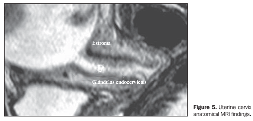
OBJECTIVE: To evaluate the uterine cervix length with magnetic resonance imaging in comparison with findings at transvaginal ultrasonography. MATERIALS AND METHODS: Twenty pregnant women between the 19th and 30th gestational weeks underwent magnetic resonance imaging and transvaginal ultrasonography for evaluation of their uterine cervix. Measurements by means of magnetic resonance imaging were performed by two specialists in imaging diagnosis for calculating the interobserver variability of the method. RESULTS: Calculation of the Pearson's correlation coefficient between measurements of the cervical length demonstrated a significant correlation between the results of both methods (r=0.628; p<0.01). The paired t test demonstrated no statistically significant difference between measurements obtained by transvaginal ultrasonography and magnetic resonance imaging (p=0.068). Interobserver agreement in cervical measurements by magnetic resonance imaging was high (a=0.96), demonstrating the reliability of the method. CONCLUSION: The comparison between both imaging methods in the evaluation of cervical biometry showed no statistically significant difference thus reinforcing the utilization of ultrasonography. However, in some cases where transvaginal ultrasonography is contraindicated, magnetic resonance imaging can be alternatively utilized for measurement of the cervical length.
Uterine cervix; Pregnancy; Magnetic resonance imaging