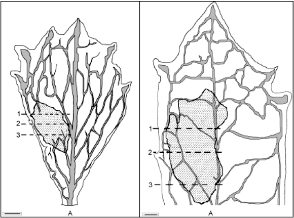
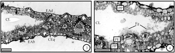
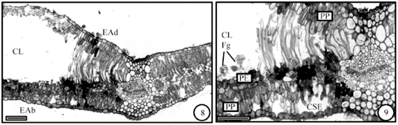
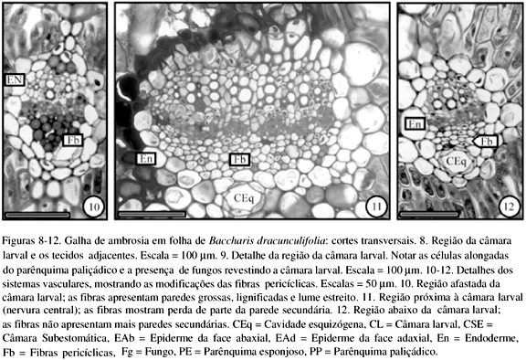
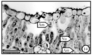
Ambrosia galls are induced by dipteran (Cecidomyiidae) and no nutritive tissue is present: fungi hyphae are the source of food to the inducer larva. Ambrosia galls in Baccharis concinna and B. dracunculifolia have only one chamber with one inducer. The fungi hyphae are observed. In B. dracunculifolia galls the hyphae are restricted to the larval chamber. The parenchyma palisade cells are elongated. In B. concinna galls hyphae spread also among chlorenchyma cells around of larval chamber. The chlorenchyma cells near to the chamber elongate slightly. In both galls, pericyclic fibers of vascular system lose their secondary walls. When the inducer is in pupal phase, the amount of hyphae increases and they fill several parts of the larval chamber. Hyphae of B. concinna galls present lipophilic globules which are not observed in the hyphae of B. dracunculifolia galls. Picnids are found only in the senescent galls of B. dracunculifolia. This paper is the first contribution to the knowledge of the ambrosia galls in the Brazilian flora.
Baccharis dracunculifolia; Baccharis concinna; Asteraceae; ambrosia galls; anatomy