
 High-Frequency Oscillatory Ventilation and High-Flow Nasal Cannula in a Child with Hepatoblastoma and Severe Respiratory Failure: Case Report
ABSTRACT
Introduction: Hepatoblastoma is the most common malignant liver neoplasm in childhood and can present with significant respiratory repercussions. In severe cases, lung-protective ventilatory strategies are essential, and the physiological mechanisms that support these interventions are discussed. The objective is to describe the ventilatory management and clinical evolution of a child with hepatoblastoma and severe respiratory failure undergoing high-frequency oscillatory ventilation (HFOV) and high-flow nasal cannula (HFNC), discussing the physiological basis and applicability of these strategies.
Case report: A 2-year-old child with a large hepatoblastoma undergoing chemotherapy developed severe respiratory failure, hypercapnia, and marked acidemia that were refractory to conventional ventilation, prompting initiation of HFOV. After stabilization, a planned transition to conventional ventilation was performed, followed by the use of HFNC after accidental extubation, without the need for reintubation, which resulted in a favorable outcome.
Conclusion: HFOV combined with the strategic use of HFNC may represent a safe and effective alternative for children with hepatoblastoma and severe respiratory impairment secondary to abdominal compression and treatment-related complications.
Key words:
Hepatoblastoma; High-Frequency Ventilation/methods; Respiratory Failure/complications; Pediatric Intensive Care Units; Child, Preschool
RESUMO
Introdução: O hepatoblastoma é a neoplasia maligna hepática mais comum na infância e pode apresentar repercussões respiratórias significativas. Nos casos graves, estratégias ventilatórias protetoras pulmonares são essenciais, sendo discutidos os mecanismos fisiológicos que sustentam essas intervenções. O objetivo é descrever o manejo ventilatório e a evolução clínica de uma criança com hepatoblastoma e insuficiência respiratória grave submetida à ventilação oscilatória de alta frequência (VOAF) e cânula nasal de alto fluxo (CNAF), discutindo os fundamentos fisiológicos e a aplicabilidade dessas estratégias.
Relato de caso: Criança de 2 anos, portadora de hepatoblastoma volumoso em quimioterapia, evoluiu com insuficiência respiratória grave, hipercapnia e acidemia acentuada, refratária à ventilação convencional, sendo iniciada VOAF. Após estabilização, realizou-se transição planejada para ventilação convencional, seguida pelo uso de CNAF após extubação acidental, sem necessidade de reintubação, resultando em desfecho favorável.
Conclusão: A VOAF combinada ao uso estratégico da CNAF pode representar uma alternativa segura e eficaz para crianças com hepatoblastoma e comprometimento respiratório grave secundário à compressão abdominal e a complicações relacionadas ao tratamento.
Palavras-chave:
Hepatoblastoma; Ventilação de Alta Frequência/métodos; Insuficiência Respiratória/complicações; Unidades de Terapia Intensiva Pediátrica; Pré-Escolar
RESUMEN
Introducción: El hepatoblastoma es la neoplasia maligna hepática más común en la infancia y puede presentar repercusiones respiratorias significativas. En los casos graves, las estrategias de ventilación protectora pulmonar son esenciales, y se analizan los mecanismos fisiológicos que sustentan estas intervenciones. El objetivo es describir el manejo ventilatorio y la evolución clínica de un niño con hepatoblastoma e insuficiencia respiratoria grave sometido a ventilación oscilatoria de alta frecuencia (VOAF) y cánula nasal de alto flujo (CNAF), discutiendo los fundamentos fisiológicos y la aplicabilidad de estas estrategias.
Informe del caso: Un niño de dos años con un hepatoblastoma voluminoso en tratamiento con quimioterapia desarrolló insuficiencia respiratoria grave, hipercapnia y acidosis marcada, refractarias a la ventilación convencional, por lo que se inició VOAF. Tras la estabilización, se realizó una transición planificada a ventilación convencional, seguida del uso de CNAF después de una extubación accidental, sin necesidad de reintubación, lo que resultó en una evolución favorable.
Conclusión: La VOAF combinada con el uso estratégico de la CNAF puede representar una alternativa segura y eficaz para niños con hepatoblastoma y deterioro respiratorio grave secundario a la compresión abdominal y a complicaciones relacionadas con el tratamiento.
Palabras clave:
Hepatoblastoma; Ventilación de Alta Frecuencia/métodos; Insuficiencia Respiratoria/complicaciones; Unidades de Cuidado Intensivo Pediátrico; Preescolar
INTRODUCTION
Hepatoblastoma (HB) is a primary liver tumor characterized by the abnormal proliferation of hepatocytes. It is a rare neoplasm, accounting for approximately 1% of pediatric cancers. HB predominantly affects children under 5 years of age, with prematurity and low birth weight identified as major risk factors1. Initially, HB may be asymptomatic; however, as the clinical condition progresses, nonspecific signs and symptoms such as nausea, abdominal discomfort, vomiting, loss of appetite, weight loss, and fatigue may emerge2.
Although HB generally presents a favorable prognosis, manifestations such as jaundice, portal hypertension, and abdominal distension leading to visceral compression may be associated with worse outcomes3. In this context, children with HB are particularly susceptible to respiratory complications, as the lungs are among the most frequently affected organs either due to drug-related toxicity or associated infections and lesions, including metastatic involvement2. Additionally, increased intra-abdominal pressure resulting from the tumor mass can impair respiratory mechanics, reduce lung compliance, and exacerbate respiratory failure.
High-flow nasal cannula (HFNC) has become an important non-invasive form of respiratory support in pediatric patients with respiratory failure. This system administers heated and humidified oxygen at high flow rates, allowing precise control of the inspired oxygen fraction (FiO2), reduction of nasopharyngeal dead space, and generation of a slight level of positive airway pressure. These factors promote improved oxygenation, decrease respiratory effort, and increase patient comfort. In children with conditions that impair respiratory mechanics, such as increased intra-abdominal pressure due to tumor burden, HFNC can be an effective strategy to optimize gas exchange and potentially avoid or assist in weaning from invasive mechanical ventilation4.
Given the susceptibility to respiratory dysfunction and the potential need for ventilatory support, the adoption of lung-protective ventilatory strategies is essential. High-frequency oscillatory ventilation (HFOV), a nonconventional ventilatory modality that employs high mean airway pressures and low tidal volumes, may help maintain alveolar recruitment and prevent atelectrauma5 in children with HB. Although HFOV is used in pediatric acute respiratory distress syndrome, reports of its use in children with hepatoblastoma and extrinsic compression from an abdominal mass remain scarce.
Therefore, the objective of this study is to describe the ventilatory management and clinical evolution of a child with hepatoblastoma and severe respiratory failure who underwent HFOV and HFNC, discussing the physiological foundations and the applicability of these strategies.
This is a case report. All available data from a single consecutive case with a complete clinical record were included. The study was approved by a Research Ethics Committee, report 7,221,384 (CAAE (submission for ethical review): 83412724.9.0000.0407), in compliance with Directive number 466/20126 of the National Health Council.
CASE REPORT
A 2-year-old female child presented to the emergency department of a countryside hospital of the state of Bahia on January 11, 2023, with abdominal distension associated with pain in the same region. She was transferred to another hospital in the same city with suspected acute obstructive abdomen.
On January 24, an abdominal computed tomography scan revealed a large expansile mass in the upper abdomen, with lobulated contours and heterogeneous contrast enhancement, measuring 15.1×14.5×10.7 cm. Two days later, she was transferred to the state capital and admitted with a history of fever and vomiting in the 24 hours preceding admission.
On February 1, she underwent an image-guided percutaneous liver biopsy and, five days later, bone marrow biopsy and myelogram. She was admitted to the intensive care unit (ICU) on February 17 with respiratory distress and tachycardia, requiring low-flow oxygen therapy. Her hemoglobin (Hb) level was 3.7 g/dL, prompting urgent transfusion of packed red blood cells (PRBC). The following day, she presented increased abdominal volume and again had Hb of 3.7 g/dL, raising concern for intratumoral bleeding, and chemotherapy was initiated. After the third PRBC transfusion, Hb increased to 6.6 g/dL.
On March 1, hepatoblastoma was diagnosed, and the second cycle of chemotherapy was initiated two days after confirmation. On March 15, she developed cardiorespiratory arrest lasting approximately 2 minutes, was intubated, and remained on invasive mechanical ventilation for seven days. After extubation, she received low-flow oxygen therapy for two days. On March 24, she was transitioned to a high-flow nasal cannula (HFNC) due to a worsening respiratory pattern (Figure 1 illustrates the chest X-ray from March 25).

After respiratory and clinical improvement, she was transferred to the ward on April 11. However, the following day, she was readmitted to the ICU due to tachycardia, tachypnea, hypoactivity, fever, and diarrheal stools. On April 19, she developed respiratory distress and oxygen desaturation, requiring intubation. One day later, significant air leak was observed around the orotracheal tube (OTT) (Figure 2A), along with hypoxemia (PaO2 55 mmHg and SaO2 60%), characterizing severe respiratory failure refractory to conventional ventilation, and severe respiratory acidosis (pH 6.88 and PaCO2 106 mmHg). The OTT was replaced (Figure 2B), but acidosis persisted (pH 6.10 and PaCO2 115 mmHg) under pressure-controlled ventilation with positive end-expiratory pressure of 10 cmH2O, inspiratory time of 0.5 seconds, respiratory rate of 38 breaths per minute, and fraction of inspired oxygen (FiO2) of 80%.

Chest radiographs showing tube position before replacement (A) and before/after replacement (B)
High-frequency oscillatory ventilation (HFOV) was therefore initiated as a rescue strategy in the context of severe hypercapnia and acidosis unresponsive to conventional ventilation, with mean airway pressure of 32 cmH2O, amplitude of 53 cmH2O, frequency of 6.0 Hz, bias flow of 30 L/min, FiO2 of 100%, and inspiratory time of 33%, resulting in progressive improvement in oxygenation. On the same day, the child developed arterial hypotension, managed with norepinephrine infusion, possibly related to the increased mean airway pressure and the underlying clinical condition.
After two days on HFOV, the patient showed significant improvement in her ventilatory status (Figure 3A). On April 22, she was transitioned back to conventional ventilation (pressure-controlled ventilation), with parameters adjusted according to a lung-protective strategy. On April 24, she experienced an accidental extubation and was subsequently placed on HFNC as post-extubation support to prevent respiratory failure, remaining on this therapy for two days. She was discharged from the ICU on April 30, breathing spontaneously with low-flow oxygen supplementation, meaning still with impaired gas exchange possibly secondary to lung injury associated with the inflammatory process resulting from the clinical picture.
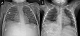
She remained hospitalized in the ward for continuation of chemotherapy. The third cycle was initiated on May 3 but interrupted the following day due to fever, and then resumed on May 5. Figure 3B illustrates the chest radiograph obtained on May 16. The fourth chemotherapy cycle occurred between May 24 and 26. The child remained in the ward while awaiting municipal authorization for enoxaparin and was discharged on July 11, 2023, breathing spontaneously in room air.
DISCUSSION
The clinical features of hepatoblastoma (HB) are often nonspecific, which can delay diagnosis and lead to the identification of an already large abdominal mass7. In the present case, the tumor caused mechanical compression of the lungs, resulting in a restrictive ventilatory pattern with decreased pulmonary compliance. This physiological alteration explains the development of hypoxemia, hypercapnia, and increased respiratory effort observed in the patient8.
Clinically, these alterations manifested as tachypnea, dyspnea, and signs of respiratory distress, characterizing restrictive respiratory failure secondary to extrinsic pulmonary compression. Understanding this pathophysiological mechanism was essential to guide ventilatory management, including the transition from conventional mechanical ventilation to high-frequency oscillatory ventilation (HFOV), aimed at protecting functional lung parenchyma.
HB is frequently associated with chemotherapy-related adverse effects, which can cause pulmonary toxicity8. These toxic effects justify the adoption of protective ventilation in children with HB, aiming to preserve lung parenchyma and minimize biotrauma9.
Children with neoplasms present several risk factors for infection; the risk goes beyond chemotherapy-induced neutropenia10. Immunological dysfunctions resulting from the underlying neoplasm, corticosteroid use, mucosal barrier damage, invasive devices, prolonged hospitalization, and nutritional deficiencies contribute to susceptibility11. These factors make the respiratory tract particularly vulnerable, as they can compromise the integrity of the alveolar-capillary barrier and, consequently, cause refractory respiratory failure requiring rescue mechanical ventilation strategies10.
The increased intra-abdominal pressure caused by the tumor mass may reduce pulmonary ventilation and affect gas exchange. The child presented severe hypoxemia and acidosis refractory to conventional ventilation, justifying the indication of HFOV. In HFOV, the use of high mean airway pressure and low tidal volumes improves lung compliance and reduces the risk of volutrauma and atelectrauma, particularly in lungs compromised by extrinsic compression12.
Despite the benefits of HFOV, the child developed arterial hypotension on the same day it was initiated. This ventilatory modality can generate hemodynamic impact by increasing intrathoracic pressure, which may raise left ventricular afterload and reduce venous return in hypovolemic children. Hemodynamic instability is a well-recognized adverse effect of HFOV, particularly in hypovolemic pediatric patients13. However, hemodynamic stability was restored with norepinephrine infusion, ensuring the safe continuation of the intervention.
Later, the child experienced accidental extubation and was transitioned to HFNC, a successful strategy in preventing reintubation. This strategy delivers heated and humidified oxygen at high flows, reducing respiratory effort, promoting the flushing of nasopharyngeal dead space to improve CO2 clearance, and facilitating alveolar ventilation. These improve oxygenation, alleviate respiratory effort, and help prevent post-extubation respiratory failure, especially in patients with large abdominal masses14.
Liberation from mechanical ventilation may have been facilitated by an assertive approach involving a planned transition from HFOV to conventional ventilation, followed by prophylactic post-extubation support using HFNC. HFNC has been established as an effective form of post-extubation support in pediatrics, reducing the risk of ventilatory failure in high-risk patients, which corroborates its use in this case15.
This case illustrates the pathophysiological application of HFOV and HFNC in restrictive respiratory failure secondary to large abdominal masses. Recent studies indicate that HFOV can act as protective ventilation, while HFNC is effective as post-extubation support, reducing reintubation16,17. However, controversies persist regarding initiation, flow adjustments, and selection criteria, especially in patients with increased intra-abdominal pressure, reinforcing the need for individualized decision-making.
CONCLUSION
This report contributes to the understanding of respiratory management in children with hepatoblastoma by documenting advanced and successful ventilatory strategies. The case suggests that HFOV may be considered a rescue strategy in children with HB and refractory severe respiratory failure, whereas HFNC appears to be a useful and safe tool for post-extubation support.
DATA AVAILABILITY STATEMENT
All the contents associated with the article are included in the manuscript.
REFERENCES
-
1 Falqueto LE, Vilar PR, Campos HG, et al. Primary malignant liver tumors: eight-year experience in a pediatric hospital in Brazil. A cross-sectional study. Rev Col Bras Cir. 2022;49:e20223273. doi: https://doi.org/10.1590/0100-6991e-20223273-en
» https://doi.org/10.1590/0100-6991e-20223273-en -
2 Hager J, Sergi CM. Hepatoblastoma. In: Sergi CM, editor. Liver Cancer. Brisbane: Exon Publications; 2021. p. 145-67. doi: https://doi.org/10.36255/exonpublications.livercancer.2021.ch8
» https://doi.org/10.36255/exonpublications.livercancer.2021.ch8 -
3 Ramos MENO, Arruda ACC, Fraga TSG. Hepatoblastoma pediátrico e seus efeitos na vida da criança. Braz J Health Rev. 2024;7(10 esp):e74941. doi: https://doi.org/10.34119/bjhrv7n10-042
» https://doi.org/10.34119/bjhrv7n10-042 -
4 Santos ACEZ, Caiado CM, Lopes AGD, et al. Comparison between high-flow nasal cannula (HFNC) therapy and noninvasive ventilation (NIV) in children with acute respiratory failure by bronchiolitis: a randomized controlled trial. BMC Pediatr. 2024;24(1):595. doi: https://doi.org/10.1186/s12887-024-05058-6
» https://doi.org/10.1186/s12887-024-05058-6 -
5 Pinzon AD, Rocha TS, Ricachinevsky C, et al. High-frequency oscillatory ventilation in children with acute respiratory distress syndrome: experience of a pediatric intensive care unit. Rev Assoc Med Bras (1992). 2013;59(4):368-74. doi: https://doi.org/10.1016/j.ramb.2013.02.007
» https://doi.org/10.1016/j.ramb.2013.02.007 -
6 Conselho Nacional de Saúde (BR). Resolução n° 466, de 12 de dezembro de 2012. Aprova as diretrizes e normas regulamentadoras de pesquisas envolvendo seres humanos [Internet]. Diário Oficial da União, Brasília, DF. 2013 jun 13 [acesso 2026 jan 16]; Edição 112; Seção I:59. Disponível em: https://bvsms.saude.gov.br/bvs/saudelegis/cns/2013/res0466_12_12_2012.html
» https://bvsms.saude.gov.br/bvs/saudelegis/cns/2013/res0466_12_12_2012.html -
7 Fan L, Na J, Shi T, et. al. Hepatoblastoma: from molecular mechanisms to therapeutic strategies. Curr Oncol. 2025;32(3):149. doi: https://doi.org/10.3390/curroncol32030149
» https://doi.org/10.3390/curroncol32030149 -
8 Ergin F, Çetingül N, Demir E, et. al. Evaluation of long erm respiratory complications in childhood and adolescent cancer survivors. J Pediatr Res. 2023;10(1):13-8. doi: https://doi.org/10.4274/jpr.galenos.2022.98216
» https://doi.org/10.4274/jpr.galenos.2022.98216 -
9 Elbahlawan L, Rains KJ, Stokes DC. Respiratory care considerations in the childhood cancer patient. Respir Care. 2017;62(6):765-75. doi: https://doi.org/10.4187/respcare.05223
» https://doi.org/10.4187/respcare.05223 -
10 Voulgaridou A, Athanasiadou KI, Athanasiadou E, et al. Pulmonary infectious complications in children with hematologic malignancies and chemotherapy-induced neutropenia. Diseases. 2020;8(3):32. doi: https://doi.org/10.3390/diseases8030032
» https://doi.org/10.3390/diseases8030032 -
11 Zembower TR. Epidemiology of infections in cancer patients. In: Safdar A, editor. Principles and practice of cancer infectious diseases. Cham: Springer; 2014. p. 43-89. doi: https://doi.org/10.1007/978-3-319-04220-6_2
» https://doi.org/10.1007/978-3-319-04220-6_2 -
12 Silva PYF, Costa MFP, Bezerra IFD, et al. Ventilação oscilatória de alta frequência em neonatologia e pediatria: uma revisão sistemática. ASSOBRAFIR Ciênc. 2019;10(1):39-48. doi: https://doi.org/10.47066/2177-9333.AC.2019.0005
» https://doi.org/10.47066/2177-9333.AC.2019.0005 -
13 Miller AG, Tan HL, Smith BJ, et al. The physiological basis of high-frequency oscillatory ventilation and current evidence in adults and children: a narrative review. Front Physiol. 2022;13:813478. doi: https://doi.org/10.3389/fphys.2022.813478
» https://doi.org/10.3389/fphys.2022.813478 -
14 Alayed T, Qadri M, Alturki A, et al. Use of high-flow nasal cannula therapy in pediatric hematology/oncology patients admitted to the pediatric intensive care unit with acute respiratory failure. Hematol Oncol Stem Cell Ther. 2025;18(1):29-34. doi: https://doi.org/10.4103/hemoncstem.HEMONCSTEM-D-24-00048
» https://doi.org/10.4103/hemoncstem.HEMONCSTEM-D-24-00048 -
15 Ramnarayan P, Richards-Belle A, Drikite L, et al. Effect of high-flow nasal cannula therapy vs continuous positive airway pressure following extubation on liberation from respiratory support in critically ill children: a randomized clinical trial. JAMA. 2022;327(16):1555-65. doi: https://doi.org/10.1001/jama.2022.3367
» https://doi.org/10.1001/jama.2022.3367 -
16 Marini JMD, Gattinoni L. Time course of evolving ventilator-induced lung injury: the "shrinking baby lung". Crit Care. 2020;48(8):1203-9. doi: https://doi.org/10.1097/CCM.0000000000004416
» https://doi.org/10.1097/CCM.0000000000004416 -
17 Goligher EC, Jonkman AH, Dianti J, et al. Clinical strategies for implementing lung and diaphragm protective ventilation: avoiding insufficient and excessive effort. Intensive Care Med. 2020;46(12):2314-26. doi: https://doi.org/10.1007/s00134-020-06288-9
» https://doi.org/10.1007/s00134-020-06288-9
Edited by
-
Scientific-editor:
Anke Bergmann. Orcid iD: https://orcid.org/0000-0002-1972-8777


