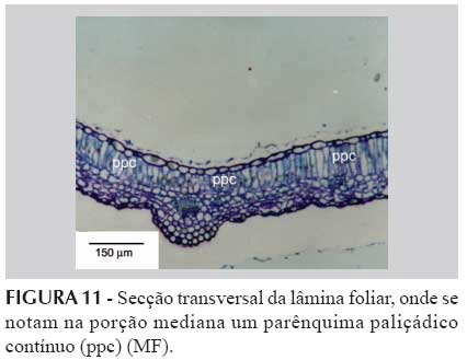
This work has focused on the structural organization analysis of Cunila microcephala leaves in photonic microscopic and electronic scanning. Conventional techniques have been used to prepare the material for obtaining semi-permanent plates. For the preparation of permanent plates, the immersion into glycol methacrylate (GMA) has been performed. The structural organization of leaves belonging to this species reveals stomata in both faces with predominance in the abaxial face (amphihypostomatic leaf). The adaxial face stomata belongs to the diacytic type. The anticlinal walls of the epidermis cells of the adaxial face are sinuous and present irregular thickening. The epidermis of both faces is single layered. Uniserial tectorial trichomes and single celled and multicelled capitate glandular trichomes are present in both faces. The mesophyll is heterogeneous dorsi-ventral. Idioblasts containing inulin crystals are present throughout the foliar plate. The central veins in cross-section, in the medium portion of the foliar plate, have shown a very simple organization, not an outstanding one, where the chlorophyllian palisade parenchyma presents a solution of continuity. The vascular fagots are collateral.
Cunila microcephala; Foliar anatomy; Glandular trichome; Inulin; Lamiaceae