Fig. 1
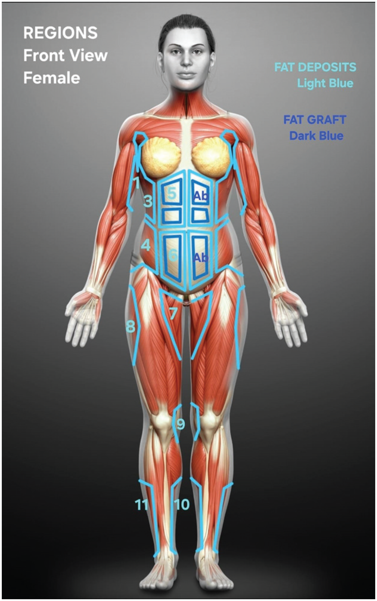
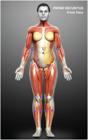
Female model, frontal view. Predefined fat deposition regions: arms (1), upper back (3), lower back (4), upper abdomen (5), lower abdomen (6), inner thighs (7), trochanteric region (8), knees (9), medial legs (10), and lateral legs (11). The figure also shows the predefined regions for fat grafting in the abdomen (Ab).
Fig. 2
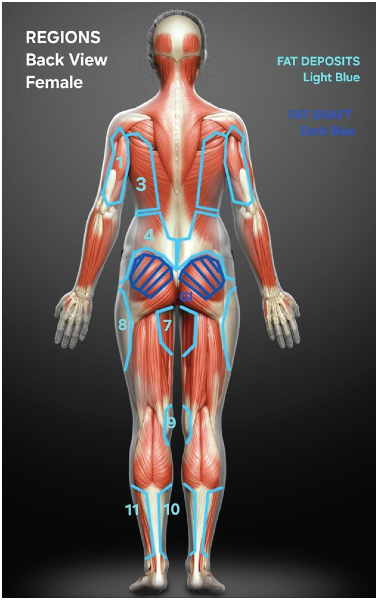
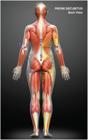
Female model, back view. Predefined fat deposition regions: upper back (3), lower Back (4), inner thighs (7), trochanteric region (8), knees (9), medial legs (10), and lateral legs (11). The figure also shows the predefined regions for fat grafting in the gluteus (Gl).
Fig. 3
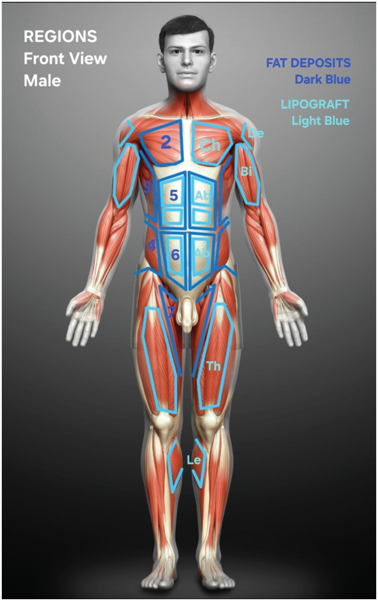
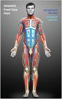
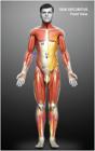
Male model, frontal view. Predefined fat deposition regions: pectoral (2), upper back (3), lower back (4), upper abdomen (5), lower abdomen (6), and inner thighs (7). The figure also shows the predefined regions for fat grafting in the abdomen (Ab), deltoids (De), biceps (Bi), chest (Ch), and thighs (Th).
Fig. 4
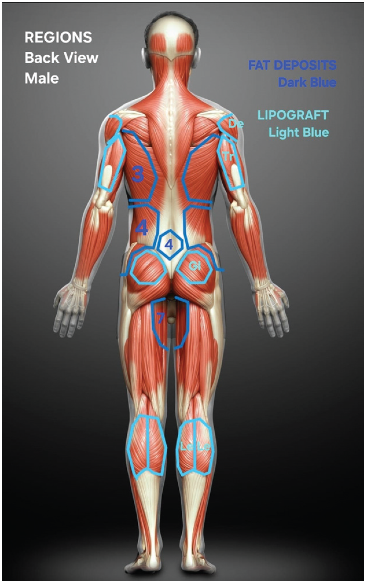
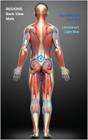
Male model, back view. Predefined fat deposition regions: upper back (3), lower back (4), and inner thighs (7). The figure also shows the predefined regions for fat grafting in the deltoids (De), triceps (Tr), and gluteus (Gl).
Fig. 5
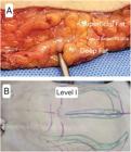
Liposuction depth levels. Level I. (A) Abdominal flap removed at abdominoplasty, with the liposuction cannula at level I, below the superficial fascia and above the muscular fascia. (B) Liposuction cannula under the superficial fascia and above the muscular fascia (Level I) in the abdominal region.
Fig. 6
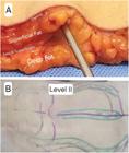
Liposuction depth levels. Level II. (A) Abdominal flap removed at abdominoplasty, with liposuction cannula at level II, above the superficial fascia and below the dermis. (B) Liposuction cannula below the dermis and above the superficial fascia (level II) in the abdominal region.
Fig. 7
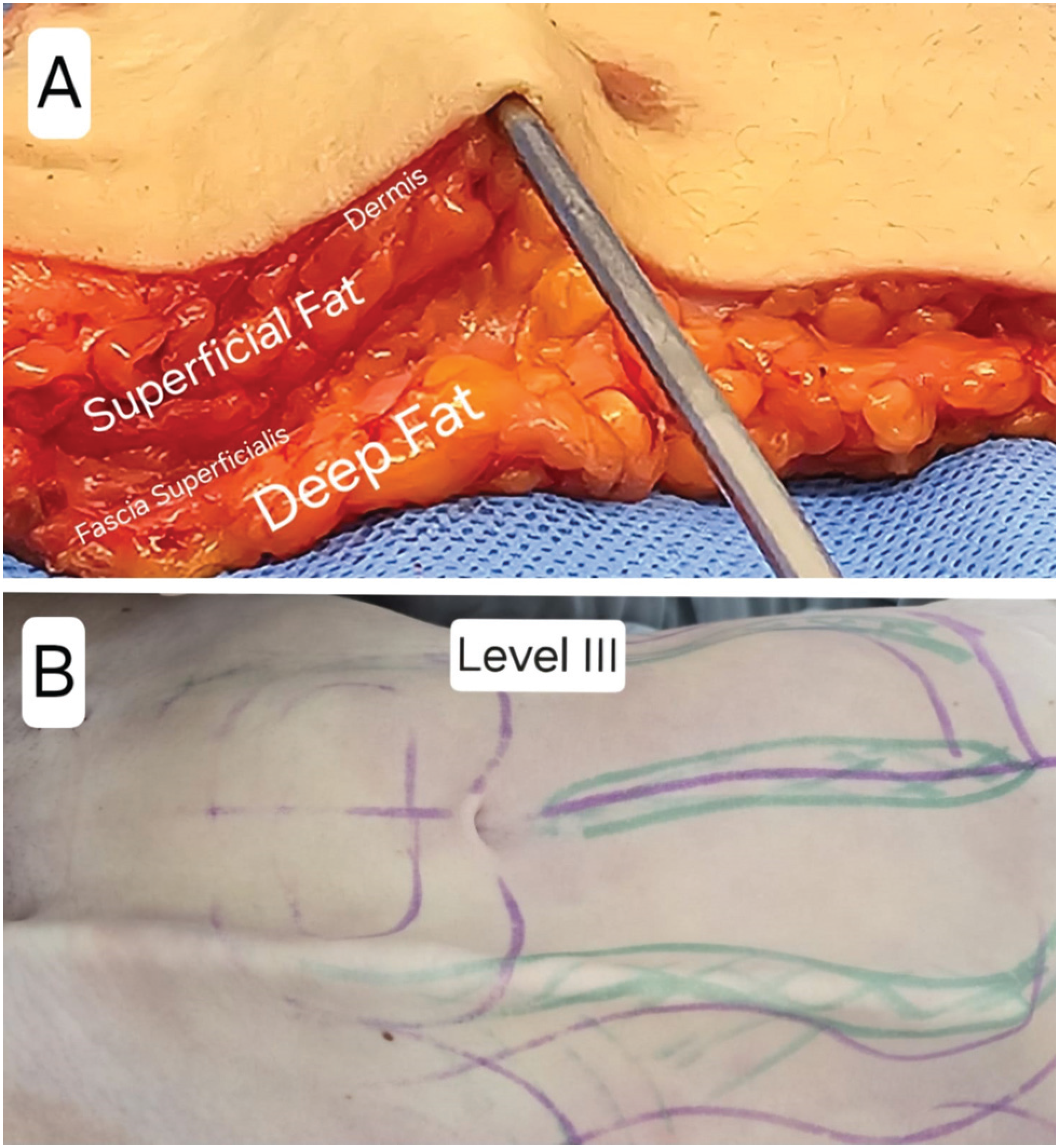
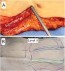
Liposuction depth levels. Level III. (A) Abdominal flap removed at abdominoplasty, with liposuction cannula at level III, just below the dermis, with the cannula holes facing downwards. (B) Juxtadermal liposuction cannula (level III) in the abdominal region.
Fig. 8
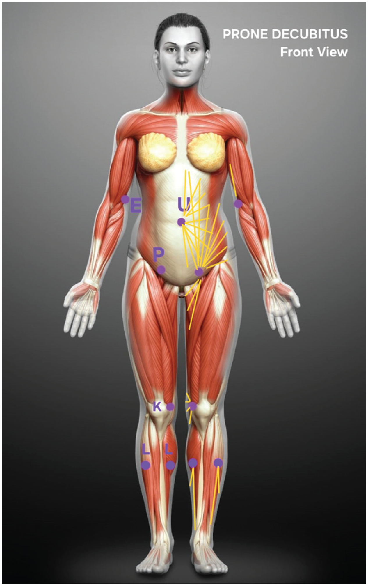
Female model, frontal view. Access portals and cannula extension (in yellow) for preferred liposuction in prone decubitus: E (elbow), U (umbilicus), P (pubis), K (knees), and L (legs).
Fig. 9
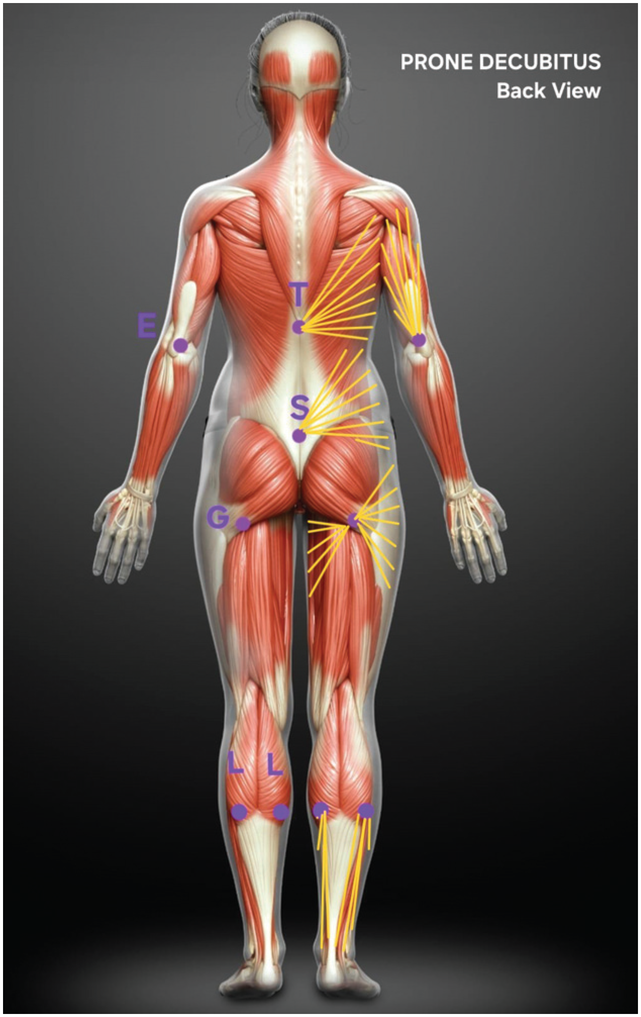
Female model, back view. Access portals and cannula extension (yellow) for preferred liposuction in prone decubitus: E (elbow), T (thoracic back), S (sacral), G (gluteal fold), and L (legs).
Fig. 10
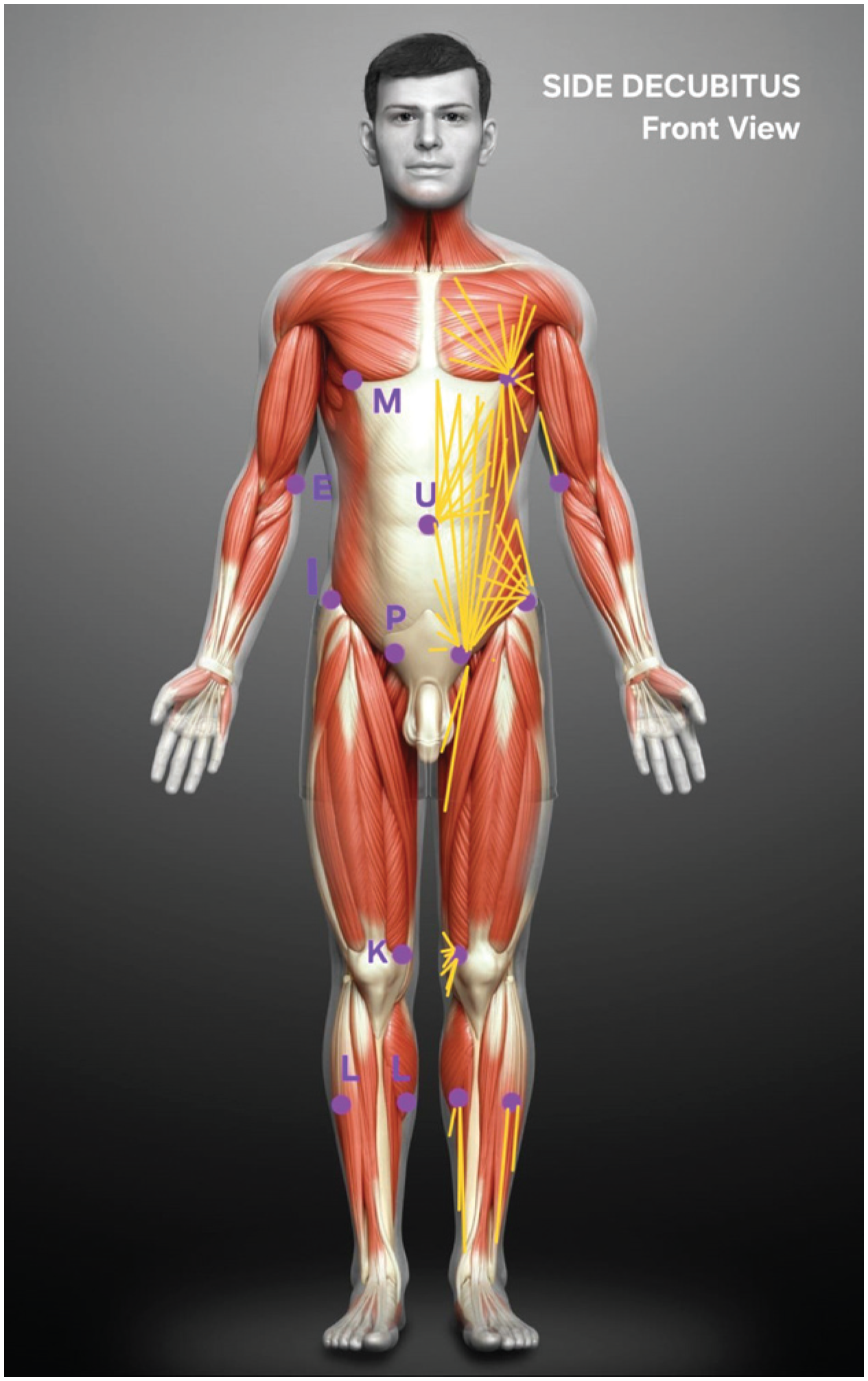
Male model, frontal view. Access portals and cannula extension (yellow) for preferred liposuction in side decubitus: E (elbow), M (mammary groove), U (umbilicus), I (iliac crest), P (pubis), K (knees), and L (legs).
Fig. 11
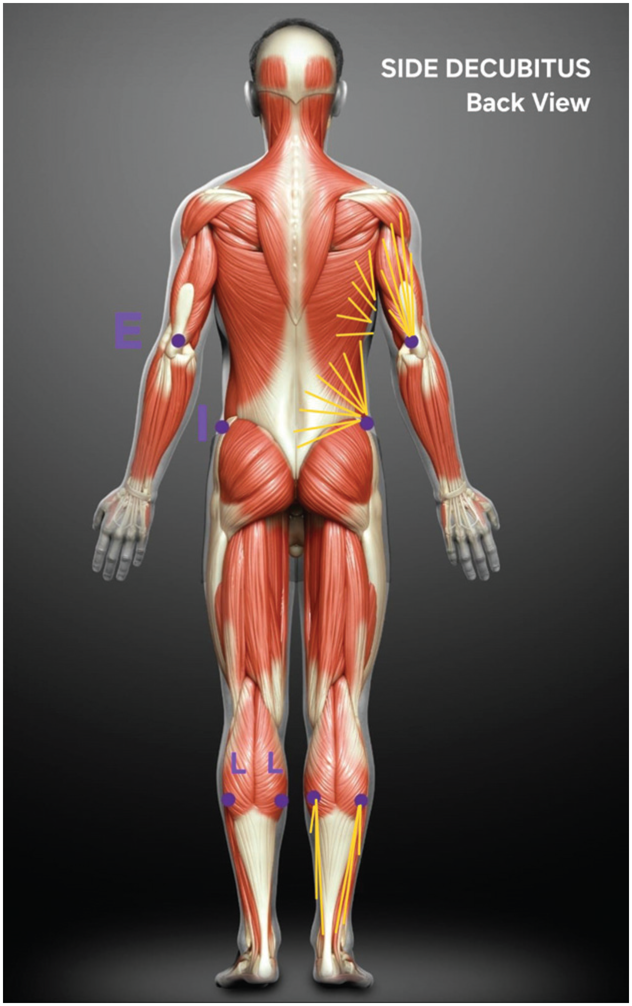
Male model, back view. Access portals and cannula extension (yellow) for preferred liposuction in side decubitus: E (elbow), I (iliac crest), and L (legs).
Fig. 12
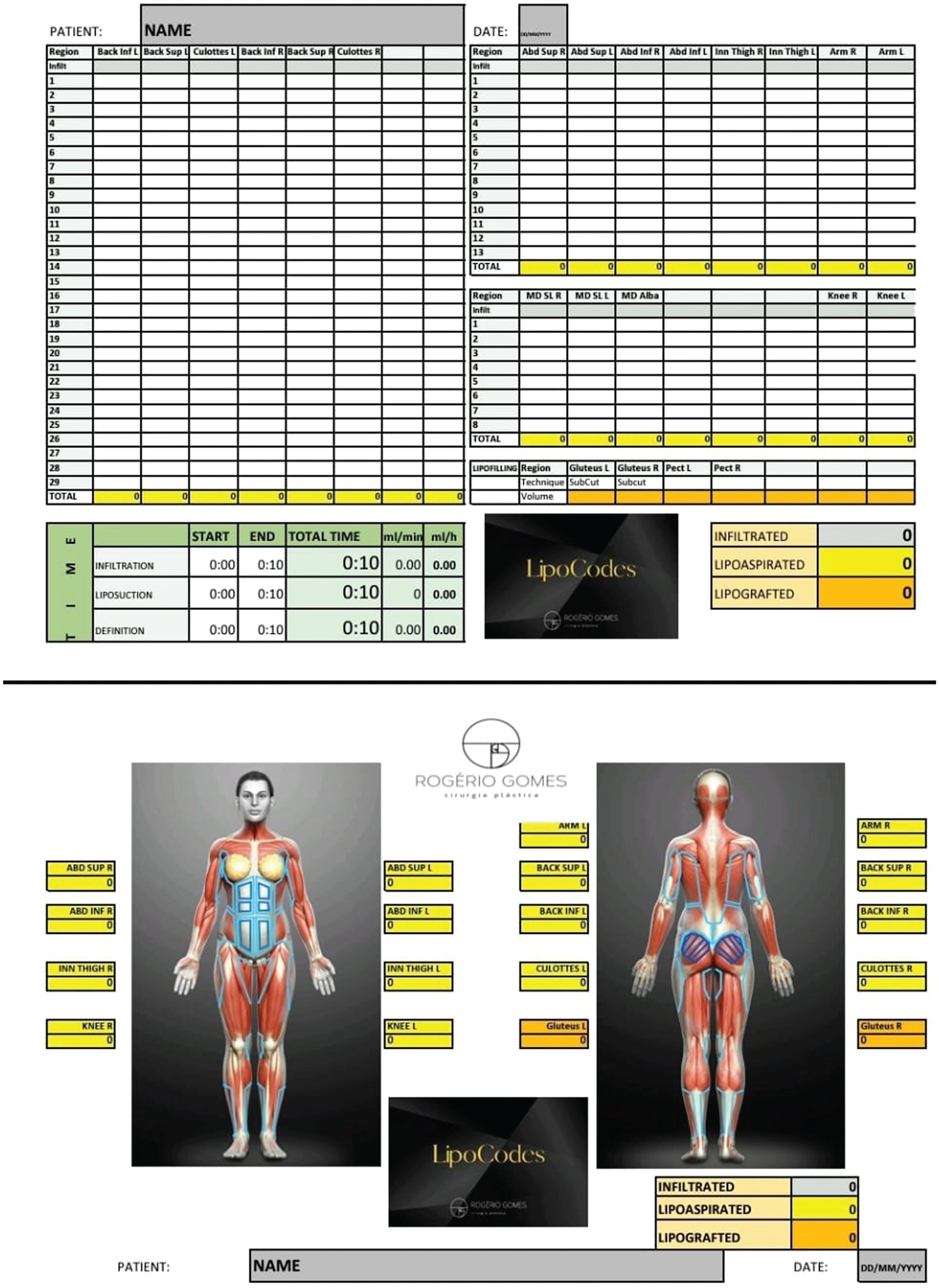
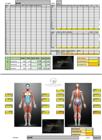
Spreadsheet for female patients. The first page has columns for each predefined aspiration and grafting region. The first line in each column, in gray, records the infiltrated volume in each region. Other lines (in white) record the aspirated and grafted volumes obtained with the intermediate device. The yellow line below each column is the total liposuction or grafting volume for each predefined region. Below and to the right, the total infiltrated, liposuctioned, and grafted volumes are calculated automatically. Below and to the left, the surgical times are recorded, including the start and end times of infiltration, aspiration, and definition liposuction. On the second page, developed to facilitate patient understanding, two female models, in front and back views, highlight the predefined aspiration and grafting regions and, next to them, the total volumes of each region at the corresponding levels. The total volumes (infiltration, aspiration, and grafting) are also at the bottom of this page.
Fig. 13
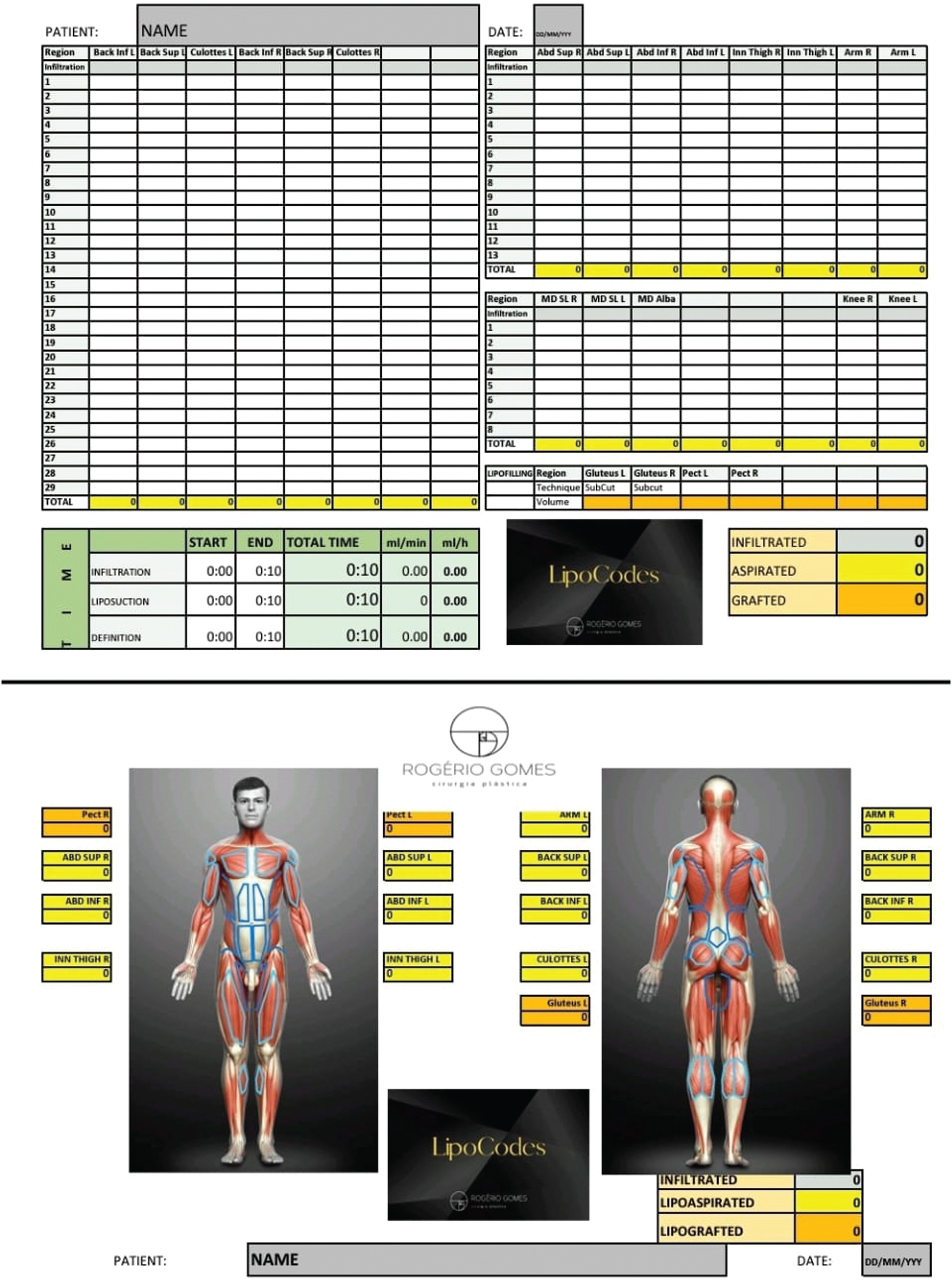
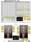
Spreadsheet for male patients. The first page has columns for each predefined aspiration and grafting region. The first line in each column, in gray, records the infiltrated volume in each region. Other lines (in white) record the aspirated and grafted volumes obtained with the intermediate device. The yellow line below each column is the total liposuction or grafting volume for each predefined region. Below and to the right, the total infiltrated, liposuctioned, and grafted volumes are calculated automatically. Below and to the left, the surgical times are recorded, including the start and end times of infiltration, aspiration, and definition liposuction. On the second page, developed to facilitate patient understanding, two female models, in front and back views, highlight the predefined aspiration and grafting regions and, next to them, the total volumes of each region at the corresponding levels. The total volumes (infiltration, aspiration, and grafting) are also at the bottom of this page.
Fig. 14
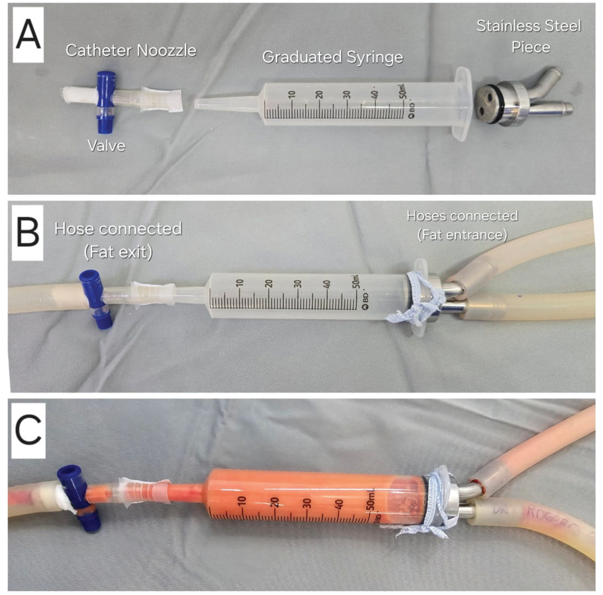
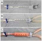
System for accurate counting of aspirated volumes, with three parts: (A) catheter tip obtained from a urine collector, with its opening and closing valve; 50mL syringe with catheter tip, graduated in milliliters, without the plunger (discarded); stainless-steel part with two outlets to connect liposuction hoses: one for liposuction and the other for exhaust. (B) Connected parts ready for use. (C) System in use, with fat in the syringe, before disposal.
Fig. 15
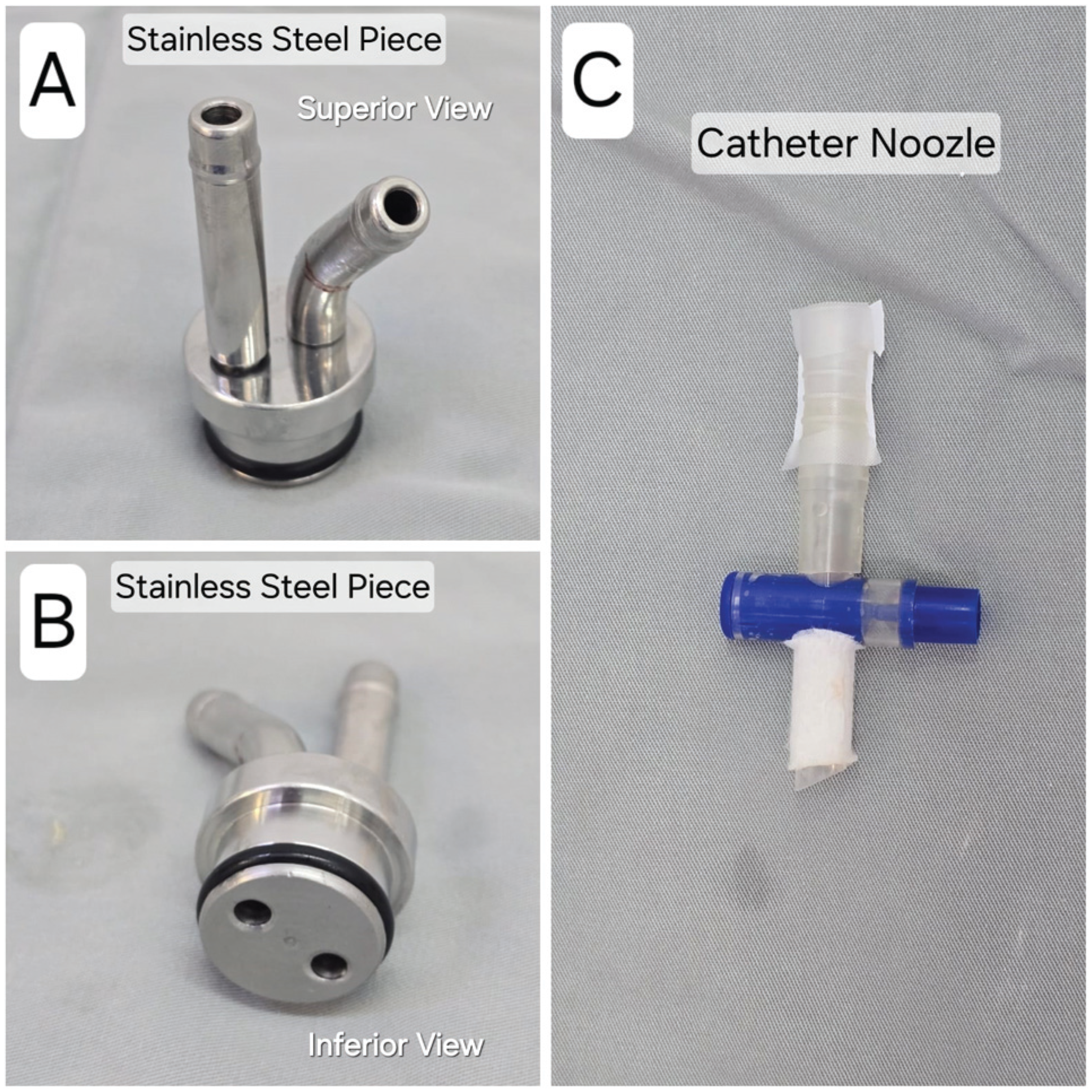
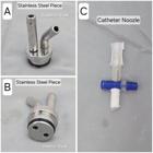
(A) Top view of the stainless-steel part with two outlets for connecting liposuction hoses. (B) Bottom view of the part. C. Catheter tip obtained from a urine collector and its opening and closing valve.
Fig. 12
Spreadsheet for female patients. The first page has columns for each predefined aspiration and grafting region. The first line in each column, in gray, records the infiltrated volume in each region. Other lines (in white) record the aspirated and grafted volumes obtained with the intermediate device. The yellow line below each column is the total liposuction or grafting volume for each predefined region. Below and to the right, the total infiltrated, liposuctioned, and grafted volumes are calculated automatically. Below and to the left, the surgical times are recorded, including the start and end times of infiltration, aspiration, and definition liposuction. On the second page, developed to facilitate patient understanding, two female models, in front and back views, highlight the predefined aspiration and grafting regions and, next to them, the total volumes of each region at the corresponding levels. The total volumes (infiltration, aspiration, and grafting) are also at the bottom of this page.
Fig. 13
Spreadsheet for male patients. The first page has columns for each predefined aspiration and grafting region. The first line in each column, in gray, records the infiltrated volume in each region. Other lines (in white) record the aspirated and grafted volumes obtained with the intermediate device. The yellow line below each column is the total liposuction or grafting volume for each predefined region. Below and to the right, the total infiltrated, liposuctioned, and grafted volumes are calculated automatically. Below and to the left, the surgical times are recorded, including the start and end times of infiltration, aspiration, and definition liposuction. On the second page, developed to facilitate patient understanding, two female models, in front and back views, highlight the predefined aspiration and grafting regions and, next to them, the total volumes of each region at the corresponding levels. The total volumes (infiltration, aspiration, and grafting) are also at the bottom of this page.
Fig. 14
System for accurate counting of aspirated volumes, with three parts: (A) catheter tip obtained from a urine collector, with its opening and closing valve; 50mL syringe with catheter tip, graduated in milliliters, without the plunger (discarded); stainless-steel part with two outlets to connect liposuction hoses: one for liposuction and the other for exhaust. (B) Connected parts ready for use. (C) System in use, with fat in the syringe, before disposal.
Fig. 15
(A) Top view of the stainless-steel part with two outlets for connecting liposuction hoses. (B) Bottom view of the part. C. Catheter tip obtained from a urine collector and its opening and closing valve.
 LipoCodes Methodology: Objective Control of Infiltrated and Aspirated Volumes in Liposuction
 Thumbnail
 Thumbnail
 Thumbnail
 Thumbnail
 Thumbnail
 Thumbnail
 Thumbnail
 Thumbnail
 Thumbnail
 Thumbnail
 Thumbnail
 Thumbnail
 Thumbnail
 Thumbnail
 Thumbnail