Abstract
Lagochile emarginata (Gyllenhal): morphology of immature and imago, and biological records (Coleoptera, Scarabaeidae, Rutelinae). The last larval instar and pupa of Lagochile emarginata are described. Pupa of the genus Lagochile Hoffmannsegg, 1817 is described for the first time. Redescription of the imago, clarifications on the morphology of immature Scarabaeoidea and biological notes are presented.
Dipropus; Insecta; Neotropical; Rutelini; Tithonia; white grub
SYSTEMATICS, MORPHOLOGY AND BIOGEOGRAPHY
Lagochile emarginata (Gyllenhal): morphology of immature and imago, and biological records (Coleoptera, Scarabaeidae, Rutelinae)
Fabiano F. AlbertoniI; Juares FuhrmannI; Sergio IdeII
IMuseu de Zoologia da Universidade de São Paulo, 04218970 São Paulo-SP, Brazil, fabianoalbertoni@gmail.com; jufuhrmann@gmail.com
IILaboratório de Entomologia Geral, Centro de Pesquisa e Desenvolvimento de Sanidade Vegetal, Instituto Biológico, 04014900 São Paulo-SP, Brazil, ide@biologico.sp.gov.br
ABSTRACT
Lagochile emarginata (Gyllenhal): morphology of immature and imago, and biological records (Coleoptera, Scarabaeidae, Rutelinae). The last larval instar and pupa of Lagochile emarginata are described. Pupa of the genus Lagochile Hoffmannsegg, 1817 is described for the first time. Redescription of the imago, clarifications on the morphology of immature Scarabaeoidea and biological notes are presented.
Keywords:Dipropus; Insecta; Neotropical; Rutelini; Tithonia; white grub.
The first taxonomic work on immatures of Neotropical Rutelini was made by Ritcher (1948; see also Ritcher 1966). After his work many authors described immatures of Rutelini, resulting in 23 genera and 34 species with described larvae, but only 6 genera and 11 species with described pupae (Table I).
The genus Lagochile Hoffmannsegg, 1817 (Scarabaeidae, Rutelinae, Rutelini) comprises 60 species distributed from Mexico to Argentina (Soula 2005). Larvae of Lagochile collaris (Blanchard, 1835) has been previously described by Jameson & Morón (2001) (as Chasmodia collaris), but the pupae of the genus remained unknown until this paper.
Larvae of Lagochile were found associated with roots and rotten woods and imagoes were reported as feeding on different kinds of fruits (Ohaus 1909; Soula 2005). Nevertheless, despite the significant amount of works on the biology of Rutelinae with economic importance, the natural history of species of Rutelinae is scarce (Ritcher 1958).
The purpose of this article is to describe the pupa and last larval instar of Lagochile emarginata (Gyllenhal, 1817). Redescription of imago and morphological notes are also made available, in order to contribute to a better comprehension of the adult morphology, adding information to the descriptions from the most recent taxonomic review by Soula (2005).
MATERIAL AND METHODS
Larvae of L. emarginata were collected in Florianópolis (Ilha de Santa Catarina), state of Santa Catarina, southern Brazil, in restinga, characterized by edaphic vegetation typical of sand dunes from lowland shore (CECCA 1997). Two larvae were collected in "Santinho", 27º47'67"S, 48º39'00"W, in a decaying trunk in a forested section of the restinga's mosaic. Four additional larvae were collected at the campus of the Universidade Federal de Santa Catarina (UFSC), 27º36'02"S, 48º31'25"W, in the roots and base of the trunk of a plant of Tithonia diversifolia (Hemsley) A. Gray (Asteraceae; margaridão). The larvae were reared in covered pots with the same rooted wood in which they had been collected and earth as substrate. The two studied pupae and two imagoes were obtained through the rearing of larvae from both places.
Larvae of Dipropus brasilianus (Germar, 1824) (Elateridae) (larva described by Costa (1977), and redescribed by Casari & Biffi (2012)) were collected associated with dead larvae of L. emarginata. The larvae were reared with fresh dead moths (Lepidoptera) and larvae of Tenebrio molitor Linnaeus, 1758 (Tenebrionidae).
Fourteen specimens from the Coleção Entomológica Adolph Hempel, Instituto Biológico, São Paulo, state of São Paulo, Brazil (IBSP) were used for the characterization of the imago. Internal morphology, mouthparts, wing, and terminalia were studied using dissected specimens by relaxing the parts in hot water. Dissected specimens and detached parts were card mounted with acid-free water-soluble glue.
The specimens were examined using a Carl Zeiss Stemi SV6 stereomicroscope and a Carl Zeiss Axioskop microscope. Illustrations were produced using a camera lucida attached to both microscopes. Photographs were taken with a Canon EOS Rebel XTi digital camera with a Canon 100mm macro lens and processed using Helicon Focus 4.2.1 software (www.heliconsoft.com/heliconfocus.html).
The terminology follows Browne and Scholtz (1994), Kukalová-Peck and Lawrence (1993, 2004) (wing), Krell (1996) (adult terminalia), and Snodgrass (1993) (general morphology). Plant names follow IPNI (2005). The immature specimens of L. emarginata and immature and adults of D. brasiliensis are housed in the Museu de Zoologia da Universidade de São Paulo, São Paulo, state of São Paulo, Brazil (MZSP, accession number: MZSP 010.254, MZSP 010.255).
RESULTS
Lagochile emarginata (Gyllenhal, 1817)
Last larval instar (Figs. 121)
Description. Body (Fig. 1) yellowish white, cranium and respiratory plates reddish brown, clypeus, labrum and mandibles dark.
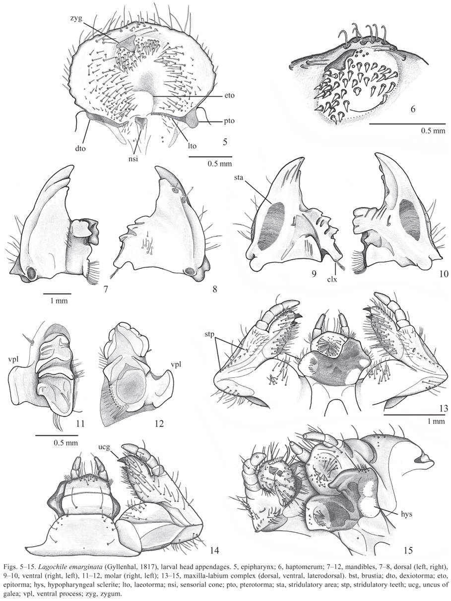
Head (Figs. 24). Epicranial and epistomal sutures present. Anterolateral stemmata present. Vertex with sparse and small punctures near frontal suture and large punctures near stemmata and on posterior area. Frons densely punctate, punctures large and irregular. Each side of epicranium with 2 dorsoepicranial setae (des), 2 lateroepicranial setae (les), 1 anteroepicranial seta (aes), 3 externoepicranial setae (ees), ventroepicranial setae irregularly distributed, 2 posterofrontal setae (pfs), 2 externofrontal setae (efs). Clypeus and labrum rugose punctate; each side with 2 lateroclypeal setae (lcs), 910 laterolabral setae (lls), and 1 mediolabral seta (mls). Epipharynx (Fig. 5) wider than long. Corypha small, with 6 setae; clithra absent. Haptomerum (Fig. 6) prominent with 38 spine-like setae and 6 sensillae; zygum beak-like; epizugum and heli absent. Acanthoparia with 7 short setae; plegmatia absent. Chaetoparia with sparse sensillae, right side with 48 setae and left with 66 setae; phobae absent. Pedium smooth and wide. Laeotorma sinuous; epitorma narrow and depressed; dexiotorma narrow and slightly curved backward; pterotorma rounded and fused to laeotorma. Haptolachus with sparse setae; crepis medially interrupted; nesium internum (sensorial cone) prominent; nesium externum (sclerotized plates) indistinct. Right mandible (Figs. 8, 9, 11) with 2 anterodorsal setae, 7 dorsal lateromolar setae, and 5 ventral brush-like setae; incisor with 2 teeth; molar (Fig. 11) without acia, brustia formed by 4 setae. Left mandible (Figs. 7, 10, 12) with similar chaetotaxy to right one, but dorsal lateromolar setae in a row; incisor with 3 teeth; molar (Fig. 12) without acia; brustia formed by 13 setae. Ventral stridulatory area of both mandibles transversally strigulate. Maxillae (Figs. 1315) symmetrical; stridulatory area with anterior tubercle and row of 68 teeth; mala with a weak separation between galea and lacinia, galea with 1 uncus, lacinia with longitudinal row of 6 spine-like setae and 1 uncus with a minute seta; palpi with 4 palpomeres, I with external seta, III with external and ventral setae. Labium (Figs. 1315): submentum with 5 short setae; mentum with 2 long setae; prementum with long setae on disc and short setae on anterior angles; palpi with 2 palpomeres, II with distal seta. Antennae glabrous, with 4 antennomeres, IV with 6 dorsal spots.
Thorax. Pronotum with 2 dorsal lobes and lateral sclerite; lobes with transversal row of long setae. Distance between the arms of the respiratory plate (Fig. 16) shorter than transversal diameter of bulla. Meso- and metathorax with 3 dorsal lobes, each of them with rows of setae similar to the pronotum. Claws of pro- and mesotarsunguli acuminated and with externodistal and internoproximal setae; claw of metatarsungulus reduced with ventral tooth-like seta.
Abdomen. Segments IVIII with spiracles (Figs. 1720); IIV with 3 dorsal lobes, each of them bearing tooth-like setae and transversal row of long setae; VIIIX without toothlike setae, with 2 rows of dorsal setae; X with sparse long setae and abundant tooth-like setae. Raster (Fig. 21): campus with 24 long setae; barbula formed by 8 long setae on each side; septula poorly defined, more distinct on ventral anal lobe; palidia formed by 1820 small tooth-like setae; tegillum formed by short setae. Anal opening transverse.
Remarks. Larvae of L. emarginata can be distinguished from those of L. collaris (the other species in the genus with described larva) by the punctures on the vertex, in the latter they are large and dense and in the former they are small and restricted to the area near the epicranial suture and posterior edge of vertex.
Male pupa (Figs. 2224)
Description. Total length: 34 mm, maximal width: 18.8 mm. Body (Figs. 2223) yellowish white, spiracles and dioneiform organs dark brown. Integument smooth, apparently glabrous but covered with a thin and short pubescence, which gives a velvety aspect (50x of magnification).
Head (Fig. 24). Vertex dorsally visible. Epistomal suture incomplete medially. Clypeus and labrum rectangular. Mandibles, malae, maxillary and labial palpi tubercle-like. Labium convex. Antennae triangular.
Thorax. Pronotum trapezoid, lateral margin rounded, posterior margin sinuous. Spiracle present in cavity formed between the anterior and medial legs and the hypomeron. Scutellum parabolic, as long as pronotum. Metanotum posteriorly projected. Metaventrite wide, anteriorly projected between pro- and mesocoxae. Medial legs rested on the anterior margin of the elytral thecae. Posterior legs with contiguous coxae, femora incompletely hidden by the posterior pterothecae.
Abdomen. Spiracles IIV oval and with sclerotized ring, I concealed by elytral thecae, spiracle VVIII represented by cuticular invagination. Segment I medially constricted and represented only by the tergite. Tergite II with anterior margin sclerotized, not forming dioneiform organs. Dioneiform organs present between segments IIIII, IIIIV, IVV, VVI. Tergite IX with large ventral lobes, lobes distally setose. Sternite IX small, with rounded genital ampulla.
Remarks. Morón (1993) characterized the pupae of Rutelini as having 54 pairs of dioneiform organs; spiracles IIV with dark scleroma and VVIII conspicuous but closed; urogomphi absent; abdomen without latero-dorsal tubercles; urotergites VIIVIII separated. The pupae of Lagochile are very similar to those of Macraspis MacLeay, 1819 (refer to Table I for known immature of Macraspis) and the pupae of both genus can be distinguished from the known pupae of other Rutelini (Paraheterosternus Moron, 1983, Rutela Latreille, 1902 and Rutelisca Bates, 1888) by the scutellum, at least as long as the pronotum and by possessing only 4 well-developed dioneiform organs. The pupae of Macraspis have the scutellum longer than pronotum and its apex extended beyond the base of urotergite I; the pupa of Lagochile have the scutellum as long as the pronotum and with its apex not extended to urotergite I.
Imago (Fig. 2575)

Redescription.Body oval (Figs. 2526, 7375). Length: 17.020.0 mm; maximal width on middle of elytra: 12.013.5 mm. Color: metallic green, eventually with red or blue (rare) reflexes.
Head (Figs. 2730) with sparse punctures. Epicranial suture absent and epistomal suture medially interrupted. Tentorium (Figs. 30, 31) with anterior arm disconnected to tentorial body; dorsal arm reduced; laminatentorium with 4 spine-like processes; tentorial bridge thin and simple. Clypeus deeply sinuated and anteriorly bordered. Labrum thick and sparsely punctured, medially divided by a large groove; epipharynx (Fig. 32) densely setose, prominent and posteriorly margined by tormae. Mandibles (Figs. 3335) with prostheca greatly developed; incisor rounded, sometimes truncate; molar small, striate. Maxillae (Figs. 3638): lacinia toothed; galea 2toothed, dorsal face densely setose; palpi with 4 palpomeres, III with peripheral seta. Labium (Figs. 29, 39) with prementum truncate and fused with mentum; mentum longer than wide, medially grooved; submentum short and fused to gula; ligula bilobed and densely setose. Hypopharynx (Figs. 29, 39) prominent, densely setose. Antennae (Fig. 40) with 10 antennomeres, clava as long as the remaining antennomeres combined.
Pronotum (Figs. 4142) glabrous, with abundant small punctures; anterior and lateral margin bordered, anterior margin medially interrupted; lateral scars slightly distinct; hypomeron rough, setose, posterior contact between hypomeron-sternum narrow. Prosternal posterior process tubercle-like. Furca short, sinuate, distally enlarged. Anterior legs (Figs. 43, 46): Tibia 3-thoothed; posterior side with longitudinal carina; spur well developed. Tarsus shorter than tibia. Male with internal claw large, bifurcate.
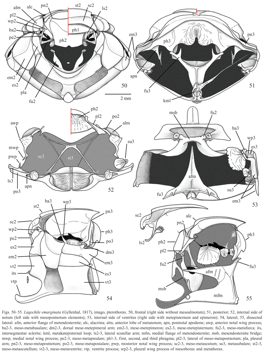
Mesothorax (Figs. 5057). Scutellum sculpture similar to that of pronotum, parabolic, longer than wide, as long as pronotum; anterior margin abruptly rounded, not bordered. Episternum smaller than epimeron, not reaching the mesocoxal cavity. Elytra smooth, with thin and sparse punctures, external stria punctate; humerus with anterior sulcus; without external membrane. Mid legs (Figs. 44, 47): Tibia cylindrical, external side with longitudinal grooves, apex with short tooth and toothlike setae; with 2 internal and contiguous spurs. Tarsus shorter than tibia, smooth, bearing distal tooth-like setae. Male with simple claws and female with bifid internal claw.
Metathorax (Figs. 5056). Episternum not reaching the mesocoxal cavity, dorsal area concealed by the mesemetepimeron contact. Metaventrite wide, densely punctate and setose; discrimen evident, posterior process short and rounded not separating the metacoxae; meso-metaventrite process prominent, free, extended between the procoxae. Metendosternite with anterior flange acuminated. Wing (Figs. 5859) with RA4+RP1 reaching RA3 distally, MP3 indistinct, MP4+CuA1 convergent to Cu. Posterior legs (Figs. 55, 58, 59). Coxae contiguous, coxa-trochanter articulation with short and sharp process.
Abdomen (Figs. 6062). Propygidium finely punctate with short setae; proximal half covered by resting elytra, separation of propygidiumventrites V indicated by depression and incomplete suture. Pygidium triangular, striated. Ventrites IV with transversal row of bristly setae; medial area smooth, laterals striated; VI striated, sparsely setose, male with a medial sinuosity that expose partially the third connective membrane (intersegmentar membrane VIIIIX). Male terminalia (Figs. 6369): Genital ring (Fig. 63, 64) of male with long, narrow and Y-shaped spiculum gastrale, sternite IX divided in 2 pieces, each piece with long setae. Aedeagus (Figs. 6567) with phalobasis as wide as long, with well developed apodeme; parameres fused, apex with short truncated process. Endophalus (Figs. 6869) with temona forming a large Yshaped structure with ventral folded arms, distal V-shaped sclerite present; raspulae (areas with basiconic sensillae; see Coca-Abia & Martín-Piera 1991) well developed on left ventral side; apex with left lobe. Female terminalia (Figs. 7072): Proximal gonocoxites superposed to distal ones. Large depression between vagina and anus present. Paraprocts triangular, large, posteriorly articulated with proctiger. Internal genitalia (Fig. 72) with 4 accessory glands; spermatheca and its gland thin and elongated; bursa copulatrix greatly enlarged.
DISCUSSION
Morphological notes
A special dilemma with the larval morphology is the interpretation of two structures of the haptomerum: helus (heli) and zygum. These structures are termed by Böving (1936) as: "Helus (-i) (Greek; from helos, a nail or pointed peg): A coarse, fixed spine without cup; belonging to region haptomerum. (Hayes: "spine")" and "Zygum (-a) (Greek; from zygon, a yoke or cross-bar): Sclerome pertaining to region haptomerum and forming its anterior margin. When typically developed, appearing as a convex cross-bar in front of sensilla and heli, but often enlarged and carrying these structures". Thus, helus sensu Böving (1936) define a haptomeral spine, if we stay in agreement with spine sensu Snodgrass (1993): "a cuticular outgrowth of the body wall formed by epidermal cells, solidly fixed to the surrounding cuticula and immovable".
When Ritcher (1948) studied an epipharynx of Rutelini larvae for first time he made the following description: "Haptomerum with a beak-like process behind which is a group of about 30 spine-like setae. Heli absent. Epizygum and zygum absent". Ritcher (1948), in agreement with Böving (1936), did not consider the spine-like setae of the posterior area of haptomerum as heli. On the other hand, disagreeing with Böving (1936), he did not consider the bike-like structure as zygum.
In addition to Ritcher (1948), other authors (e.g., Micó et al. 2001; Table I) described Rutelini larvae and considered the spine-like setae of haptomerum as heli, and kept it maintained in concordance with Ritcher (1948) about the absence of zygum, while other works maintain Böving's (1936) definition of heli (e.g., Micó et al. 2008).
We prefer to be in strict accordance with Böving (1936) and apply helus as a concept of spine of haptomerum, and zygum as the anterior sclerome of the same region. In fact, zygum can have intermediary forms between a thin long cross-bar and a beak-like structure, or another form with helus or sensilla incorporated in it (see fig. 1 in Böving (1936); fig. 14 and 15 in Ritcher (1948); fig. 10 in Micó et al. (2001) for aberrant zygum with sensillae and fig. 22 for zygum absent, and fig. 2 in Neita-Moreno & Morón (2008) for large and bidentate zygum: "proceso haptomeral prominente y bidentado"). According to this view all Rutelini larvae lack heli and some possess a beak-like zygum.
Biological notes
Life cycle. The larvae of Lagochile emarginata were found in roots of Tithonia diversifolia, which is an exotic species (Souza & Lorenzzi 2005), and within a decaying tree trunk in a restinga site. Ritcher (1966) refers to the larvae of some species of Rutelinae as feeding on the roots of different plant species, or on decaying wood, or in the soil in the vicinity of decaying wood. Following Ritcher (1958) the studied species belongs to the Sacarabaeidae group of decaying wood and living root feeders.
One observed larva showed an interruption of feeding to construct the pupal chamber, with compacted soil walls; 21 days after the larva had started to build the pupal chamber the pupa emerged, staying inside the open larval exuvia. The pupal instar lasted 24 days.
In addition, one imago was collected by D. C. Bená and J. Fuhrmann in the area of Instituto Biológico, São Paulo, state of São Paulo, in March 13th, 2009. It was eating pieces of a ripe fruit of Artocarpus heterophyllus Lam. (Moraceae, jaqueira, jackfruit tree) on the ground.
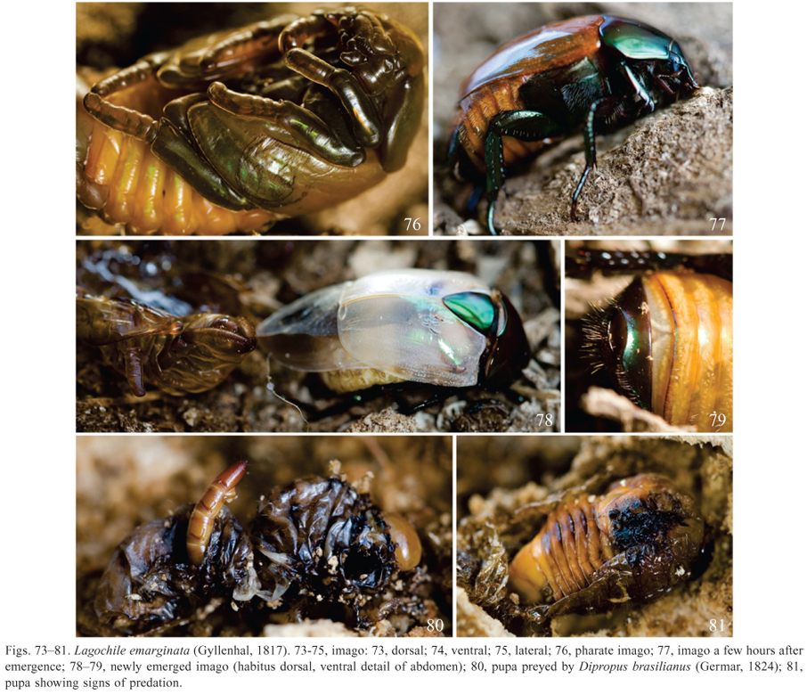
Development (Figs. 7681). The imago of L. emarginata has metallic color tending to green. The color is visible yet on the pharate imago, even on the elytra and ventrites IIV which were milky white when it just emerged (Fig. 7678).
The imago emerged with head, thorax, scutellum, legs, pygidium and ventrite V completely sclerotized and pigmented, being just a slightly softer than it will be (Figs. 7778). As soon as the beetle emerged, the elytra and the membranous wings were extended backward acquiring the shape expected for a mature beetle's wings. The membranous wings grew stretched (Fig. 78) and the folding under the elytra happened about 6 hours after it has emerged (Fig. 77).
Associated species. Larvae of Dipropus brasilianus were collected feeding on dead larvae of L. emarginata (Figs. 80) and in the compressed material left by the ruteline in the tunnel it made through the wood. Among this compressed material approximately 15 larvae of Elateridae were found. A ruteline larva was found dead in the tree trunk with some larvae of D. brasilianus inside and next to it. A pupa of L. emarginata was found with marks of predation (Fig. 81) and a larva of D. brasilianus was seen nearby. Also another dead ruteline larva was found decomposing into the same trunk. Here we report larvae of D. brasilianus preying on larvae of L. emarginata and wonder whether there is any role of the compressed material left by the saproxylophagous ruteline larvae in this interaction.
Ritcher (1966) points Elateridae as one of the insects reported as predators of Scarabaeidae, but no Rutelinae species has been previously reported as prey. Other beetles pointed as predators are adults Carabidae, which prey on larvae and imagoes of scarabs, and larvae and imagoes of Histeridae, that prey on larvae of Scarabaeidae. Some larvae of Elateridae are known to be predators or scavengers, but with no indication of specificity (Costa et al. 1988).
ACKNOWLEDGMENTS
We would like to thank Sônia A. Casari (MZUSP) for the Elateridae identification; André G. Martins, Daniela C. Bená and Mônica A. Ulysséa for helping on field work. This study was supported by grants from Fundação de Amparo à Pesquisa do Estado de São Paulo (FAPESP) (2012/024411 to FFA and 2011/200016 to JF)
Received 11 September 2013;
Accepted 13 December 2013
Associate Editor: Marcela L. Monné
- Böving, A.G. 1936. Description of the larva of Plectris aliena Chapin and explanation of new terms applied to the epipharynx and raster. Proceedings of the Entomological Society of Washington 38: 169185.
- Browne, D.J. & Scholtz, C.H. 1994. The morphology and terminology of the windwing articulation and wing base of the Coleoptera, with specific reference to the Scarabaeoidea. Systematic Entomology 19: 133143.
- Casari, S.A. & Biffi, G. 2012. Immatures of Dicrepidius Eschscholtz, 1829 and Dipropus Germar, 1839 (Elateridae, Elaterinae, Ampedini, Dicrepidiina). Zootaxa 3587: 6577.
- CECCA Centro de Estudos Cultura e Cidadania. 1997. Unidades de conservação e áreas protegidas da Ilha de Santa Catarina: caracterização e legislação Florianopolis, Editora Insular, 158 p.
- Coca-Abia, M. & Martín-Piera, F. 1991. Anatomy and Morphology of the genitalia in the subtribe Rhizotrogina (Col., Melolonthidae, Melolonthini): Taxonomic implications, p. 6178. In: Zunino, M., Blas, M. & Belles, X. (Eds.). Advances in Coleopterology Barcelona, European Association of Coleopterology, 323 p.
- Costa, C. 1977. Studies on Elateridae (Coleoptera). Biological notes on neotropical larvae. Papéis Avulsos de Zoologia 31: 718.
- Costa, C., Vanin, S.A. & Casari-Chen, S.A. 1988. Larvas de Coleoptera do Brasil São Paulo, Museu de Zoologia, Universidade de São Paulo, 282 p.
- Dam, M.V. & Dam, A.V. 2006. Description of the larva of Pseudocotalpa sonorica Hardy (Scarabaeidae: Rutelinae, Rutelini) with notes on life history. The Coleopterists Bulletin 60: 3136.
- INPI. 2005. The International Plant Names Index Available at: http://www.ipni.org/ (accessed 9 May 2011).
- Jameson, M.L. & Morón, M.A. 2001. Descriptions of the larvae of Chlorota cincicollis Blanchard and Chasmodia collaris (Blanchard) (Scarabaeidae: Rutelinae: Rutelini) with a key to the larvae of the American genera of Rutelini. The Coleopterists Bulletin 55: 385396.
- Jameson, M.L. 1996. Revision and Phylogeny of the Neotropical genus Cnemida (Coleoptera: Scarabaeidae: Rutelinae). Insecta Mundi 10: 285315.
- Jameson, M.L. 1999. Phylogenetic analysis of the subtribe Rutelina and revisions of the Rutela generic groups (Coleoptera: Scarabaeidae: Rutelini). Bulletin of the University of Nebraska State Museum 14: l184.
- Jameson, M.L., Ratcliffe, B.C. & Morón, M.A. 1994. A Synopsis of the Neotropical Genus Calomacraspis Bates with a Key to Larvae of the American Genera of Rutelini (Coleoptera: Scarabaeidae: Rutelinae). Annals of the Entomological Society of America 87: 4358.
- Krell, F.T. 1996. Die Kopulationsorgane des Maikäfers Melolontha melolontha (Insecta: Coleoptera: Scarabaeidae). Ein Beitrag zur vergleichenden und funktionellen Anatomie der ektodermalen Genitalien der Coleoptera. Stuttgarter Beiträge zur Naturkunde, Serie A (Biologie) 537: 1101.
- KukalováPeck, J. & Lawrence, J.F. 1993. Evolution of the hind wing in Coleoptera. Canadian Entomologist 125: 181258.
- KukalováPeck, J. & Lawrence, J.F. 2004. Relationships among coleopteran suborders and major endoneopteran lineages: Evidence from hind wing characters. European Journal of Entomology 101: 95144.
- Micó, E., Hall, W.E. & Ratcliffe, B.C. 2001. Descriptions of the larvae of Hoplopyga singularis (Gory and Percheron) and Hologymnetis cinerea (Gory and Percheron) with a revised key to the larvae of New Word Gymnetini (Coleoptera: Scarabaeidae: Cetoniinae). The Coleopterists Bulletin 55: 205217.
- Micó, E., Morón, M.A., ípek, P. & Galante, E. 2008. Larval morphology enhances phylogenetic reconstruction in Cetoniidae (Coleoptera: Scarabaeoidea) and allows the interpretation of the evolution of larval feeding habits. Systematic Entomology 33: 128144.
- Monné, M.A. 1969. Descripción del ultimo estadio larval de "Macraspis dichroa cribrata" Waterh., "Blaesia atra" Burm. y "Marmarina tigrina" (Gory and Perch.) (Coleoptera, Scarabaeidae). Revista Brasileira de Biologia 29: 367376.
- Morón, M.A. & Deloya, C. 1991. Los coleópteros lamelicornios de la Reserva de la Biosfera "La Michilía", Durango, México. Folia Entomologica Mexicana 81: 209283.
- Morón, M.A. & Nogueira, G. 2000. Third stage and pupa of Paraheterosternus luedeckei (Becker) (Coleoptera: Melolonthidae: Rutelinae). Journal of the Kansas Entomological Society 73: 6267.
- Morón, M.A. & Paucar-Cabrera, A. 2003. Larvae and pupae of species of the genus Macraspis (Coleoptera: Rutelinae: Rutelini). Canadian Entomologist 135: 467491.
- Morón, M.A. 1976a. Descripción de las larvas de tres especies mexicanas de pelidnotinos (Coleoptera; Melolonthidae, Rutelinae) y algunas observaciones sobre su biología. Anales del Instituto de Biología, Serie Zoología 47: 718.
- Morón, M.A. 1976b. Descripción de las larvas de tres especies mexicanas de melolonthinos (Coleoptera; Melolonthidae, Dynastinae y Rutelinae). Anales del Instituto de Biología, Serie Zoología 47: 119134.
- Morón, M.A. 1983. A revision of the subtribe Heterosternina (Coleoptera: Melolonthidae, Rutelinae). Folia Entomologica Mexicana 55: 31101.
- Morón, M.A. 1993. Observaciones comparativas sobre la morfología pupal de los Coleoptera Melolonthidae neotropicales. Giornale Italiano di Entomologia 6: 249255.
- Neita-Moreno, J.C. & Morón, M.A. 2008. Estados inmaduros de Ancognatha ustulata (Coleoptera: Melolonthidae: Dynastinae: Cyclocephalini). Revista Mexicana de Biodiversidad 79: 355361.
- Ohaus, F. 1909. Bericht über eine entomologische Studienreise in Südamerika. Stettiner Entomologische Zeitung 70: 3145.
- Pardo-Locarno, L.C. & Morón, M.A. 2007. Larva and pupa of Chrysophora chrysochlora (Coleoptera: Scarabaeidae: Rutelinae: Rutelini). Canadian Entomologist 139: 8086.
- Ritcher, P.O. 1948. Descriptions of the larvae of some ruteline beetles with keys to tribes and species (Scarabaeidae). Annals of the Entomological Society of America 41: 206212.
- Ritcher, P.O. 1958. Biology of Scarabaeidae. Annual Review of Entomology 3: 311334.
- Ritcher, P.O. 1966. White grubs and their allies.A study of NorthAmerican scarabaeoid larvae Covallis, Oregon State University Press, 219 p.
- Snodgrass, R.E. 1993. Principles of Insect Morphology Ithaca, Cornell University Press, xiv+667 p.
- Solis, A. & Morón, M.A. 1998. Neotropical genus Platyrutela Bates (Coleoptera: Melolonthidae). Annals Entomological Society of America 3: 269278.
- Soula, M. 2005. Les coleopteres du monde 26, 3: Rutelini 2, Revision dês Anthicheirina 3 Canterbury, Hillside Books, 409 p.
- Souza, V.C. & Lorenzi, H. 2005. Botânica Sistemática. Guia ilustrado para identificação das famílias de angiospermas da flora brasileira, baseado em APG II Nova Odessa, Instituto Plantarum de Estudos da Flora LTDA, 640 p.
- Vanin, S.A. & Costa, C. 1980. Larvae of Neotropical Coleoptera. III. Scarabaeidae, Rutelinae. Papéis Avulsos de Zoologia 33: 275282.
Publication Dates
-
Publication in this collection
09 Apr 2014 -
Date of issue
Mar 2014
History
-
Accepted
13 Dec 2013 -
Received
11 Sept 2013