 Descriptive analysis of genital colonization across gestational trimesters in pregnant women with different obstetric histories
ABSTRACT
Objective: The aim of this study was to assess the Ureaplasma parvum variation of genital colonization during pregnancy with an exploratory approach among women with term and preterm birth histories.
Methods: Prospective case-control study of 24 pregnant women with history of full-term pregnancy and 26 pregnant women with history of prematurity whose endpoint was to assess the colonization pattern of these agents with a vaginal/cervical sample collected at the beginning of each trimester of gestation. Collected data: sociodemographic characteristics, previous diseases, gynecological and obstetric history, and microorganisms in vaginal secretion samples by real-time polymerase chain reaction (PCR).
Results: The overall number of cases positive for at least one microorganism was 18 (36.0%). A significantly higher positivity rate was observed in the second and third trimesters in both study groups compared with the first trimester (p<0.001). The microorganisms identified in the vaginal environment were Ureaplasma parvum (66.6%), Mycoplasma hominis (16.7%), and both (16.7%), and the frequency of both these microorganisms was comparable in both groups at each pregnancy trimester (p for all = nonsignificant), but with a difference between the trimesters. Prematurity in the current pregnancy occurred in a single case (6.7%) in a pregnant woman without history of prematurity in the previous pregnancy (p=1.00).
Conclusion: The most frequently identified microorganism was Ureaplasma parvum, and the overall incidence of positive cases including all microorganisms was 36.0%. Although Ureaplasma parvum was the most frequently identified microorganism, the sample was not powered to assess its association with prematurity.
Keywords
Vaginal microbiome; Preterm delivery; Ureaplasma; Mycoplasma hominis; Pregnant women
Introduction
Premature birth is one of the leading causes of neonatal morbidity and mortality worldwide. Ureaplasma spp. and Mycoplasma spp. have been frequently isolated from amniotic fluid and placenta in preterm births and are associated with reports of infertility, stillbirth, chorioamnionitis, and neonatal and perinatal morbidity.(1)
Although almost 50 years have passed since these microorganisms have been associated with perinatal and neonatal complications, the pathological effects, and distinctions between the various species and biovars of both Mycoplasma and Ureaplasma remain unclear. The emergence of molecular-based investigations has revitalized the research on these microorganisms, allowing a better understanding of their pathogenesis, especially regarding their transition from commensalism to infection.(2)
Mycoplasma spp. and Ureaplasma spp. were initially studied using enzyme-linked immunosorbent assay (ELISA) techniques, but it was only after advancements in DNA amplification techniques by real-time PCR that these microorganisms became better detected and identified.(2)
Studies reveal that the rates of genital colonization by these microorganisms are comparable worldwide, ranging from 18–51% and 51–80%, respectively,(2,3) and although many studies have found associations between colonization/infection and unfavorable pregnancy and neonatal, perinatal, and postpartum outcomes, systematic reviews, and meta-analyses published in the last 12 years(4,5) and recent studies(6,7) have shown conflicting results in this regard. The challenges in establishing such cause-effect relationship reside in the following factors: 1) need for accuracy in differentiating the presence of the bacteria as commensal or a pathogen, 2) occurrence of a polymicrobial nature in the vaginal microbiota, 3) difficulty in differentiating Ureaplasma spp. from other microorganisms, and 4) presence of other factors involved in the pathophysiology of this complex disorder.(8)
This study was conducted to assess the variation of genital colonization during pregnancy with an exploratory approach among women with term and preterm birth histories.
Methods
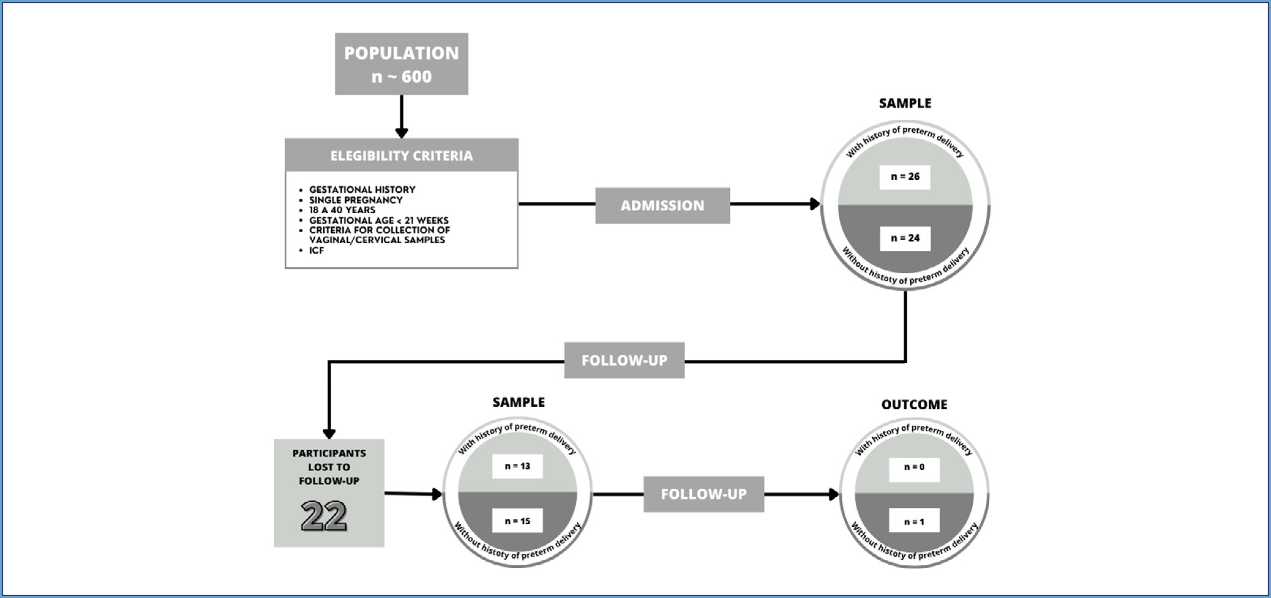
Case-control study conducted at a low-risk prenatal service in a basic public health care unit in southern Brazil from July 2018 to February 2020. 50 pregnant women were enrolled according to their subsequent order of admission to the service for routine prenatal consultations. Inclusion criteria: a) with or without history of preterm delivery/abortion, b) age between 18 and 40 years, c) single pregnancy, d) gestational age of up to 20 weeks at the first prenatal visit, e) fulfillment of criteria for collection of vaginal/cervical material (absence of antimicrobial therapy in the last 30 days, 72 hours of sexual abstinence, 72 hours of any vaginal procedure, such as vaginal examination and vaginal ultrasound), and f) signed free and informed consent form. Exclusion criteria: comorbidities, and a positive test for Chlamydia trachomatis, Trichomonas vaginalis, or Neisseria gonorrhoeae. Patients diagnosed with any infection were managed according to the national prenatal care protocol. The participants without history of preterm delivery were allocated to the full-term (FT) group (n = 24), while those with history of preterm delivery comprised the preterm (PT) group (n = 26). In all, 28 pregnant women were followed up during the three pregnancy trimesters, 15 in the FT group and 13 in the PT group. Two patients tested positive for Chlamydia trachomatis by nucleic acid amplification tests (NAATs) performed on vaginal/cervical swabs collected at study enrollment and were excluded as they required treatment, while 20 patients did not return for further evaluation (Figure 1).
All participants answered a questionnaire collecting information about sociodemographic data, lifestyle habits, gynecological and obstetric history, type of delivery, and newborn weight. Also underwent routine obstetric evaluation and gynecological examination for collection of vaginal/cervical samples over three prenatal consultations, one in each pregnancy trimester. Vaginal pH was measured, and samples were collected from the middle third of the vaginal wall using sterile swabs and from the cervix. The collected material was used for preparation of vaginal smears for classification of the vaginal microbiota and detection of infections by Trichomonas vaginalis, Chlamydia trachomatis, and Neisseria gonorrhoeae, and the presence of the microorganisms, Mycoplasma genitalium, Mycoplasma hominis, Ureaplasma urealyticum, and Ureaplasma parvum using nucleic acid amplification tests (NAATs), as recommended for high-sensitivity screening.(10)
The vaginal microbiota was classified into one of the following categories: a) normal (scores 0 to 3), b) intermediate (scores 4 to 6, represented by the presence of morphotypes compatible with Lactobacillus concomitant with an expressive accessory microbiota), and c) bacterial vaginosis (scores 7 to 10, represented by depletion of Lactobacillus and replacement by granular microbiota). A search for morphotypes compatible with pseudohyphae or hyphae of Candida spp. was also performed.(11)
The results are reported following the STrengthening the Reporting of Observational Studies in Epidemiology (STROBE) guidelines.(9)
Measures of central tendency and dispersion are expressed as mean and standard deviation (SD) for continuous variables with symmetric distribution and as median and interquartile range (IQR) for those with asymmetric distribution. Categorical variables are expressed as absolute and relative frequency.
The Student's t test was applied to estimate differences between continuous variables with symmetric distribution, while the Mann-Whitney test was used for those with asymmetric distribution. Fisher's exact and Pearson's chi-square tests were applied to estimate differences between categorical variables. A significance level of 5% was considered for all tests.
To determine the required sample size to detect a 45% difference between trimesters, we use the formula for comparing proportions:
Confidence level: 95% → Zα/2=1.96; statistical power: 80% → Zβ=0.84
Expected proportions: p1=0.50 (initial assumed proportion); p2=0.05
The required sample size was 15 participants per group without adjusting for losses. Considering a 20% loss rate, the adjusted sample size increases to 19 participants per group. For the follow-up, the estimated sample was 15 pregnant women in each group.
The study was approved by the Human Research Ethics Committee of the Health Sciences Sector of the Federal University of Paraná with the number #CAAE 57341416.3.3002.0096.
Results
The 50 participants included in the study had a mean age of 29.4±5.9 years and a mean gestational age of 13.6±4.5 weeks. Participants in the PT group had a younger maternal age than those in the FT group (p<0.001). The groups were similar in terms of tobacco, alcohol, and drug use, number of partners, and number of sexual intercourses per week (Table 1).
History of sexually transmitted infection, genital complaints, vaginal pH, and classification of the genital microbiota were also comparable between the groups (p for all=nonsignificant). The PT group had a higher gestational age (p=0.02), a greater number of pregnancies (p=0.03), and history of more abortions (p<0.001), which was expected considering that previous abortion was a criterion for inclusion of the participant in the PT group. No difference was observed between groups regarding the type of delivery (p=1.00), newborn weight (p=0.60), and frequency of colonization by Candida spp, Chlamydia trachomatis, Mycoplasma hominis, or Ureaplasma parvum (p for all = nonsignificant). There was also no difference regarding Nugent scores or frequency of positivity for Mycoplasma hominis or Ureaplasma parvum in the three trimesters (p for all=nonsignificant) (Table 2).
History of sexually transmitted infection, cervical cancer screening, genital complaints, and classification of vaginal microbiota
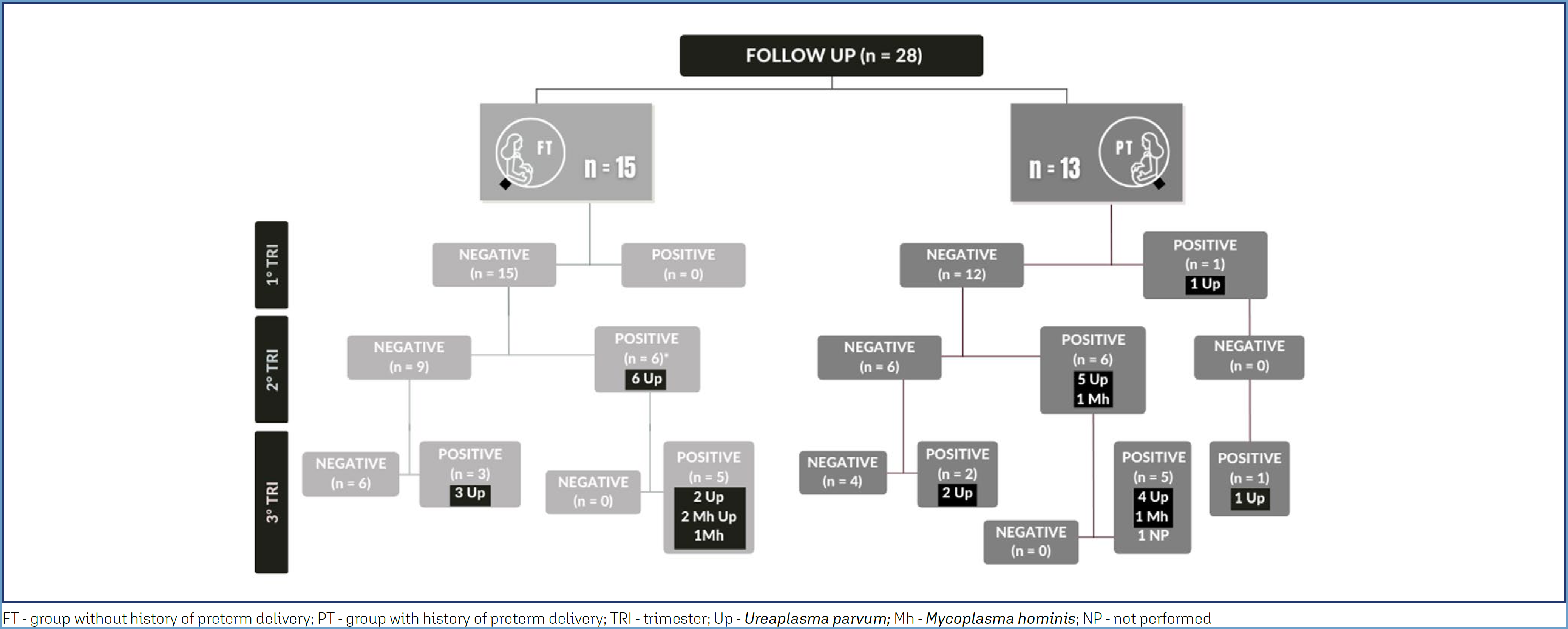
Between the participants who completed all three follow-up visits (n=28) and those who were lost to follow up (n=22), no significant differences were observed in the analyzed variables (p for all = nonsignificant). The overall incidence of positivity for the microorganisms – considering all participants with a positive result in the three trimesters and two participants who were positive in the 2nd trimester but negative in the 3rd trimester – was 36.0% (n=18). Among the participants in the PT group, only one was positive for Ureaplasma parvum in the first trimester; this participant had, for the same microorganism, a negative result in the second trimester and, again, a positive result in the third trimester. The other participants in the PT group had a negative test in the first trimester; six of them tested positive in the second trimester (five for Ureaplasma parvum and one for Mycoplasma hominis). Of the five participants who tested positive for Ureaplasma parvum in the second trimester, four remained positive and one was lost to follow up in the third trimester. The only patient who tested positive for Mycoplasma hominis in the second semester remained positive for this microorganism in the third trimester (Figure 2).
There was a significant increase in tests positive for microorganisms in the vaginal environment, from one case in the first trimester (3.6%) to 12 cases in the second trimester (3.6% versus 42.8%, p<0.001) and 16 cases in the third trimester (3.6% versus 61.5%; p<0.001) (Table 3).
Number and total of positive cases for microorganisms according to the trimester of pregnancy
The frequency of tests positive in all three trimesters was similar in both groups, and no significant differences were observed based on the presence or absence of history of preterm delivery (p for all = nonsignificant) (Table 3). Only one case of prematurity was observed among all 28 participants who were followed up across all three trimesters (3.6%), and it was recorded in a participant in the FT group (0.0% in the PT group versus 3.6% in the FT group; p=1.00), which had a positive test for Ureaplasma parvum in the second trimester and for Ureaplasma parvum and Mycoplasma hominis in the third trimester. Another case with positivity for Ureaplasma parvum and Mycoplasma hominis was observed, but in the FT group.
Discussion
Although differences between groups were explored, the sample size limits the ability to draw conclusions regarding associations with preterm delivery. The highest incidence was observed for Ureaplasma parvum, and there was a significant increase in positivity for Mycoplasma hominis and Ureaplasma parvum throughout pregnancy, particularly in the last two trimesters. The overall incidence of positivity was 36.0% (n=18).
No differences were observed between the groups regarding Nugent scores or frequency of tests with a positive result, and the most frequently detected microorganism was Ureaplasma spp., with an accumulated frequency of 86.7% in the FT group and 100.0% in the PT group. Breugelmans et al.(12) also found no association between abnormal vaginal microbiota and preterm birth. Changes in the vaginal microbiota because of the host's immune response, the virulence of the microorganism, or presence of local factors in the lower genital tract may favor the invasion of the genital tract by Ureaplasma spp., but the reason why some pregnant women present premature birth when colonized with these microorganisms (albeit asymptomatic) while others do not remain unknown.(12)
Another recent study also found no consistent association between infection by these microorganisms and prematurity. Tétu et al.(7) conducted a cohort study including 821 pregnant women who underwent amniocentesis between 14 and 24 weeks and observed preterm delivery (<35 weeks) in 26 women (3.2%), all of whom had negative PCR results for Mycoplasma spp.
Although colonization was detected, the limited sample size prevents assessment of a potential relationship with preterm birth.(12-16) The only case of prematurity recorded in the present study was in a participant with a positive result for Ureaplasma spp. in the second trimester and for Ureaplasma spp. and Mycoplasma spp. in the third trimester. The association of several microorganisms, such as bacterial vaginosis with Ureaplasma spp., has been cited as a possibility of triggering the mechanism of uterine contractions, although we did not find the citation of the association of Ureaplasma spp. and Mycoplasma spp. could have a similar effect.(17)
Ureaplasma parvum was also the most incident in both study groups, which agrees with the findings by Payne et al.(16) and Albert et al.(18) The combination of change in vaginal microbiota and presence of Ureaplasma spp. has been found at a frequency of 17.5% and suggested to be an independent factor for prematurity.(12) Notably, the association between Ureaplasma spp. and prematurity was not observed in the present study.
Another important aspect was that these microorganisms increased in frequency in the second and third trimesters and were not detected in the first trimester. This suggests that certain characteristics (immunological, hormonal, or other) in each trimester may prevent these microorganisms from developing in early pregnancy but facilitate their colonization or infection in late pregnancy. Chan et al.(19) have shown that depletion of Lactobacillus species and greater bacterial diversity leads to increased levels of mannose binding lectin and certain immunoglobulins (IgM and IgG), complement (C3b and C5), and interleukins (IL8, IL6, and IL-1β), increasing the risk of premature birth. These and other authors have proposed that the dysregulation of the response of the innate and adaptive immune system, through the effect of the complement cascade, may be one of the triggers of the microbiota related to preterm delivery.(20)
The reasons for the increasing incidence of Mycoplasma spp. over the two final trimesters of pregnancy are unclear. Future studies are needed to elucidate the present study's findings and clarify whether this increasing incidence may influence the gestational outcome, particularly in relation to prematurity. This study reveals new insight into the Mycoplasma spp. vaginal pattern during the pregnancy and could bring an important implication factor for future studies exploring relationships between the vaginal microbiome, vaginal host health, and pregnancy outcomes.
The main systematic reviews published to date show inconsistent evidence of association between colonization/infection by Mycoplasma genitalium and preterm delivery. These reviews recommend studies using DNA amplification by PCR to elucidate the role of this microorganism in pregnancy and its outcomes since the absence of correct typing could be the actual reason for the existing conflicting results.(4,5)
This case-control study carried out to test the hypothesis that colonization of the genital environment by Ureaplasma spp. and Mycoplasma spp. is related to prematurity in the current pregnancy. The most frequent microorganism identified in our study was Ureaplasma parvum. The overall incidence of the microorganisms of interest was 36.0%, with a higher positivity rate in the second and third trimesters. This study was not powered to assess associations between colonization and preterm birth. The reasons for the increasing incidence of Mycoplasma spp. over the two final trimesters of pregnancy are unclear. Future studies are needed to elucidate the findings of the present study and clarify whether this increasing incidence may influence the gestational outcome, particularly in relation to prematurity.
Conclusion
The most frequent microorganism identified in our study was Ureaplasma parvum. The overall incidence of the microorganisms was 36.0%, with a higher positivity rate in the second and third trimesters. Although Ureaplasma parvum was the most frequently identified microorganism, no definitive conclusions regarding its relationship with prematurity can be drawn due to the limited sample size.
Data availability:
The research data are described in the article presented.
References
-
1 Sprong KE, Mabenge M, Wright CA, Govender S. Ureaplasma species and preterm birth: current perspectives. Crit Rev Microbiol. 2020;46(2):169-81. doi: 10.1080/1040841x.2020.1736986
» https://doi.org/10.1080/1040841x.2020.1736986 -
2 Murhta AP, Edwards JM. The role of Mycoplasma and Ureaplasma in adverse pregnancy outcomes. Obstet Gynecol Clin North Am. 2014;41(4):615-27. doi: 10.1016/j.ogc.2014.08.010
» https://doi.org/10.1016/j.ogc.2014.08.010 -
3 Capoccia R, Greub G, Baud D. Ureaplasma urealyticum, mycoplasma hominis, and adverse pregnancy outcomes. Curr Opin Infect Dis. 2013;26(3):231-40. doi: 10.1097/qco.0b013e328360db58
» https://doi.org/10.1097/qco.0b013e328360db58 -
4 Vallely LM, Egli-Gany D, Pomat W, Homer CS, Guy R, Wand H, et al. Adverse pregnancy and neonatal outcomes associated with neisseria gonorrhoeae, Mycoplasma genital, Mycoplasma hominis, ureaplasma urealyticum and ureaplasma parvum: a systematic review and meta-analysis protocol. BMJ Open. 2018;8(11):e024175. doi: 10.1136/bmjopen-2018-024175
» https://doi.org/10.1136/bmjopen-2018-024175 -
5 Ma C, Du J, Dou Y, Chen R, Li Y, Zhao L, et al. The associations of genital Mycoplasmas with female infertility and adverse pregnancy outcomes: a systematic review and meta-analysis. Reprod Sci. 2021;28(11):3013-31. doi: 10.1007/s43032-020-00399-w
» https://doi.org/10.1007/s43032-020-00399-w -
6 Takakura S, Kodama Y, Yamashita R, Kino E, Kawano N, Tomimori K, et al. Characteristics and influence of Mycoplasma/Ureaplasma cultures in amniotic fluid on perinatal outcomes. J Obstet Gynaecol Res. 2020;46(3):389-95. doi: 10.1111/jog.14183
» https://doi.org/10.1111/jog.14183 -
7 Tétu A, Guerby P, Rallu F, Duperron L, Morin V, Bujold E. Mid-trimester microbial invasion of the amniotic cavity and the risk of preterm birth. J Matern Fetal Neonatal Med. 2022;35(21):4071-4. doi: 10.1080/14767058.2020.1846704
» https://doi.org/10.1080/14767058.2020.1846704 -
8 DiGiulio DB, Romero R, Amogan HP, Kusanovic AP, Bik EM, Gotsch F, et al. Microbial prevalence, diversity and abundance in amniotic fluid during preterm labor: a molecular and culture-based investigation. PLoS One. 2008;3(8):e3056. doi: 10.1371/journal.pone.0003056
» https://doi.org/10.1371/journal.pone.0003056 -
9 STROBE Checklists. Strengthening the reporting of observational studies in epidemiology. 2024 [cited 2024 Jun 26]. Available from: https://www.strobe-statement.org/checklists/
» https://www.strobe-statement.org/checklists/ -
10 GEN Instruções de uso. XGEN MULTI UP. 2019 [cited 2024 Jul 1]. Available from: https://consultas.anvisa.gov.br/api/consulta/produtos/25351958460201622/anexo/T15354834/nomeArquivo/IU_Uretrite+Plus_Rev.04.pdf?Authorization=Guest
» https://consultas.anvisa.gov.br/api/consulta/produtos/25351958460201622/anexo/T15354834/nomeArquivo/IU_Uretrite+Plus_Rev.04.pdf?Authorization=Guest -
11 Nugent RP, Krohn MA, Hillier SL. Reliability of diagnosing bacterial vaginosis is improved by a standardized method of gram stain interpretation. J Clin Microbiol. 1991;29(2):297-301. doi: 10.1128/jcm.29.2.297-301.1991
» https://doi.org/10.1128/jcm.29.2.297-301.1991 -
12 Breugelmans M, Vancutsem E, Naessens A, Laubach M, Foulon W. Association of abnormal vaginal flora and Ureaplasma species as risk factors for preterm birth: a cohort study. Acta Obstet Gynecol Scand. 2010;89(2):256-60. doi: 10.3109/00016340903418769
» https://doi.org/10.3109/00016340903418769 -
13 Kataoka S, Yamada T, Chou K, Nishida R, Morikawa M, Minami M, et al. Association between preterm birth and vaginal colonization by mycoplasmas in early pregnancy. J Clin Microbiol. 2006;44(1):51-5. doi: 10.1128/jcm.44.1.51-55.2006
» https://doi.org/10.1128/jcm.44.1.51-55.2006 -
14 Vogel I, Thorsen P, Hogan VK, Schieve LA, Jacobsson B, Ferre CD. The joint effect of vaginal Ureaplasma urealyticum and bacterial vaginosis on adverse pregnancy outcomes. Acta Obstet Gynecol Scand. 2006;85(7):778-85. doi: 10.1080/00016340500442423
» https://doi.org/10.1080/00016340500442423 -
15 Romero R, Yoon BH, Mazor M, Gomez R, Gonzalez R, Diamond MP, et al. A comparative study of the diagnostic performance of amniotic fluid glucose, white blood cell count, interleukin-6, and gram stain in the detection of microbial invasion in patients with preterm premature rupture of membranes. Am J Obstet Gynecol. 1993;169(4):839-51. doi: 10.1016/0002-9378(93)90014-a
» https://doi.org/10.1016/0002-9378(93)90014-a -
16 Payne MS, Ireland DJ, Watts R, Nathan EA, Furfaro LL, Kemp MW, et al. Ureaplasma parvum genotype, combined vaginal colonisation with Candida albicans, and spontaneous preterm birth in an Australian cohort of pregnant women. BMC Pregnancy Childbirth. 2016;16(1):312-25. doi: 10.1186/s12884-016-1110-x
» https://doi.org/10.1186/s12884-016-1110-x -
17 Taylor-Robinson D. Mollicutes in vaginal microbiology: mycoplasma hominis, Ureaplasma urealyticum, Ureaplasma parvum, and Mycoplasma genitalium. Res Microbiol. 2017;168(9-10):875-81. doi: 10.1016/j.resmic.2017.02.009
» https://doi.org/10.1016/j.resmic.2017.02.009 -
18 Albert AY, Chaban B, Wagner EC, Schellenberg JJ, Links MG, van Schalkwyk J, et al., VOGUE Research Group. A study of the vaginal microbiome in healthy Canadian women utilizing cpn60. Based on molecular profiling reveals distinct Gardenerella subgroup community state types. PLoS One. 2015;10(8):e0135620. doi: 10.1371/journal.pone.0135620
» https://doi.org/10.1371/journal.pone.0135620 -
19 Chan D, Bennett PR, Lee YS, Kundu S, Teoh TG, Adan M, et al. Microbial-driven preterm labour involves crosstalk between the innate and adaptive immune response. Nat Commun. 2022;13(1):975. doi: 10.1038/s41467-022-28620-1
» https://doi.org/10.1038/s41467-022-28620-1 -
20 Marconi C, Andrade-Ramos BR, Peraçoli JC, Donders GG, da Silva MG. Amniotic fluid interleukin-1 beta and interleukin-6, but not interleukin-8 correlate with microbial invasion of the amniotic cavity in preterm labor. Am J Reprod Immunol. 2011;65(6):549-56. doi: 10.1111/j.1600-0897.2010.00940.x
» https://doi.org/10.1111/j.1600-0897.2010.00940.x
Edited by
-
Associated Editor
Silvana Maria Quintana (https://orcid.org/0000-0002-9311-786X) Universidade de São Paulo, Ribeirão Preto, São Paulo/SP, Brazil

 Source: Flow diagram constructed according to STROBE recommendations.(