
 Choroideremia initially misdiagnosed as retinitis pigmentosa: a multimodal and genetic diagnostic reappraisal
ABSTRACT
Choroideremia is a rare X-linked chorioretinal dystrophy that may closely resemble retinitis pigmentosa (RP) in its early stages. We describe two brothers who were initially diagnosed with RP and later found to have choroideremia after multimodal imaging and genetic testing. The proband, a 49-year-old man, presented with progressive visual loss, diffuse chorioretinal atrophy, and bone spicule pigmentation. His 51-year-old brother had advanced disease with no light perception. Both displayed fundus autofluorescence and OCT findings typical of choroideremia. A hemizygous nonsense variant in the CHM gene (c.619C>T; p.Arg207*) confirmed the diagnosis. This case highlights the persistent challenge of differentiating early choroideremia from RP and reinforces the importance of genetic analysis in male patients with suspected X-linked retinal dystrophies. Genetic confirmation enables accurate counseling, prognosis, and consideration for emerging gene-based therapies.
Keywords:
Choroideremia; Retinal dystrophies; Retinitis pigmentosa; Genetic testing
RESUMO
A coroidemia é uma distrofia coriorretiniana rara ligada ao cromossomo X que pode se assemelhar muito à retinite pigmentosa em seus estágios iniciais. Descrevemos dois irmãos que foram inicialmente diagnosticados com retinite pigmentosa e, posteriormente, descobriu-se que tinham coroidemia, após exames de imagem multimodais e testes genéticos. O probando, um homem de 49 anos, apresentava perda visual progressiva, atrofia coriorretiniana difusa e pigmentação em espículas ósseas. Seu irmão de 51 anos tinha a doença em estágio avançado, sem percepção de luz. Ambos apresentavam autofluorescência do fundo do olho e achados de tomografia de coerência óptica típicos da coroidemia. Uma variante hemizigótica sem sentido no gene CHM (c.619C>T; p.Arg207*) confirmou o diagnóstico. Este caso destaca o desafio persistente de diferenciar a coroidemia precoce da retinite pigmentosa e reforça a importância da análise genética em pacientes do sexo masculino com suspeita de distrofias retinianas ligadas ao cromossomo X. A confirmação genética permite aconselhamento preciso, prognóstico e consideração de terapias emergentes baseadas em genes.
Descritores:
Coroideremia; Distrofias retinianas; Retinose pigmentar; Testes genéticos
INTRODUCTION
Choroideremia (CHM) is a progressive, X-linked retinal dystrophy caused by mutations in the CHM gene, which encodes the Rab escort protein-1 (REP1).(1-5) REP1 plays a critical role in intracellular vesicle trafficking within photoreceptors, the retinal pigment epithelium (RPE), and choroidal cells.(2-4) Loss of REP1 function results in progressive degeneration of these tissues.(2,3) Clinically, CHM typically manifests in adolescence with night blindness (nyctalopia), followed by peripheral visual field constriction and, eventually, central vision loss.(3,5-7) The prevalence of CHM is estimated to range from 1 in 50,000 to 1 in 100,000 males.(3,5)
In its early stages, CHM can closely resemble retinitis pigmentosa (RP), leading to diagnostic delays.(3,5,6) Advances in multimodal imaging techniques, such as color fundus photography, fundus autofluorescence (FAF), optical coherence tomography (OCT), and microperimetry, alongside next-generation sequencing (NGS) for inherited retinal dystrophy genes, have significantly improved the diagnostic accuracy of these conditions.(8,9) Early genetic confirmation of CHM provides essential information for genetic counseling, prognosis, and potential inclusion in gene therapy trials, which are increasingly offering therapeutic options for patients with inherited retinal diseases.(10,11)
A case series of two male siblings, aged 49 and 51, previously diagnosed with RP, was re-evaluated and subsequently diagnosed with CHM following genetic testing. Both patients underwent a comprehensive ophthalmic assessment, including color fundus photography, FAF, OCT, and microperimetry. Additionally, targeted NGS for inherited retinal dystrophy genes was performed. The family pedigree was analyzed to determine the mode of inheritance. Written informed consent was obtained from both patients.
The study was approved by the Research Ethics Committee of the Hospital Oftalmológico de Brasília (CEP-HOB, Brazil) under protocol number CAAE 89671925.7.0000.5667, in accordance with the principles of the Declaration of Helsinki.
CASE REPORT
The proband, aged 49, reported progressive nyctalopia and peripheral vision loss since his late twenties. His visual acuity was 20/80 in the right eye and counting fingers at one meter in the left. Anterior segment examination and intraocular pressures were normal. Fundus examination revealed optic disc pallor, vascular attenuation, bone spicule pigmentation, and extensive atrophy of the RPE and choroid. OCT showed severe loss of the outer retinal layers and diffuse choroidal thinning. Fundus autofluorescence revealed a central area of preserved autofluorescence surrounded by extensive hypoautofluorescence, indicative of advanced retinal degeneration.
His 51-year-old brother, who had no light perception in both eyes, showed similar but more advanced findings. Fundus imaging demonstrated diffuse chorioretinal atrophy with scleral exposure, and OCT confirmed complete loss of the outer retinal layers.
A summary of the clinical, imaging, and genetic findings in both siblings is provided in table 1.
Genetic testing identified a hemizygous nonsense mutation in the CHM gene (c.619C>T; p.Arg207*) in both siblings, confirming the diagnosis of X-linked CHM. The family pedigree revealed a pattern of maternal transmission, and female relatives were subsequently referred for carrier testing and genetic counseling (Figure 2).
Representative multimodal imaging findings from both siblings, including wide-field fundus photography, FAF, and OCT, are shown in figure 1.
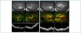
(A-C) Multimodal imaging of the proband (49 years). (A) Wide-field fundus photograph showing optic disc pallor, vascular attenuation, peripheral bone spicule pigmentation, and diffuse chorioretinal atrophy. (B) Fundus autofluorescence demonstrates a preserved central island surrounded by extensive peripheral hypoautofluorescence, typical of choroideremia. (C) Spectral-domain optical coherence tomography through the fovea reveals thinning of the outer retina and retinal pigment epithelium with residual foveal structure. (D-F) Multimodal imaging of the affected brother (51 years). (D) Advanced diffuse chorioretinal atrophy with bare scleral exposure on fundus photograph. (E) Autofluorescence showing loss of central signal and absence of peripheral preservation. (F) Optical coherence tomography confirming total outer retinal and choroidal atrophy.

Pedigree of the affected family showing an X-linked recessive inheritance pattern. Circles denote females and squares denote males. Dotted circles indicate heterozygous female carriers; filled squares represent affected males; and open symbols indicate unaffected individuals. The maternal transmission and lack of father-to-son inheritance are characteristic of X-linked retinal dystrophies such as choroideremia preservation. Optical coherence tomography confirming total outer retinal and choroidal atrophy.
DISCUSSION
Choroideremia can be clinically misdiagnosed as RP in its early stages due to overlapping symptoms, including night blindness and peripheral vision loss.(3,5-7) However, in contrast to RP, where degeneration of the photoreceptors is typically the earliest event, RPE and choroidal atrophy are often the initial pathological features in CHM.(2,3,5) Multimodal imaging techniques play a crucial role in distinguishing between these two entities.(8,11) In CHM, FAF typically shows a central area of preserved autofluorescence surrounded by hypoautofluorescent regions, reflecting the pattern of retinal degeneration.(6,8,11) OCT provides further confirmation by demonstrating significant outer retinal loss and choroidal thinning.(6,8)
Although the clinical presentation of early CHM can be indistinguishable from RP, the definitive diagnosis is often made through genetic testing, which allows for the identification of mutations in the CHM gene.(2,4,5) In this case, targeted NGS revealed a hemizygous nonsense mutation (c.619C>T; p.Arg207*), confirming the diagnosis of CHM.(2,4) This underscores the importance of genetic testing in providing a definitive diagnosis, particularly in cases where the clinical presentation is ambiguous.(1-5) As an X-linked disorder, CHM predominantly affects males, while female carriers may present with patchy fundus changes or remain asymptomatic.(3-5) Recognizing this inheritance pattern is essential for appropriate family counseling and carrier detection.(3,4)
The identification of CHM mutations is not only crucial for diagnosis but also has significant implications for family planning and genetic counseling.(3-5) In this case, the family pedigree suggested a maternal inheritance pattern, with several female relatives potentially being carriers of the mutation. Therefore, carrier testing was offered to other family members to assess the risk of transmission to future generations.(3-5)
In addition, molecular diagnosis is essential for determining eligibility for emerging therapeutic options.(1,10,11) Gene replacement therapies using adeno-associated virus (AAV) vectors have shown promising results in early-phase clinical trials, with evidence of structural and functional stabilization of the retina in patients with early-stage CHM.(1,10) Genetic diagnosis is a prerequisite for inclusion in these trials, offering hope for a therapeutic breakthrough that could alter the disease course.(1,10,11)
This report underscores the value of revisiting historical RP diagnoses using modern genetic tools. Molecular confirmation of CHM not only corrects longstanding diagnostic errors but also provides families with essential information for counseling and reproductive planning.(3-5) Early identification further enables timely inclusion in gene therapy trials, offering hope for patients with inherited retinal dystrophies where precision molecular diagnosis remains the cornerstone for accurate classification and treatment eligibility.(1,10,11)
Financial support:
Institution:
Data Availability Statement:
The datasets generated and/or analyzed during the current study are included in the manuscript.
REFERENCES
- 1 McClements ME, MacLaren RE. Gene therapy for retinal disease. Transl Res. 2013;161(4):241-54.
- 2 Preising M, Ayuso C. Rab escort protein 1 (REP1) in intracellular traffic: a functional and pathophysiological overview. Ophthalmic Genet. 2004;25(2):101-10.
- 3 Coussa RG, Traboulsi EI. Choroideremia: a review of general findings and pathogenesis. Ophthalmic Genet. 2012;33(2):57-65.
- 4 van den Hurk JA, van de Pol DJ, Wissinger B, van Driel MA, Hoefsloot LH, de Wijs IJ, et al. Novel types of mutation in the choroideremia (CHM) gene: a full-length L1 insertion and an intronic mutation activating a cryptic exon. Hum Genet. 2003;113(3):268-75.
- 5 Mura M, Sereda C, Jablonski MM, MacDonald IM, Iannaccone A. Clinical and functional findings in choroideremia due to complete deletion of the CHM gene. Arch Ophthalmol. 2007;125(8):1107–13.
- 6 Aleman TS, Han G, Serrano LW, Fuerst NM, Charlson ES, Pearson DJ, et al. Natural history of the central retina in choroideremia: a prospective cross-sectional study. Ophthalmology. 2017;124(3):359–73.
- 7 Simunovic MP, Jolly JK, Xue K, Edwards TL, Groppe M, Downes SM, et al. The spectrum of CHM gene mutations in choroideremia and their relationship to clinical phenotype. Invest Ophthalmol Vis Sci. 2016;57(14):6033–9.
- 8 Jolly JK, Xue K, Edwards TL, Groppe M, MacLaren RE. Characterizing the Natural History of Visual Function in Choroideremia Using Microperimetry and Multimodal Retinal Imaging. Invest Ophthalmol Vis Sci. 2017;58(12):5575–83.
- 9 Georgiou M, Fujinami K, Michaelides M. Retinal imaging in inherited retinal diseases. Ann Eye Sci. 2020;5:25.
- 10 Xue K, Jolly JK, Barnard AR, Rudenko A, Salvetti AP, Patrício MI, et al. Beneficial effects on vision in patients undergoing retinal gene therapy for choroideremia. Nat Med. 2018;24(10):1507–12.
- 11 MacDonald IM, Russell L, Chan CC. Choroideremia: new findings from ocular pathology and review of recent literature. Surv Ophthalmol. 2009;54(3):401–7.


