Dubowitz syndrome palpebral ptosis: a case report
ABSTRACT
To report a case of Dubowitz syndrome with eyelid ptosis in the right eye, successfully treated with surgical correction. A 9-year-old child, previously diagnosed with Dubowitz syndrome, presented with congenital ptosis in the right eye. Surgical correction of eyelid ptosis was performed. The postoperative outcome showed significant functional and aesthetic improvement, with satisfactory eyelid position and absence of complications. Dubowitz syndrome is a rare autosomal recessive disorder characterized by multiple congenital anomalies, including ophthalmologic manifestations such as eyelid ptosis. Surgical correction may provide excellent functional and cosmetic results in selected cases.
Keywords:
Blepharoptosis; Eye diseases/genetics; Multiple abnormalities/ genetics; Chromosome aberrations; Growth disorders/genetics; Syndrome
RESUMO
O objetivo desse trabalho foi relatar um caso de síndrome de Dubowitz com ptose palpebral em olho direito, tratada com sucesso por correção cirúrgica. Criança de 9 anos, previamente diagnosticada com síndrome de Dubowitz, apresentando ptose palpebral congênita em olho direito. Foi realizada correção cirúrgica da ptose palpebral. Observou-se melhora funcional e estética significativa no pós-operatório, com posicionamento adequado da pálpebra e ausência de complicações. A síndrome de Dubowitz é uma doença autossômica recessiva rara, caracterizada por múltiplas anomalias congênitas, incluindo manifestações oftalmológicas como a ptose palpebral. A correção cirúrgica pode proporcionar excelentes resultados funcionais e estéticos em casos selecionados.
Descritores:
Blefaroptose; Oftalmopatias/genética; Anormalidades múltiplas/genética; Aberrações cromossômicas; Transtornos do crescimento/genética; Síndrome
INTRODUCTION
Dubowitz syndrome was first described by Victor Dubowitz in 1965(1) and later classified as Dubowitz syndrome by Gorlin and Opitz in 1971.(2) The clinical findings of this condition affect multiple systems, including neurological, cardiovascular, musculoskeletal, dermatological, and ocular systems. Ocular manifestations may include strabismus, eyelid ptosis, dacryocystitis, hypertelorism, telecanthus, epicanthus, and retinal abnormalities, among others.(3,4) It is a rare autosomal recessive disorder. We report the case of a child diagnosed with Dubowitz syndrome who showed a good surgical outcome following correction of eyelid ptosis.
CASE RESPORT
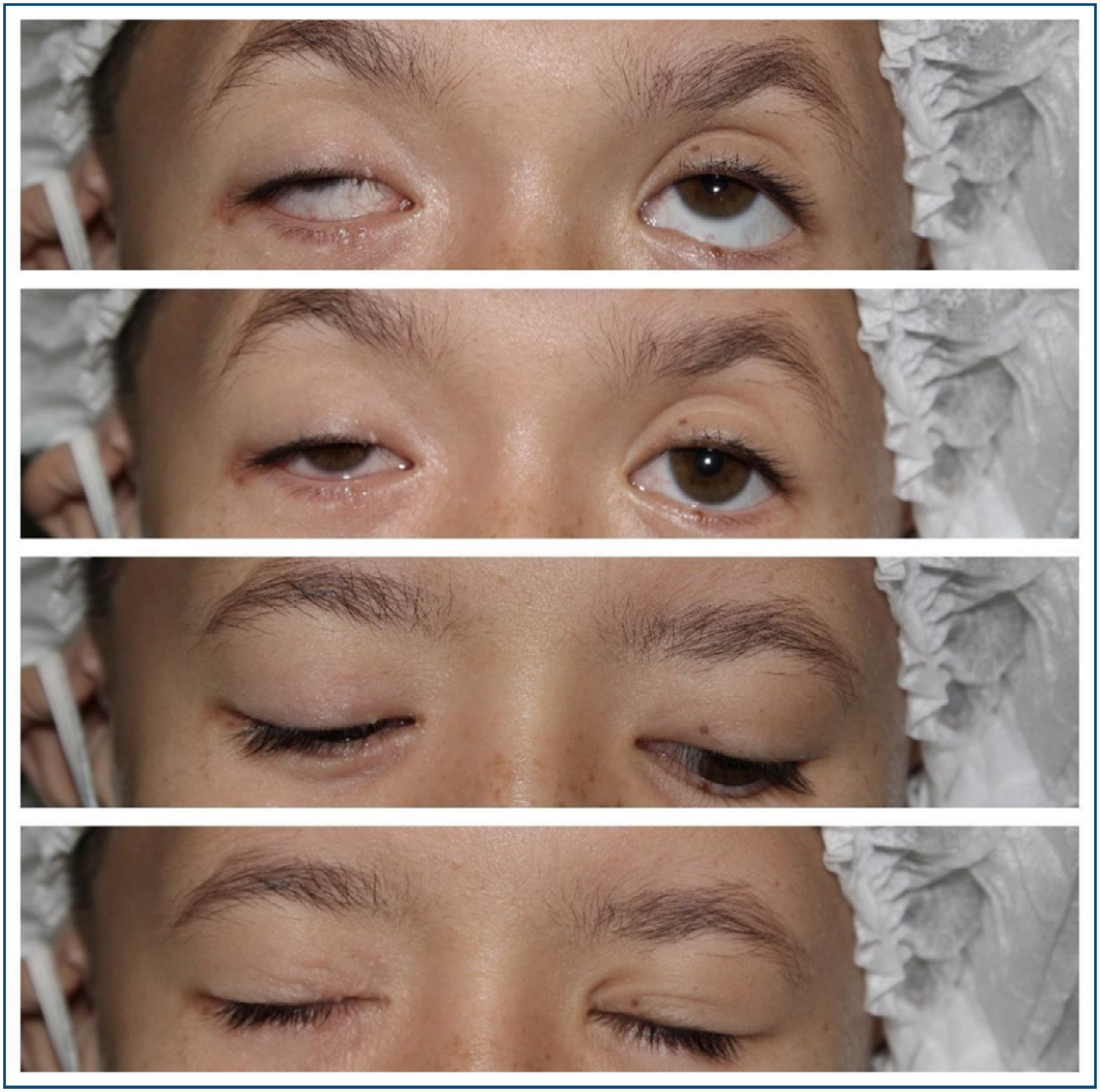
Male, 9-year-old child, was born at full term with low birth weight, cleft palate, low-set ears, saddle nose, and a triangular facial shape. The ophthalmologic examination revealed an uncorrected visual acuity of 20/50 in the right eye (OD) and 20/25 in the left eye (OS). Refraction OD: -0.50 sph -1.00 cyl × 15 (20/30) and OS: plane (20/25). The patient was orthophoric, with eyelid ptosis in the right eye. Eyelid fissure measurements were 6 mm in OD and 8 mm in OS. The margin reflex distance (MRD1) measured 1 mm in OD and 3 mm in OS. The distance from the upper eyelid margin to the limbus was +4 mm in OD and +2 mm in OS. The eyelid crease was absent in OD and measured 6 mm in OS. Levator muscle function was 6 mm in OD and 12 mm in OS.
Surgical correction of right upper eyelid ptosis was indicated, consisting of aponeurotic shortening of the levator palpebrae superioris muscle. The procedure was performed under general anesthesia.
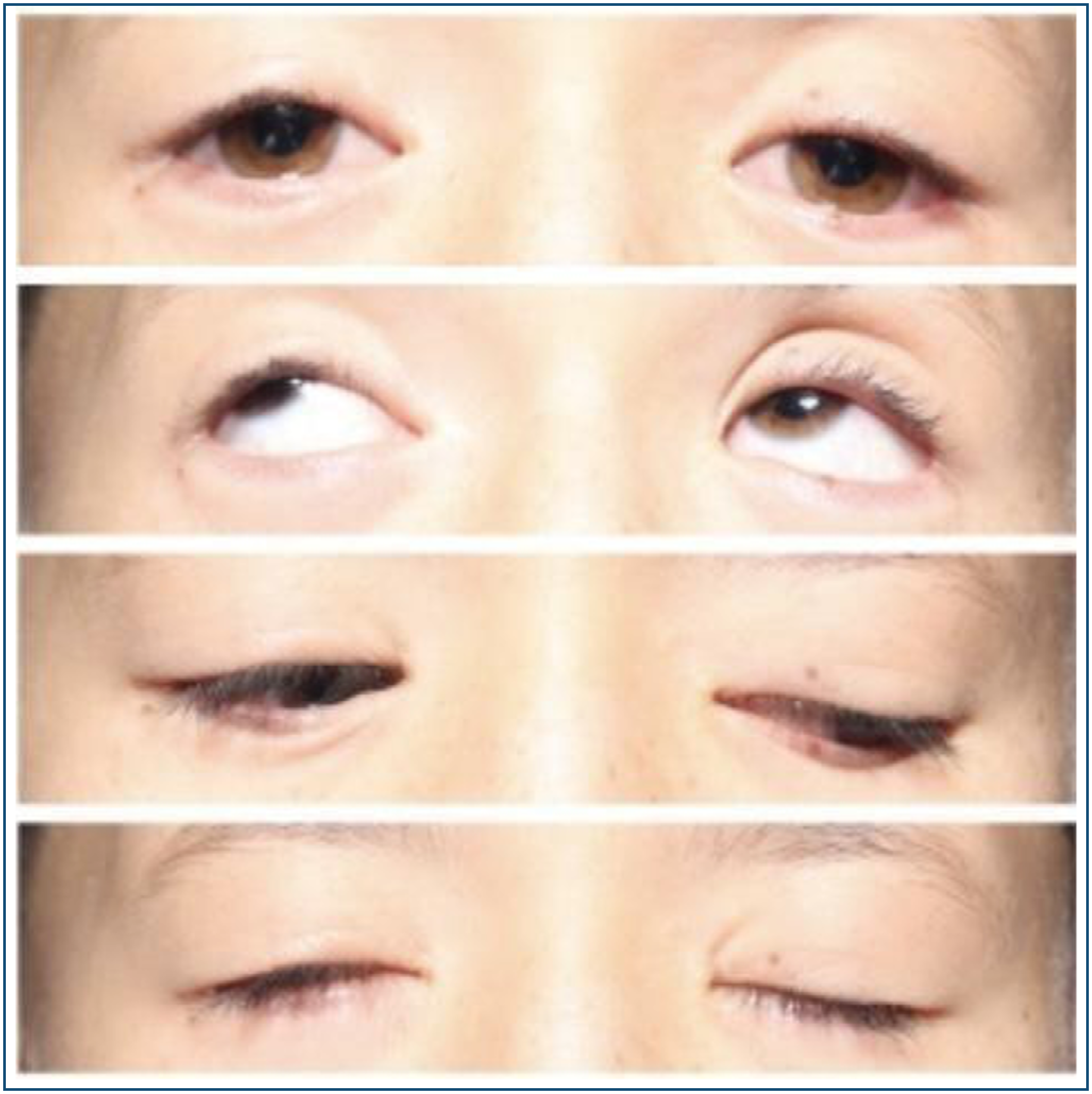
Postoperative evaluation demonstrated excellent eyelid symmetry, with an eyelid fissure height of 8 mm in both eyes and a margin reflex distance of 3 mm bilaterally. The patient's parents reported high satisfaction with the aesthetic outcome (Figures 1 and 2).


This study was approved by Ethical Committee under # CAAE : 46012721.3.0000.5103.
DISCUSSION
Dubowitz syndrome, first described in 1965(¹), is a rare congenital disorder characterized by multiple developmental anomalies. As of 2017, just over 150 cases had been reported in Europe.(4) The syndrome appears to affect both sexes and all ethnic groups equally4. Syndromic genetic alterations are often associated with facial and ocular abnormalities; therefore, the ophthalmologist plays a key role in identifying ocular findings, which can raise suspicion of the condition and lead to referral for genetic evaluation. Our patient was referred to us by his geneticist with a confirmed clinical diagnosis of Dubowitz syndrome. It is important to emphasize that genetic testing in this syndrome has limited diagnostic relevance, as the diagnosis is primarily based on clinical criteria.(4) Early recognition is important for the proper diagnosis and multidisciplinary management of these children, as the syndrome may be associated with severe and sometimes undiagnosed malformations of other organ systems.(2-4) The main clinical features include microcephaly (100%), sparse hair (70%), sloping forehead (80%), telecanthus (60%), ptosis or blepharophimosis (usually asymmetric) (65%), epicanthal folds (50%), broad or prominent nasal bridge (55%), palatal abnormalities (50%), micrognathia (80%), thin or high-pitched voice (55%), and low-set ears (75%).(4)
Data Availability Statement:
The datasets generated and/or analysed during the current study are included in the manuscript.
REFERENCES
- 1 Dubowitz V. Familial low birthweight dwarfism with an unusual facies and a skin eruption. J Med Genet. 1965;2(1):12-7.
- 2 Grosse R, Gorlin J, Opitz JM. The Dubowitz syndrome. Z Kinderheilkd. 1971;110(3):175-87.
- 3 Dias VG, Mendonca Filho DR, Vargas MA, Goncalves FF, Gigante E, Valerio FJ. Síndrome de Dubowitz: relato de caso. Arq Bras Oftalmol. 2004;67(2):337-40.
- 4 Soares A, Ribeiro S. Ophthalmological abnormalities in Dubowitz Syndrome. Oftalmologia. 2017;41(2):1-6.
Edited by
-
Associate Editor:
Renato Wendell Ferreira Damasceno Universidade Estadual de Ciências da Saúde de Alagoas, Maceió, AL, Brazil https://orcid.org/0000-0002-9380-0335