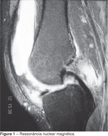
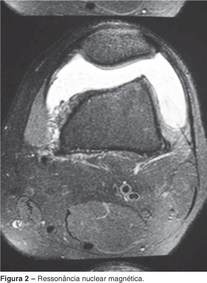
Male patient, 34 years old, had severe pain in his left knee in association with functional incapacitation, with no apparent triggering factor. He sought medical attention in December 2006, at which time he was prescribed NSAIDS. After a year, reported increased swelling and pain at the site. He was referred to a knee specialist with a suspected meniscal injury. Upon examination, we detected severe swelling of the joint with limitation of motion, pain exacerbated, and negative joint aspiration. Since simple radiographic results were normal, an MRI of the knee was requested. The MRI revealed massive accumulation of synovial fluid, together with marked synovial proliferation, especially focal thickening clumps with intermediate signal on T1 and T2, a hypointense signal on T2, and discreet suggestive of pigmented villonodular synovitis with intact meniscus and ligaments. The patient underwent arthroscopy of the left knee, which revealed whitish irregular fragments, and underwent arthrotomy with removal of the lesion and extensive synovectomy. The material was submitted to pathological examination, which showed the presence of synovial chondromatosis. Eight months after surgery, the patient presents with no complaints, with a 130° range in the left knee without joint bleeding or signs of inflammation. Synovial chondromatosis is a rare benign metaplasia of the synovial membrane, leading to the formation of cartilaginous loose bodies in the joint space. It is difficult to diagnose because 95% of the nodules, when not calcified, can be overlooked radiologically.
Knee; Metaplasia; Chondromatosis