
 New freshwater Ceratomyxa species, Ceratomyxa affinis n. sp. (Myxozoa: Ceratomyxidae) in the gallbladder of Leporinus affinis from Brazilian Amazon
Abstract
The genus Ceratomyxa is composed of approximately 300 described species, most of which are found parasitizing hosts in marine aquatic environments. The present study, through phylogenetic, molecular and morphological analyses, described a new species of Ceratomyxa found parasitizing Leporinus affinis specimens from the Tartarugalzinho River, in municipality of Tartarugalzinho, state of Amapá, Brazil. The new species was found parasitizing the urinary bladder of L. affinis. The myxospores of Ceratomyxa affinis n. sp. are 7.2 µm long and 43.2 µm thick with a posterior angle of 170º. The polar capsules measure 3.9 µm long and 4.05 µm wide. Based on morpho-molecular analyses, it was possible to prove that Ceratomyxa affinis n. sp. is a new species, contributing to the knowledge of the parasitic fauna of fish in the Amazon region.
Keywords:
Myxozoan; fish; parasite; Amazon; parasitology
Resumo
O gênero Ceratomyxa é composto por cerca de 300 espécies descritas encontradas em sua maioria parasitando hospedeiros de ambientes aquáticos marinhos. O presente estudo, usando análises filogenéticas, moleculares e morfológicas, descreveu um uma nova espécie de Ceratomyxa encontrado parasitando Leporinus affinis oriundos do Rio Tartarugalzinho, município de Tartarugalzinho, no estado do Amapá, Brasil. A nova espécie foi encontrada parasitando a vesícula urinária de L. affinis. Os myxosporos de Ceratomyxa affinis n. sp. possuem 7.2 µm de comprimento e 43.2 µm de espessura com ângulo posterior de 170º. As capsulas polares medem 3.9 µm de comprimento e 4.05 µm de largura. Baseada nas análises morfo-molecular, foi possível comprovar que Ceratomyxa affinis n. sp. é uma nova espécie, contribuindo para o conhecimento da parasitofauna de peixes da região amazônica.
Palavras-chave:
Myxozoários; peixe; parasito; Amazônia; parasitologia
Introduction
Parasites belonging to the class Myxozoa are classified as obligate eukaryotic endoparasites and present a complex life cycle that involves vertebrate and invertebrate hosts. This group is considered one of the most abundant in terms of species richness, with approximately 2,600 described species, frequently found in aquatic hosts. Myxozoan spores have simple morphological structures, with a rounded, oval or ellipsoid-shaped body, which has two valves that surround two polar capsules, which in some cases have different sizes. (Atkinson et al., 2018; Okamura et al., 2018; Sindeaux-Neto, et al., 2021; Eiras et al., 2023; Cardoso et al., 2025).
The genus Ceratomyxa Thélohan, 1892 is composed of about 300 described species that are mostly found parasitizing hosts in marine aquatic environments. However, there are also records of species described parasitizing freshwater fish mainly, in South America. Myxospores of this genus exhibit an elongated or arcuate-shaped spore body with two polar capsules located at the top of the spore, parallel to the suture line, and they are commonly found parasitizing the host's gallbladder (Zatti et al., 2018, 2023 ; Eiras et al., 2018; Martel et al., 2024; Carvalho et al., 2024).
Among the fish that make up the Anastomidae family, there is the genus Leporinus Agassiz 1829, which houses the largest number of described species, and is widely distributed in all river basins in the country. The fish belonging to this group are of great commercial importance and are popularly known in Brazil as “aracus” and “piaus.” Leporinus affinis Günther 1864 is an anastomid belonging to the genus Leporinus, known as “piau-flamengo”. Specimens of this species have a fusiform body structure with a coloration composed of dark and transversal bars on the body and can reach 30 cm in total length. These fish have herbivorous habits and are commonly found in streams with rocky and sandy bottoms. (Santos & Zuanon, 2008; Birindelli & Britski, 2013; Zacardi et al., 2017; Froese & Pauly, 2024).
Studies focused on the parasitic fauna of fish are of utmost importance as they can be used to understand the functioning of the parasite-host-environment ecological triad. In the context of parasites infecting aquatic animals, fish found in lakes and rivers are the most common vectors for a wide variety of parasitic organisms, with myxozoa being one of the most common. With that in mind, therefore, the present study uses phylogenetic, molecular and morphological analyses, to describe a new species of Ceratomyxa found parasitizing the gallbladder of specimens of L. affinis from the Tartarugalzinho River, Municipality of Tartarugalzinho, state of Amapá, in eastern Amazon.
Material and Methods
Study area and fish collection
The municipality of Tartarugalzinho is an important component of the Amapá fishing sector. It is part of a lake region and has an extensive biodiversity found in its vegetation, waterfalls, lakes and rivers. The Tartarugalzinho River (Figure 1) is the most important river in the municipality. Draining art of its waters into the Duas Bocas Lake, it has extensive vegetation cover and a unique ecosystem that enables a rich ichthyofauna (Bidone et al., 1997; Yokomizo, 2012; Silveira et al., 2015).

Location map of Leporinus affinis collections in Tartarugalzinho River (red dots), in municipality of Tartarugalzinho, state of Amapá: Pedreira River, Brazilian Amazon.
The specimens of Leporinus affinis (n= 13) were collected between August 14, 2023 and October 9, 2024. The specimens were collected by the team from the Laboratory of Morphophysiology and Animal Health at the State University of Amapá (LABMORSA) with the help of a local fisher. After being collected, the specimens were transported alive to the Laboratory of Morphophysiology and Animal Health (LABMORSA), in thermal boxes containing water from the environment itself and artificial aeration. The collections were approved by the Animal Use Committee (CEUA) and registered in the Biodiversity Authorization and Information System (SISBIO/ICMBIO).
Dissection of host fish
In LABMORSA, the fish were anesthetized with tricaine methanesulfonate (MS222 Sigma), and subsequently desensitized by means of neural myelotomy with the aid of sharp forceps. To measure the biometric data, the specimens were weighed (g) and measured (cm). With the anesthetized specimens, macroscopic analysis was performed on the animal's body surface, under a binocular stereomicroscope, with the aim of observing the existence of cysts in the tegument, lesions or loss of lining. Then, an incision was made in the ventral region of the specimens so that the internal organs were exposed. Afterwards, with the help of sharp forceps, the gallbladder was removed without leaking the internal liquid, placed between a slide and a coverslip and observed fresh using light microscopy.
Morphometry and histological analysis
All gallbladder that showed the presence of parasites were placed between slide and coverslip and photographed using a digital camera (Moticam 2300 3.0 M), for fresh morphological observation of the parasite. The analysis of the morphometric characteristics of the spores found was performed according to the methodology proposed by Lom & Arthur (1989), and consisted of spore length (SL), spore thickness (ST), polar capsule length (PCL), polar capsule width (PCW) and posterior angle (PA), dimensions were obtained by averaging all measurements of 30 spores. The calculation of parasite prevalence was tabulated according to Bush et al. (1997).
The fragments of gallbladder parasitized were collected and fixed in Davidson (95% alcohol, formaldehyde, acetic acid and water) for a period of 24 h and subsequently dehydrated in an increasing battery of alcohols (70%, 80%, 90%, 100% I, 100% II and 100% III), and were then subjected to diaphanization in xylene and impregnated in paraffin blocks. After cooling, the blocks were cut by a microtome into 5 µm-thick ribbons. The cut material was fixed on a glass slide and stained with hematoxylin-eosin (H&E) (Luna, 1968).
DNA extraction and sequencing
To perform molecular biology, gallbladder infected with microparasite were collected and stored in Eppendorf tubes with 80% ethyl alcohol. DNA from each sample was extracted using the ReliaPrep gDNA Tissue Miniprep System kit (Promega, USA) according to the protocol provided by the manufacturer. The samples were sized by spectrometry (Biodro Duo) with a wavelength of 250nm; for the amplification of the 18sDNA gene fragment the polymerase chain reaction (PCR) was performed in a thermocycler (MyGene MG96G).
The primers 18E (5'-CTGGTTGATCCTGCCAGT-3') (Hillis & Dixon, 1991) and 18R (5'- CTACGGAAACCTGTTACG-3') (Whipps et al., 2003) were used for the first stage of amplification. Cycling was performed under the following conditions: initial denaturation at 95°C for 15 min, followed by 35 cycles of 95°C for 1 min, 48°C for 1.5 min, 72°C for 2 min and final extension at 72°C for 10 min. For the second round of amplification, primers 18E - MC3 (5'-GATTAGCCTGACGATCACTCCACGA-3') and 18R-MC5 were used (5'-CCTGAGAAACGGCTACCACATCCA-3') (Molnár et al., 2002). The cycle with initial denaturation at 95°C for 15 min was applied, followed by 35 cycles of 95°C for 30 seconds, 56°C for 30 seconds, 72°C for 1 min andmin and the final extension at 72°C for 10 min.
The PCR results were subjected to electrophoin resis 1.5% agarose gel in a Tris-borate-EDTA (TBE) buffer solution, then stained with UniSafe Dye (UniScience, Brazil) and subsequently visualized using the Bluegel Electrophoresis System. All samples that presented positive results were sent to ACTGene (Alvorada, RS, Brazil) for the sequencing process.
Phylogenetic analyses
The sequences obtained were edited in the Geneious® 7.1.3 software. After BLASTn searches (Altschul et al., 1997), the result generated was compared with sequences deposited in GenBank through the Basic Local Alignment Search Tool (BLASTn) of the National Center for Biotechnology Information (NCBI), to determine the similarity between the sequences.
A database was aligned using the ClustalW algorithm with its default parameters, in the Geneious 7.1.3 software (Kearse et al., 2012). Maximum parsimony Bayesian inference (BI) analyses were performed, with the aid of the MrBayes software 3.2.7a (Ronquist & Huelsenbeck, 2003), through the CIPRES platform based on the evolution model (GTR + I + G), chosen by the jModelTest evaluation, based on the lowest classification of the Bayesian information criterion (BIC). The following probabilities were based on 10 million generations using Markov Chain Monte Carlo algorithms (MCMC). A tree was generated from the BI products and was evaluated using the topologies (Miller et al., 2010). With the help of the MEGA11 program, it was possible to observe the genetic distance of myxozoans and thus define the connections between species. FigTree 1.3.1 software was used to generate the tree and CorelDraw 2019 was used to adjust and format it.
Results
Morphological description of spores
Thirteen specimens of L. affinis were examined, of which 10 (76.9%) were parasitized by Ceratomyxa affinis n. sp., present in plasmodia in the gallbladder and as loose myxospores in the bile. The plasmodia were elongated and vermiform-like (Figures 2C-D), and the myxospores were slightly arched (Figures 2A-B) and had rounded ends.
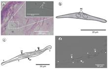
(A) Histological section of the fish gallbladder showing the spores of Ceratomyxa affinis n. sp. stained with eosin and hematoxylin. Highlighting: Single myxopore of Ceratomyxa affinis n. sp.; (B) Schematic drawing of Ceratomyxa affinis n. sp.; (C) Schematic drawing of plasmodium of Ceratomyxa affinis n. sp.; (D) Photomicrography of plasmodium (*) with fresh spores of Ceratomyxa affinis n. sp., in the gallbladder of Leporinus affinis. PC: polar capsule; SP: spore.
The myxospores of Ceratomyxa affinis n. sp. are 7.2 µm long and 43.2 µm thick with a posterior angle of 170º. The polar capsules are 3.9 µm long and 4.05 µm wide. Due to the conditions of the processed material, it was not possible to observe the turns of the polar filament. Regarding the motility of the plasmodia, they had undulating movements when observed fresh under light microscopy.
Remarks
In the state of Amapá, five species of Ceratomyxa spp. have been described, namely Ceratomyxa gracillima Zatti et al., 2018, Ceratomyxa macapaensis Bittencourt et al., 2022, Ceratomyxa matosi Martel et al., 2024, Ceratomyxa edilsonis Carvalho et al., 2024, Ceratomyxa tavariensis Araújo et al., 2024 and Ceratomyxa tessaloniensis Cardoso et al., 2025 in Mesonauta festivus, Boulengerella cuvieri, Pimelodella cristata, Pterophyllum scalare and Astyanax mexicanus, respectively. This makes Ceratomyxa affinis n. sp. described in L. affinis the seventh species of the genus Ceratomyxa occurring in the state of Amapá and the third species described from the Tartarugalzinho River, in the municipality of Tartarugalzinho.
When comparing the morphometric data of the myxosporean Ceratomyxa affinis n. sp. with the other species of Ceratomyxa spp. described in the state of Amapá, it is possible to observe that all the measurements of Ceratomyxa affinis n. sp. are larger than for all Ceratomyxa spp. described not only in the state of Amapá, but also in the Amazon region. Regarding the motility of the plasmodia and myxospore formation of Ceratomyxa spp. described in Amapá, the plasmodia and myxospores of Ceratomyxa affinis n. sp. resemble those described for C. edilsonis, C. matosi, C. tavariensis and C. macapaensis, which present vermiform plasmodia and slightly arched myxospores. This differs from the plasmodia of C. tessaloniensis, which had amoeboid-like motility and strongly arched myxospores.
Taxonomic summary
Kingdom: Animalia Linnaeus, 1758
Class Myxozoa Grassé, 1970 (Kyger et al., 2021)
Subclass Myxosporea Bütschli, 1881
Order Bivalvulida Shulman, 195
Family Ceratomyxidae Doflein, 1899
Genus Ceratomyxa Thélohan, 1892
Species Ceratomyxa affinis n.sp.
Host:Leporinus affinis Günther 1864
Site of infection: Gallbladder
Prevalence: 10/13 (76.9%)
Locality: Tartarugalzinho River, municipality of Tartarugalzinho, state of Amapá, Brazil (01º30’32.5” N; 050º55’10.3” W)
Type material: Glass slide with spores stained with Hematoxylin and Eosin (H&E) was deposited in the collection of the National Amazonas Research Institute (INPA), Manaus, Amazonas, Brazil (accession number: CND 000111).
GenBank accession number: PV765332
Etymology: The species-specific epithet refers to the species name of the host fish.
Molecular data and phylogenetic analysis
The partial SSU rDNA sequence for Ceratomyxa affinis n. sp. obtained in the present study had 870 base pairs (GenBank accession number: PV765332), which were G + C (A = 0.2718, C = 0.2007, G = 0.2849, T = 0.2426). Assuming a GTR + G model of nucleotide substitution, estimated nucleotide substitution rates were A - C = 0, 9048, A - G = 2.1973, A - T = 1.4964, C - G = 0.6508, C - T = 3.8475, G - T = 1.0000, with a gamma distribution of G = 0.5390.
To construct the phylogenetic tree, 16 sequences of species from the Ceratomyxidae family that are available in GenBank were used, and Ellipsomyxa tucujuensis Ferreira et al., 2021 and Myxodavisia bulani (Fiala et al., 2015) were used as outgroups. BLASTn search showed that the sequence of Ceratomytxa affinis n. sp. did not match any other sequence deposited in GenBank. In this analysis, no sequence had greater than 85.25% similarity with Ceratomyxa affinis n. sp. The analysis of the degree of coverage in BLASTn with the SSU 18S rDNA analysis from other Ceratomyxa spp., showed an approximate average similarity of 99% with the target sequence.
The analysis of the p distance showed a large genetic divergence between the other species of Ceratomyxa spp. (Table 1). The smallest genetic distance (p) found between Ceratomyxa affinis n. sp. with another species of Ceratomyxa was 6.8% with Ceratomyxa vermiformis Adriano and Okamura, 2017 and no more than 10.7% with Ceratomyxa mandii Araujo et al., 2022 (Table 1).
The phylogenetic tree showed two main clades, A and B, as shown in Figure 3, with strong nodal support. Clade A was formed by hosts found in the Brazilian Amazon and some marine species, whereas Clade B was formed by species that parasitized hosts of the order Siluriforms from the Brazilian Amazon region.

Maximum likelihood phylogenetic tree based on SSU-rDNA sequences of Ceratomyxa affinis n. sp. and other myxozoans. Nodal supports are indicated for Bayesian inference with posterior probabilities. GenBank accession numbers are presented after each myxozoans species name.
Ceratomyxa affinis n. sp. did not group with any species of Ceratomyxa spp. in clade A1 (nodal support = 0.8), while in clade A2 (nodal support = 0.7) all other species of Ceratomyxa spp. were organized, from both the Amazon region and from the marine environment. In the phylogenetic arrangement presented in Figure 3, a species of Ceratomyxa, Ceratomyxa ALTERADO Silva et al., 2020, described in Hemiodus unimaculatus Bloch, 1794, grouped in isolation, showing the same behavior as Ceratomyxa affinis n. sp. in clade A1.
Discussion
The genus Ceratomyxa is characterized by slightly arched myxospores, with two elongated valves that are generally wider than they are long (Gunter et al., 2009). Normally, myxospores of this genus are found inside plasmodia, and may have a shape and movement similar to worms or amoeboids (Zatti et al., 2023; Müller et al., 2025).
The myxospores of Ceratomyxa affinis n. sp. were found in vermiform plasmodia, as were the myxospores of C. edilsonis and C. tavariensis described in P. cristata and P. scalare, respectively (Araújo et al., 2024; Carvalho et al., 2024). Furthermore, the aforementioned species of Ceratomyxa spp. were captured in the same study area of this work, in the Tartarugalzinho River. There is thus a pattern of Ceratomyxidae plasmodia for the studied area. The plasmodia of Ceratomyxa affinis n. sp. differ from the plasmodia of C. tessaloniensis found in A. mexicanus, in the Flexal River, in the municipality of Macapá, which had movement and had amoeboid-like shapes. (Cardoso et al., 2025).
Müller et al. (2025) state that the motility of Ceratomyxa spp. plasmodia is restricted to the freshwater lineage of South America and to a few marine species, and that there is a large gap in the observation of the vegetative stages of coelozoic Ceratomyxidae, since the plasmodia structures are delicate and easily degraded post-mortem in the host, leaving only free myxospores in the parasitized organ.
Adriano et al. (2021) state that plasmodia of Ceratomyxa spp. with undulating motility similar to nematodes are exclusive to freshwater, that is, the plasmodia of Ceratomyxa affinis n. sp. confirm what was previously described, as also happens with the plasmodia of C. macapensis in M. festivus (Bittencourt et al., 2022), Ceratomyxa fonsecai in Hemiodus unimaculatus (Silva et al., 2020), C. matosi in B. cuvieri (Martel et al., 2024) and Ceratomyxa raniculiformis in Plagioscion squamosissimus (Zatti et al., 2023). All species of Ceratomyxa spp. from the Amazon region have polysporic plasmodia, as does Ceratomyxa affinis n. sp., which according to Silva et al. (2020), Okamura et al. (2015) and Bartošová-Sojková et al. (2018), is considered a more efficient strategy for dispersing parasites in the environment.
In the Amazon region, all species of Ceratomyxa spp. were found only in the gallbladder of their hosts, meaning that they showed tropism for this organ, a characteristic for freshwater fish. Nonetheless, there a report of the occurrence of Ceratomyxa spp. in the urinary bladder of marine fish (Eiras, 2006; Eiras et al., 2018).
Regarding the morphometric measurements of the myxospores of Ceratomyxa affinis n. sp., it was possible to observe that this presents the largest measurements of myxospores of Ceratomyxa spp. in the Amazon region. The myxospore length of Ceratomyxa affinis n. sp. (7.2 µm) was close to the length of Ceratomyxa amazonensis Mathews et al., 2016 with 7.0 µm. All other measurements were not close to any other species of Ceratomyxa spp. described in the Brazilian Amazon region (Table 2).
Comparative morphometric table of measurements (µm) of Ceratomyxa affinis n. sp. with other species of Ceratomyxa spp. described in brazilian amazon.
The shape of the myxospores of Ceratomyxa affinis n. sp. was slightly arched, as was also the case with Ceratomyxa deformis, Ceratomyxa anomala, C. matosi, C. macapaensis, C. edilsonis and C. tavariensis (Müller et al., 2025; Martel et al., 2024; Carvalho et al., 2024; Araújo et al., 2024). In South America, only three species of Ceratomyxa spp. have strongly aqueous myxospores, these being Ceratomyxa vermiformis in Colossoma macropomum (Adriano & Okamura, 2017), Ceratomyxa gracillima in Brachyplatystoma rousseauxii (Zatti et al., 2018) and C. tessaloniensis in A. mexicanus (Cardoso et al., 2025).
According to Fiala et al. (2015), Ceratomyxa leatherjacketi and Ceratomyxa tunisiensis are basal Ceratomyxidae for the phylogenetic arrangement of this family, and as demonstrated in the phylogenetic tree (Figure 3), these Ceratomyxa species mentioned above were found in the same clade (A) as Ceratomyxa affinis n. sp., corroborating the authors. These authors state that the long arms of the phylogenetic trees of Ceratomyxidae indicate rapid evolution of this group in freshwater environments.
Ceratomyxa affinis n. sp. behaved as an “outlier” species, due to its isolation in clade A1, that is, not grouping with any other Ceratomyxa species. Baum (2008) suggest that “outlier” species are closer to the root of the tree, indicating that they are considered to be the oldest or ancestral branches within the phylogenetic tree, or that they diverged earlier from the common ancestor of the group.
Conclusions
Based on morpho-molecular analyses, it was possible to prove that Ceratomyxa affinis n. sp. is a new species. This contributes to knowledge of the parasitic fauna of fish in the Amazon region, in which this was the seventh species of this genus described in the state of Amapá, and the third described in fish from the municipality of Tartarugalzinho. That may indicate a rapid evolution of the Ceratomyxidae family in the Brazilian Amazon region.
Acknowledgements
The authors would like to thank the all members of research grupo “Sanidade de Organismos Aquáticos da Amazônia” (SOAA) for all support in this study, to thank the fishermen, Nelson, for their local knowledge, support and availabity and to thank to Adria Sales for all the digital support for creating the schematic drawing. Also thank to Fundação Coordenação de Aperfeiçoamento de Pessoal de Nível Superior (CAPES) for financial support with scholarship to Luize Cristine Pantoja dos Reis.
Data availability
The DNA sequences are deposited in Genbank (PV765332); a glass slide with spores stained with hematoxylin and eosin (H&E) is deposited in the collection of the National Amazonas Research Institute (INPA), Manaus, Amazonas, Brazil (accession number: CND 000111). The data generated during the study are included in this article.
-
How to cite: Reis LCP, Carvalho AA, Ferreira RLS, Morais SC, Souza IWC, Araujo RF, et al. New freshwater Ceratomyxa species, Ceratomyxa affinis n. sp. (Myxozoa: Ceratomyxidae) in the gallbladder of Leporinus affinis from Brazilian Amazon. Rev Bras Parasitol Vet 2025; 34(4); e0096. https://doi.org/10.1590/S1984-29612025054
-
Ethics declaration
This study was performed in line with the principles of the Animal Use Ethics Committee of the Federal Rural University of the Amazon: nº 8323110522; and Biodiversity Authorization and Information System: licence 50376-1.
References
-
Adriano EA, Okamura B. Motility, morphology and phylogeny of the plasmodial worm, Ceratomyxa vermiformis n. sp. (Cnidaria: Myxozoa: Myxosporea). Parasitology 2017; 144(2): 158-168. http://doi.org/10.1017/S0031182016001852 PMid:27821209.
» http://doi.org/10.1017/S0031182016001852 -
Adriano EA, Zatti SA, Okamura B. How to build single-celled cnidarians with worm-like motility: lessons from Myxozoa. J Anat 2021; 240(3): 475-488. http://doi.org/10.1111/joa.13566 PMid:34643951.
» http://doi.org/10.1111/joa.13566 -
Altschul SF, Madden TL, Schäffer AA, Zhang J, Zhang Z, Miller W, et al. Gapped BLAST and PSI-BLAST: a new generation of protein database search programs. Nucleic Acids Res 1997; 25(17): 3389-3402. http://doi.org/10.1093/nar/25.17.3389 PMid:9254694.
» http://doi.org/10.1093/nar/25.17.3389 -
Araújo BL, Adriano EA, Franzolin GN, Zatti SA, Naldoni J. A novel Ceratomyxa species (Myxozoa: Cnidaria) infecting an Amazonian catfish. Parasitol Int 2022; 89: 102582. http://doi.org/10.1016/j.parint.2022.102582 PMid:35395395.
» http://doi.org/10.1016/j.parint.2022.102582 -
Araújo RF, Carvalho AA, Ferreira RLS, Morais SC, Reis LCP, Oliveira PLS, et al. A new Ceratomyxa (Cnidaria: Myxosporea) infecting the ornamental fish species Pterophyllum scalare from the Amazon Region, Brazil. Rev Bras Parasitol Vet 2024; 33(4): e018324. http://doi.org/10.1590/s1984-29612024075 PMid:39699488.
» http://doi.org/10.1590/s1984-29612024075 -
Atkinson SD, Bartholomew JL, Lotan T. Myxozoans: ancient metazoan parasites find a home in phylum Cnidaria. Zoology 2018; 129: 66-68. http://doi.org/10.1016/j.zool.2018.06.005 PMid:30170750.
» http://doi.org/10.1016/j.zool.2018.06.005 -
Azevedo C, Rocha S, Casal G, São Clemente SC, Matos P, Al-Quraishy S, et al. Ultrastructural description of Ceratomyxa microlepis sp. nov. (Phylum Myxozoa): a parasite infecting the gall bladder of Hemiodus microlepis, a freshwater teleost from the Amazon River. Mem Inst Oswaldo Cruz 2013; 108(2): 150-154. http://doi.org/10.1590/0074-0276108022013004 PMid:23579792.
» http://doi.org/10.1590/0074-0276108022013004 -
Bartošová-Sojková P, Lövy A, Reed CC, Lisnerová M, Tomková T, Holzer AS, et al. Life in a rock pool: radiation and population genetics of myxozoan parasites in hosts inhabiting restricted spaces. PLoS One 2018; 13(3): e0194042. http://doi.org/10.1371/journal.pone.0194042 PMid:29561884.
» http://doi.org/10.1371/journal.pone.0194042 - Baum D. Reading a phylogenetic tree: the meaning of monophyletic groups. Nat Ed 2008; 1(1): 190.
-
Bidone ED, Castilhos ZC, Santos TJS, Souza TMC, Lacerda LD. Fish contamination and human exposure to mercury in Tartarugalzinho River, Amapa State, Northern Amazon, Brazil: a screening approach. Water Air Soil Pollut 1997; 97(1-2): 9-15. http://doi.org/10.1023/A:1018383502637
» http://doi.org/10.1023/A:1018383502637 -
Birindelli JLO, Britski HA. Two new species of Leporinus (Characiformes: Anostomidae) from the Brazilian Amazon, and redescription of Leporinus striatus Kner 1858. J Fish Biol 2013; 83(5): 1128-1160. http://doi.org/10.1111/jfb.12206 PMid:24580659.
» http://doi.org/10.1111/jfb.12206 -
Bittencourt LS, Silva DT, Hamoy I, Carvalho AA, Silva MF, Videira M, et al. Morphological and phylogenetic features of Ceratomyxa macapaensis n. sp. (Myxozoa: Ceratomyxidae) in Mesonauta festivus Heckel, 1840 (Cichliformes: Cichlidae) from the Eastern Amazon Region. Acta Parasitol 2022; 67(1): 322-329. http://doi.org/10.1007/s11686-021-00460-x PMid:34426891.
» http://doi.org/10.1007/s11686-021-00460-x -
Bush AO, Lafferty KD, Lotz JM, Shostak AW. Parasitology meets ecology on its own terms: Margolis et al. revisited. J Parasitol 1997; 83(4): 575-583. http://doi.org/10.2307/3284227 PMid:9267395.
» http://doi.org/10.2307/3284227 -
Cardoso S, Carvalho AA, Ferreira RLS, Oliveira JEF, Araújo RF, Nascimento LSO, et al. Morphological and Phylogenetic Analyses of Ceratomyxa tessaloniensis n. sp. In Astyanax mexicanus De Fillipi, 1853 (Characiformes: Characidae) from the Flexal River, Tessalônica Community, Amapá, Amazon, Brazil. Acta Parasitol 2025; 70(1): 45. http://doi.org/10.1007/s11686-024-00958-0 PMid:39870884.
» http://doi.org/10.1007/s11686-024-00958-0 -
Carvalho AA, Ferreira RLS, Nascimento LSO, Morais SC, Araujo RF, Costa MA, et al. A gallbladder Ceratomyxidae (Myxozoa: Bivalvulida) parasite described in Pimelodella cristata (Müller & Troschel, 1848) from the Eastern Amazon. Acta Parasitol 2024; 69(4): 2006-2015. http://doi.org/10.1007/s11686-024-00930-y PMid:39388054.
» http://doi.org/10.1007/s11686-024-00930-y -
Eiras JC. Synopsis of the species of Ceratomyxa Thélohan, 1892 (Myxozoa: Myxosporea: Ceratomyxidae). Syst Parasitol 2006; 65(1): 49-71. http://doi.org/10.1007/s11230-006-9039-5 PMid:16676229.
» http://doi.org/10.1007/s11230-006-9039-5 -
Eiras JC, Cruz C, Saraiva A. Synopsis of the species of Ceratomyxa Thélohan, 1892 (Cnidaria, Myxosporea, Ceratomyxidae) described between 2007 and 2017. Syst Parasitol 2018; 95(5): 427-446. http://doi.org/10.1007/s11230-018-9791-3 PMid:29594910.
» http://doi.org/10.1007/s11230-018-9791-3 -
Eiras JC, Barman GD, Chanda S, Panigrahi AK. An update of the species of Myxosporea (Cnidaria, Myxozoa) described from Indian fish. J Parasit Dis 2023; 47(1): 12-36. http://doi.org/10.1007/s12639-022-01558-3 PMid:36910326.
» http://doi.org/10.1007/s12639-022-01558-3 -
Fiala P, Bartošová-Sojková CM, Whipps CM. Classification and phylogenetics of myxozoa. In: Okamura B, Gruhl A, Bartholomew JL, editors. Myxozoan evolution, ecology and development Cham: Springer; 2015. p. 85-110. http://doi.org/10.1007/978-3-319-14753-6_5
» http://doi.org/10.1007/978-3-319-14753-6_5 -
Franzolin GN, Araújo BL, Zatti SA, Naldoni J, Adriano EA. Occurrence of the host-parasite system Rhaphiodon vulpinus and Ceratomyxa barbata n. sp. in the two largest watersheds in South America. Parasitol Int 2022; 91: 102651. http://doi.org/10.1016/j.parint.2022.102651 PMid:35998817.
» http://doi.org/10.1016/j.parint.2022.102651 -
Froese R, Pauly D. FishBase [online]. Kiel: FishBase Team. 2024 [cited 2025 May 15]. Available from: http://www.fishbase.org
» http://www.fishbase.org -
Gunter NL, Whipps CM, Adlard RD. Ceratomyxa (Myxozoa: Bivalvulida): robust taxon or genus of convenience? Int J Parasitol 2009; 39(12): 1395-1405. http://doi.org/10.1016/j.ijpara.2009.04.008 PMid:19460381.
» http://doi.org/10.1016/j.ijpara.2009.04.008 -
Hillis DM, Dixon MT. Ribosomal DNA: molecular evolution and phylogenetic inference. Q Rev Biol 1991; 66(4): 411-453. http://doi.org/10.1086/417338 PMid:1784710.
» http://doi.org/10.1086/417338 -
Kearse M, Moir R, Wilson A, Stones-Havas S, Cheung M, Sturrock S, et al. Geneious basic: an integrated and extendable desktop software platform for the organization and analysis of sequence data. Bioinformatics 2012; 28(12): 1647-1649. http://doi.org/10.1093/bioinformatics/bts199 PMid:22543367.
» http://doi.org/10.1093/bioinformatics/bts199 -
Kyger R, Luzuriaga-Neira A, Layman T, Milkewitz Sandberg TO, Singh D, Huchon D, et al. Myxosporea (Myxozoa, Cnidaria) lack DNA cytosine methylation. Mol Biol Evol 2021; 38(2): 393-404. http://doi.org/10.1093/molbev/msaa214 PMid:32898240.
» http://doi.org/10.1093/molbev/msaa214 -
Lom J, Arthur JR. A guideline for the preparation of species descriptions in Myxosporea. J Fish Dis 1989; 12(2): 151-156. http://doi.org/10.1111/j.1365-2761.1989.tb00287.x
» http://doi.org/10.1111/j.1365-2761.1989.tb00287.x - Luna LG. Manual of histologic staining methods of the Armed Forces Institute of Pathology 3rd ed. New York: McGraw‐Hill; 1968.
-
Martel CS, Souza FA, Vidal SC, Carvalho AA, Hamoy IG, Nascimento LSO, et al. Ceratomyxa matosi n. sp. (Myxozoa: Ceratomyxidae) parasitizing the gallbladder of Boulengerella cuvieri (Characiformes: Ctenoluciidae) State of Amapá, Brazilian Amazon. Rev Bras Parasitol Vet 2024; 33(3): e011024. http://doi.org/10.1590/s1984-29612024058 PMid:39383387.
» http://doi.org/10.1590/s1984-29612024058 -
Mathews PD, Naldoni J, Maia AA, Adriano EA. Morphology and small subunit rDNA-based phylogeny of Ceratomyxa amazonensis n. sp. parasite of Symphysodon discus, an ornamental freshwater fish from Amazon. Parasitol Res 2016; 115(10): 4021-4025. http://doi.org/10.1007/s00436-016-5173-4 PMid:27314232.
» http://doi.org/10.1007/s00436-016-5173-4 -
Miller MA, Pfeiffer W, Schwartz T. Creating the CIPRES Science Gateway for inference of large phylogenetic trees New York: IEEE; 2010. http://doi.org/10.1109/GCE.2010.5676129
» http://doi.org/10.1109/GCE.2010.5676129 -
Molnár K, Eszterbauer E, Székely C, Dán Á, Harrach B. Morphological and molecular biological studies on intramuscular Myxobolus spp. of cyprinid fish. J Fish Dis 2002; 25(11): 643-652. http://doi.org/10.1046/j.1365-2761.2002.00409.x
» http://doi.org/10.1046/j.1365-2761.2002.00409.x -
Müller MI, Atkinson SD, Bartholomew JL, Adriano EA. Novel Amazonian Ceratomyxa species (Cnidaria: Myxozoa) with amoeboid plasmodial motility. Microb Pathog 2025; 199: 107235. http://doi.org/10.1016/j.micpath.2024.107235 PMid:39716650.
» http://doi.org/10.1016/j.micpath.2024.107235 -
Okamura B, Gruhl A, Bartholomew JL. Myxozoan evolution, ecology and development Cham: Springer International Publishing; 2015. http://doi.org/10.1007/978-3-319-14753-6
» http://doi.org/10.1007/978-3-319-14753-6 -
Okamura B, Hartigan A, Naldoni J. Extensive uncharted biodiversity: the parasite dimension. Integr Comp Biol 2018; 58(6): 1132-1145. http://doi.org/10.1093/icb/icy039 PMid:29860443.
» http://doi.org/10.1093/icb/icy039 -
Ronquist F, Huelsenbeck JP. MrBayes 3: bayesian phylogenetic inference under mixed models. Bioinformatics 2003; 19(12): 1572-1574. http://doi.org/10.1093/bioinformatics/btg180 PMid:12912839.
» http://doi.org/10.1093/bioinformatics/btg180 -
Santos GM, Zuanon J. Leporinus amazonicus, a new anostomid species from the amazon lowlands, Brazil (Osteichthyes: characiformes). Zootaxa 2008; 1815(1): 35-42. http://doi.org/10.11646/zootaxa.1815.1.3
» http://doi.org/10.11646/zootaxa.1815.1.3 -
Silva MF, de Carvalho AEFB, Hamoy I, Matos ER. Coelozoic parasite of the family Ceratomyxidae (Myxozoa, Bivalvulida) described from motile vermiform plasmodia found in Hemiodus unimaculatus Bloch, 1794. Parasitol Res 2020; 119(3): 871-878. http://doi.org/10.1007/s00436-019-06505-5 PMid:31897794.
» http://doi.org/10.1007/s00436-019-06505-5 - Silveira OFM, Silveira OT, Neto SVC. História natural das regiões dos Lagos e Sucuriju Amapá, Brasil. In: Costa SV No, editor. Inventário biológico das áreas do Sucuriju e região dos Lagos, no Amapá: relatório final PROBIO. Macapá: IEPA; 2015.
-
Sindeaux-Neto JL, Velasco M, Silva DT, Matos P, Silva MF, Gonçalves EC, et al. Myxobolus freitasi n. sp. (Myxozoa: Bivalvulida), a parasite of the brain of the electric knifefish in the Brazilian Amazon region. Rev Bras Parasitol Vet 2021; 30(1): e020920. http://doi.org/10.1590/s1984-296120201081 PMid:33729315.
» http://doi.org/10.1590/s1984-296120201081 -
Whipps CM, Adlard RD, Bryant MS, Lester RJG, Findlav V, Kent ML. First report of three Kudoa Species from Eastern Australia: Kudoa thyrsites from Mahi mahi (Coryphaena hippurus), Kudoa amamiensis and Kudoa minithyrsites n. sp. from Sweeper (Pempheris ypsilychnus). J Eukaryot Microbiol 2003; 50(3): 215-219. http://doi.org/10.1111/j.1550-7408.2003.tb00120.x PMid:12836879.
» http://doi.org/10.1111/j.1550-7408.2003.tb00120.x - Yokomizo GKI. Produtividade da soja na região do Município de Tartarugalzinho-AP: comunicado técnico (INFOTECA-E) Macapá: Embrapa Amapá; 2012.
-
Zacardi DM, Bittencourt SCS, Nakayama L, Queiroz HL. Distribution of economically important fish larvae (Characiformes, Prochilodontidae) in the Central Amazonia, Brazil. Fish Manag Ecol 2017; 24(4): 283-291. http://doi.org/10.1111/fme.12222
» http://doi.org/10.1111/fme.12222 -
Zatti SA, Atkinson SD, Bartholomew JL, Maia AAM, Adriano EA. Amazonian waters harbour an ancient freshwater Ceratomyxa lineage (Cnidaria: myxosporea). Acta Trop 2017; 169: 100-106. http://doi.org/10.1016/j.actatropica.2017.02.006 PMid:28185825.
» http://doi.org/10.1016/j.actatropica.2017.02.006 -
Zatti SA, Atkinson SD, Maia AAM, Bartholomew JL, Adriano EA. Ceratomyxa gracillima n. sp. (Cnidaria: Myxosporea) provides evidence of panmixia and ceratomyxid radiation in the Amazon basin. Parasitology 2018; 145(9): 1137-1146. http://doi.org/10.1017/S0031182017002323 PMid:29338808.
» http://doi.org/10.1017/S0031182017002323 - Zatti SA, Araújo BL, Adriano EA, Maia AAM. A new freshwater Ceratomyxa species (Myxozoa: Ceratomyxidae) parasitizing a sciaenid fish from the Amazon Basin, Brazil. Parasitol Int 2023; 97: 102796. http://doi.org/10.1016/j.parint.2023.102796. PMid:37595832.
Edited by
-
Assistant Editor:
Marcos Tavares Dias


