ABSTRACT
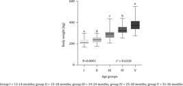
The present study aimed to correlate the age and weight of Girolando heifers with uterine and ovarian development. Sixty heifers between 12 and 36 months old were weighed and subjected to ultrasound assessment to measure the diameters of each uterine horn and ovaries, monthly. These measures continued until their first ovulation. The animals were divided, for data analysis, according to their age into five groups: GI (12 to 14 months), GII (15 to 18 months), GIII (19 to 24 months), GIV (25 to 30 months), and GV (31 to 36 months). The dispersion diagram was used to assess the correlation between data collected monthly from uterus/ovaries and weight/age. An average daily growth of 0.0032 mm was recorded for the right ovary (RO) and 0.0051 mm for the left ovary (LO). The average size of the RO in GI was 17.58 mm and the LO was 15.28 mm; in GII, the RO was 16.72 mm and the LO was 15.89 mm; in GIII, the RO was 18.37 mm and the LO was 16.55 mm; in GIV, the RO was 19.09 mm and the LO was 17.49 mm; and in GV, the RO was 19.89 mm and the LO was 18.96 mm. The uterine horns showed an average daily growth of 0.0064 mm and 0.0071 mm for the right (RU) and left (LU) uterine horn, respectively. The average sizes of the uterine horns per group were: RU, 11.46 mm and LU, 11.48 mm; RU, 12.41 mm and LU, 12.63 mm; RU, 13.16 mm and LU, 13.19 mm; RU 14.30 mm and LU, 14.58 mm; and RU, 16.12 mm and LU, 16.68 mm for the groups GI, GII, GIII, GIV, and GV, respectively. The heifers showed an average daily weight gain of 0.224 kg and the groups GI, GII, GIII, GIV, and GV had an average weight of 220.5, 239.3, 287.6, 332.9, and 380.1 kg, respectively. Therefore, weight growth correlates with the growth of the ovaries and uterine horns; furthermore, these data correlate positively as the animals age.
Keywords:
morphometry; puberty; weight growth
1. Introduction
The Girolando breed is one of the most popular crossbreds in Brazil, due to high dairy production and appropriate rusticity to develop well in a tropical climate. Replacement heifers play a crucial role in milk production system in Brazil, since the age at which they enter into reproductive life is an important factor for the reproductive efficiency of the property, as well as for the profitability and recovery of the capital invested in these animals. Puberty is influenced by not only genetic factors, breed, and heredity, but also by external factors, directly linked to nutritional management and their environment. Puberty can be studied from a physiological or animal performance point of view. Physiological puberty is the age when the animal comes into its first heat followed by ovulation, which is usually when the animal weighs around 40 to 50% of its adult body weight (Stevenson, 2007Stevenson, J. S. 2007. Clinical reproductive physiology of the cow. p.258-270. In: Current therapy in large animal theriogenology. 2nd ed. Youngquist, R. S. and Threlfall, W. R., eds. Saunders Elsevier, Missouri, USA. https://doi.org/10.1016/B978-072169323-1.50038-6
https://doi.org/10.1016/B978-072169323-1...
). While, animal performance puberty is characterized as the moment when the heifer reaches 65 to 70% of the average live weight of an adult female in its herd and can begin to reproduce without any consequences related to its physiological development (Gonçalves et al., 2008Gonçalves, P. B. D.; Figueiredo, J. R. and Freitas, V. J. F. 2008. Biotécnicas aplicadas à reprodução animal. 2.ed. Roca, São Paulo.). Besides age and weight, the development of the reproductive tract is of fundamental importance for the fertility of bovine females. The evaluation and monitoring of the reproductive tract can determine whether the internal reproductive organs are able to conceive, gestate, and achieve parturition. Thus, with the assessment and monitoring of the factors mentioned above, the producer is able to plan managements that will provide an increase in the reproductive efficiency of the herd and, consequently, greater productivity of the property.
Research related to this theme is especially important to a country with a vocation for agribusiness. Thus, the objective of this study was to analyze the influence of age and weight on uterine and ovarian development of Girolando heifers, from twelve months old to their first ovulation, in a descriptive manner, to propose the hypothesis that these variables are related.
2. Material and Methods
This experiment was conducted on a farm in Seropédica, RJ, Brazil (22°44'29" S, 43°42'19" W, and 682 m altitude). All the experimental procedures adopted were approved by the local ethics committee on animal use (case no. 23083.025555/2017-43), in accordance with the ethical principles of animal experimentation established by the National Council for the Control of Experimentation Animal (CONCEA) and with the current legislation.
Sixty Girolando heifers from 12 to 36 months old were used from different genetic groups (ranging from ¼ to ¾ Holstein × Gir). The information obtained monthly from the animals was recorded separately according to five age groups – 12 to 14, 15 to 18, 19 to 24, 25 to 30, and 31 to 36 months (GI, GII, GIII, GIV, and GV, respectively) – to be able to count on the largest number of animals (data) available for this study. Data collection started with the 12-month-old heifers that, as they grew older, passed through the different age groups, and, throughout the study period, new animals came into the GI for evaluation. All heifers were kept in the same pasture area (Panicum maximum cv. Tanzânia) and with access to good quality water ad libitum. Sorghum silage was provided in a trough once a day, at the rate of approximately 2 kg per animal, between July and September.
The heifers were weighed monthly on a digital scale, from 12 months until the moment their first ovulation was detected (presence of corpus luteum in one of the ovaries). On the same day that the heifers were weighed, they were evaluated by rectal palpation and by ultrasound examination (Mindray DP 2200-Vet, 7.5 MHz rectal transducer) until the first ovulation was detected. The ultrasound evaluation of the uterus recorded the diameter of the right and left horns with a cross-section measure, immediately after the uterine bifurcation when the two uterine horns could be observed separately, as described by Silenciato et al. (2020)Silenciato, L. N.; Ferreira, J. E.; Couto, S. R. B.; Silva, O. R.; Resende, O. A.; Silva, A. F.; Palhano, H. B. and Mello, M. R. B. 2020. Ultrasonographic evaluation of the reproductive tract as predictors of pregnancy in Girolando heifers submitted to Timed Artificial Insemination. Brazilian Journal of Veterinary Medicine 42:e104220. https://doi.org/10.29374/2527-2179.bjvm104220
https://doi.org/10.29374/2527-2179.bjvm1...
. Two measures of the largest cross-sectional diameters of each horn were made, and the average was recorded. The right and left ovaries were also evaluated according to their diameters, as well as to the presence of antral follicles and corpus luteum. To calculate the average diameter of the ovaries and uterine horns of each heifer in the experiment, the total number of measurements made was used. For this reason, the “n” value in some situations was greater than 60. Because the corpus luteum remains in the ovary for approximately 17 days in non-pregnant females, every fifteen days, the heifers were examined with the aid of ultrasound equipment for ovarian evaluation and detection of the corpus luteum.
Heifers that reached puberty were inseminated after natural estrus observation or synchronized for embryo transfer (used as recipients). Thus, age and weight at puberty (first ovulation) and age at first service (artificial insemination or embryo transfer) were recorded. Heifers that became pregnant were followed up to calving, and the pregnancy rate at the first service and the average age at the first calving were calculated.
Data on reproductive structures from 12 months to the insemination of heifers were subjected to analysis of variance and used for distribution in five different age groups (13, 16, 22, 27, and 35 months). The experimental design was completely randomized, with different numbers of repetitions, with repeated measures. The data were subjected to analysis of variance and the F test, according to the following model:
in which Yij = dependent variable associated with the h-th animal, μ = average of all observations, αi = fixed effect of organ i (body weight, diameter of the uterus and ovary), and eij = random error associated with each observation. The data were subjected to analysis of variance at the 5% significance level, using GraphPadPrism (version 5.0). When the differences were detected, the means were compared by the Fisher test, at the 5% significance level, or by the Bonferroni test, at the 10% significance level.
3. Results
When analyzing the development of the genital system, an increase in the ovaries was observed (Table 1) with the evolution of age according to age groups (GI, GII, GIII, GIV, and GV). The diameter of the right ovary (RO) showed an average daily growth of 0.0032 mm. When analyzing the differences between the respective groups, the analysis of variance did not find any significant difference in diameters of the right ovaries between GI and GII, GI and GIII, GIII and GIV, and GIV and GV (P>0.05) (Figure 1).
Evolution of weights, ovaries, and uterine horns of Girolando heifers according to age groups
Diameter of the right (A) and left (B) ovaries of Girolando heifers according to age group.
Regarding the diameter of the left ovary (LO), the average daily growth observed was 0.0051 mm. The differences between the diameters of the left ovaries in the age groups I and II, I and III, II and III, III and IV, and IV and V again were not significant, according to the analysis of variance (P>0.05) (Figure 1).
The analysis of the relationship between variables using Pearson's correlation model (P<0.05) demonstrated a growth relationship between the right and left ovaries (Figure 2). An increase in the uterine horns was observed with evolution of age. Table 1 shows the average diameter of this variable according to each group, as well as the number of evaluations performed during the data collection period, standard deviation, maximum and minimum values, and the coefficient of variation of the right (RU) and left (LU) uterine horns, respectively.
Relationship in the diameter growth between the right (RO) and left (LO) ovaries of Girolando heifers according to age groups.
When analyzing the diameter of the right and left uterine horns in the groups, no significant differences were found in the diameters of the right uterine horn between GI and GII and between GII and GIII (P>0.05), and for the left uterine horn, there were also no significant differences between GI and GII and GII and GIII (P>0.05) (Figure 3).
Diameter of the left (A) and right (B) uterine horns of Girolando heifers according to age groups.
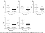
The analysis of the relationship between the variables using Pearson's correlation model (P<0.05) demonstrated a growth relationship between the right and left uterine horns (Figure 4). Regarding weight growth, the analysis of variance showed a significant difference (P<0.05) among all the evaluated groups (Figure 5). Through the unpaired T test, the differences in groups were evaluated, regarding the diameters between right and left ovaries, right and left uterine horns, right ovary and right uterine horn, right ovary and left uterine horn, left ovary and right uterine horn, and left ovary and left uterine horn. All variables analyzed showed significant differences in all groups (Figures 6, 7, 8, 9, 10, and 11).
Relationship in the growth of the left (LU) and right (RU) uterine horns of Girolando heifers according to age groups.
Weight development (body weight) of Girolando heifers belonging to age groups during the data collection period.
Difference in diameter between right (RO) and left (LO) ovaries of Girolando heifers according to age groups.
Difference in diameter between right (RU) and left (LU) uterine horns of Girolando heifers according to age groups.
Difference in diameter between the right ovary (RO) and the right (RU) uterine horn of Girolando heifers according to age groups.
Difference in diameter between the right ovary (RO) and the left (LU) uterine horn of Girolando heifers according to age groups.
Relationship between the growth of the diameter of the left ovary (LO) and the right (RU) uterine horn with the age of Girolando heifers according to age groups.
Relationship between the growth of the diameter of the left ovary (LO) and the left (LU) uterine horn with the age of Girolando heifers according to age groups.
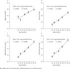
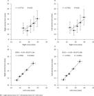
Using Pearson's correlation model, the relationships between the diameter of the right ovary and age, left ovary and age, right uterine horn and age, and left uterine horn and age were evaluated, which made it possible to assess the relationship of these variables with weight gain, as it is contextualized with the age groups (Figure 12). Still using the same statistical model, the relationship between the diameters of right and left ovaries in the groups, diameters of the right ovaries and right uterine horns, diameters of the right ovaries and left uterine horns, diameters of the left ovaries and right uterine horns, and diameters of the left ovaries and left uterine horns were evaluated. All variables analyzed showed a positive relationship (Figure 13).
Relationship between the growth of the diameter of the right (A) and left (B) ovaries and right (C) and left (D) uterine horns with the age of Girolando heifers.
Relationship of growth of the diameter of the right ovaries with right (A) and left (B) uterine horns, left ovary with right (C) and left (D) uterine horns with the age of Girolando heifers.
Out of the 60 animals that participated in the data collection of this study, 34 (56.7%) reached puberty during the development of the study. The average weight and age at first ovulation were 354.2 kg and 29.67 months, respectively (Table 2). The pregnancy rate (PR) at the first service was 64.7%; out of 34 animals that reached puberty, 16 females were conceived through conventional artificial insemination (PR = 75%) and 18 animals through timed embryo transfer (PR = 55.6%).
4. Discussion
In the present study, ovary growth was observed as the age of the heifers increased, reflecting the development of the ovarian stroma and an increase in the quantity and size of the antral follicles, which, according to Honaramooz et al. (2004)Honaramooz, A.; Aravindakshan, J.; Chandolia, R. K.; Beard, A. P.; Bartlewski, P. M.; Pierson, R. A. and Rawlings, N. C. 2004. Ultrasonographic evaluation of the pre-pubertal development of the reproductive tract in beef heifers. Animal Reproduction Science 80:15-29. https://doi.org/10.1016/S0378-4320(03)00136-2
https://doi.org/10.1016/S0378-4320(03)00...
, tend to follow the growth of the ovaries. The increase in ovarian activity is characteristic of the transition from the prepubertal to the peri-pubertal period due to the progressive reduction in the negative feedback exerted by estradiol, resulting in the establishment of the LH release pattern, which allows the follicles to reach the final stages of growth and maturation through to ovulation.
The growth of the right and left ovaries showed a positive relationship between them (P<0.0001; r2: RO = 0.07783 and LO = 0.09453), despite having different sizes. The right ovaries had a lower daily growth rate than the left ovaries, despite being slightly larger than the group averages. Such results disagree with those found by Honaramooz et al. (2004)Honaramooz, A.; Aravindakshan, J.; Chandolia, R. K.; Beard, A. P.; Bartlewski, P. M.; Pierson, R. A. and Rawlings, N. C. 2004. Ultrasonographic evaluation of the pre-pubertal development of the reproductive tract in beef heifers. Animal Reproduction Science 80:15-29. https://doi.org/10.1016/S0378-4320(03)00136-2
https://doi.org/10.1016/S0378-4320(03)00...
, who observed that the size of heifer ovaries grew with age, but did not differ and there was no interaction between them. On the other hand, Monteiro et al. (2008)Monteiro, C. M. R.; Perri, S. H. V.; Carvalhal, R. and Carvalho, R. G. 2008. Estudo morfológico comparativo dos ovários de vacas e novilhas da raça Nelore (Bos taurus indicus). Ars Veterinaria 24:122-126. observed that the right ovaries, both of cows and heifers, weighed more and were larger than the left ovaries, similar to the results of the present study. The right (P<0.0001; r2 = 0.07318) and the left (P<0.0001; r2 = 0.09453) ovaries were also positively related to weight gain. This result corroborates those found by Št'astná and Št'astný (2018)Št'astná, D. and Št'astný, P. 2018. The morphological changes of uterus in postnatal development of heifers. Acta Fytotechnica et Zootechnica 21:52-59. https://doi.org/10.15414/afz.2018.21.02.52-59
https://doi.org/10.15414/afz.2018.21.02....
, who, by inducing subclinical hypoglycemia by reducing the portions of their diet from the postnatal phase until the pubescent period, observed a decrease in the weight of the ovaries of the animals undergoing treatment, and obtained a strong relationship between ovarian development and carcass weight.
A decrease in the mean size of the right ovary (Table 1 and Figure 1) was observed in animals in GII (15 to 18 months of age). This fact can be explained by the entry of some new animals to the group. Some heifers were unable to enter GI at 12 months as the evaluator could not reach through the rectum for rectal palpation and, consequently, for the ultrasound evaluation. These animals only joined the evaluation groups after they had reached an appropriate size. These differences in size between animals of the same age range may be related to the different genetic groups of Girolando heifers and may have resulted in a decrease in the average size of this organ. Further studies on a descriptive research bases in the area of anatomical and physiological events are necessary to elucidate the respective observation.
Like that of the ovaries, the growth of the uterine horns was also related to the age of the animals in the study and there was a positive relationship between the right and left horns (P<0.0001; r2: RU = 0.09453 and LU = 0.2257). The left uterine horns obtained an average daily growth and an average size in the groups greater than the right uterine horns. Bilateral asymmetry has been documented in the reproductive tract of cattle, although many aspects of its physiology remain unknown. Similar data were found by Parkale and Hukeri (1989)Parkale, D. D. and Hukeri, V. B. 1989. Study of biometry of buffalo (Bubalus bubalis) ovaries. The Indian Journal of Animal Reproduction 10:17-19. in a study carried out with the genital tract of buffaloes. However, Correia et al. (2018)Correia, P. B. C.; Baron, E. E.; Pavani, K.; Pacheco-Lima, J.; Lopes, S.; Nunes, H.; Lourenço, J.; Furnas, S. and Silva, F. M. 2018. Morphometric characterization of lidia cow (Bos taurus) reproductive apparatus. Spanish Journal of Agricultural Research 16:e04SC03. https://doi.org/10.5424/sjar/2018163-12833
https://doi.org/10.5424/sjar/2018163-128...
concluded that the right and left horns are practically symmetrical in a morphometric study of the reproductive tract of Lidia cows (Bos taurus taurus).
The results of the present study demonstrated a positive relationship between the growth of uterine horns and ovarian development. More specifically the results were: between the growth of the right uterine horn and the right ovary (P<0.0001; r2: RU = 0.2032 and RO = 0.07783), between the right uterine horn and the left ovary (P<0.0001; r2: RU = 0.2032 and LO = 0.09453), between the left uterine horn and the left ovary (P = 0.0001; r2: RU = 0.2257 and LO = 0.09453), as well as between the left uterine horn and the right ovary (P<0.0001; r2: LU = 0.2257 and RO = 0.07783). Honaramooz et al. (2004)Honaramooz, A.; Aravindakshan, J.; Chandolia, R. K.; Beard, A. P.; Bartlewski, P. M.; Pierson, R. A. and Rawlings, N. C. 2004. Ultrasonographic evaluation of the pre-pubertal development of the reproductive tract in beef heifers. Animal Reproduction Science 80:15-29. https://doi.org/10.1016/S0378-4320(03)00136-2
https://doi.org/10.1016/S0378-4320(03)00...
observed a strong relationship between the development of the segments of the reproductive tract (vagina, cervix, and body of the uterus) with the pattern of changes in the maximum diameter of the ovarian follicle of two-week-old heifers until puberty. Larger follicles have greater steroidogenic capacity, suggesting that periods of accelerated follicle growth, influenced by gonadotropins, resulted in increases in the growth of the reproductive tract. Št'astná and Št'astný (2018)Št'astná, D. and Št'astný, P. 2018. The morphological changes of uterus in postnatal development of heifers. Acta Fytotechnica et Zootechnica 21:52-59. https://doi.org/10.15414/afz.2018.21.02.52-59
https://doi.org/10.15414/afz.2018.21.02....
also found a highly significant influence of ovarian growth on the number and size of follicles between 4 and 13 mm and in the changes that occur in uterine weight and diameter during the development of heifers from three to 15 months old. However, Silenciato et al. (2020)Silenciato, L. N.; Ferreira, J. E.; Couto, S. R. B.; Silva, O. R.; Resende, O. A.; Silva, A. F.; Palhano, H. B. and Mello, M. R. B. 2020. Ultrasonographic evaluation of the reproductive tract as predictors of pregnancy in Girolando heifers submitted to Timed Artificial Insemination. Brazilian Journal of Veterinary Medicine 42:e104220. https://doi.org/10.29374/2527-2179.bjvm104220
https://doi.org/10.29374/2527-2179.bjvm1...
found a negative relationship between ovarian and uterine diameters when assessing the degree of development of the reproductive tract of crossbred Girolando heifers ranging in age from 19 to 37 months old, subjected to a timed artificial insemination protocol. In this work, the average diameter of the right and left horns was calculated, and from these two values, the average was calculated, reaching a single value to represent the uterine diameter of each animal.
During the aforementioned study by Št'astná and Št'astný (2018)Št'astná, D. and Št'astný, P. 2018. The morphological changes of uterus in postnatal development of heifers. Acta Fytotechnica et Zootechnica 21:52-59. https://doi.org/10.15414/afz.2018.21.02.52-59
https://doi.org/10.15414/afz.2018.21.02....
, the ovarian influence on uterine development was greater than the influence of body weight, but the carcass weight also had a strong significant association with the uterine diameter. The study also concluded the importance of sufficient energy to support development. Progressive changes have been observed in the development of mucous structures, especially the mucous component of the uterine epithelium, as well as a different development and quantitative changes in the superficial and glandular cells of the uterus that are under the control of prepubertal steroids. Heifers induced to subclinical hypoglycemia showed a delay in reproductive development, presenting uterine structures of prepubertal animals, which were not consistent with their age that was of animals already able to reproduce. Our results corroborate those found by Št'astná and Št'astný (2018)Št'astná, D. and Št'astný, P. 2018. The morphological changes of uterus in postnatal development of heifers. Acta Fytotechnica et Zootechnica 21:52-59. https://doi.org/10.15414/afz.2018.21.02.52-59
https://doi.org/10.15414/afz.2018.21.02....
, when we found a positive relationship between weight gain and the growth of the right (P<0.0001; r2 = 0.2032) and left (P<0.0001; r2 = 0.2257) uterine horns.
In a study carried out with 488 beef heifers, Holm et al. (2016)Holm, D. E.; Nielen, M.; Jorritsma, R.; Irons, P. C. and Thompson, P. N. 2016. Ultrasonographic reproductive tract measures and pelvis measures as predictors of pregnancy failure and anestrus in restricted bred beef heifers. Theriogenology 85:495-501. https://doi.org/10.1016/j.theriogenology.2015.09.031
https://doi.org/10.1016/j.theriogenology...
stated that the development of the reproductive system is influenced by both age and live weight. Therefore, post-weaning nutrition is important to influence age at puberty.
Another study with dairy heifers also showed that the feeding strategy had a clear influence on weight and age at puberty. Furthermore, a high level of cholesterol during the initial stages of growth, due to the nutritional scheme, was related to the precocity of reaching puberty. This finding has important implications for the commercial management of heifers to reduce the cost of replacing dairy herds (Abeni et al., 2019Abeni, F.; Petrera, F. and Le Cozler, Y. 2019. Effects of feeding treatment on growth rates, metabolic profiles and age at puberty, and their relationships in dairy heifers. Animal 13:1020-1029. https://doi.org/10.1017/S1751731118002422
https://doi.org/10.1017/S175173111800242...
). According to Kenny et al. (2018)Kenny, D. A.; Heslin, J. and Byrne, C. J. 2018. Early onset of puberty in cattle: implications for gamete quality and embryo survival. Reproduction, Fertility and Development 30:101-117. https://doi.org/10.1071/RD17376
https://doi.org/10.1071/RD17376...
, there is an agreement among most authors in the field that the influence of nutrition on the sexual maturation of bulls and heifers is greater the earlier it is applied in postnatal life. Heifers showed a decrease of one to three months in age at puberty when they received improved nutrition before they were six months old compared with heifers that received better feed after they were six months old (Gasser, 2013Gasser, C. L. 2013. Joint Alpharma-Beef Species Symposium: Considerations on puberty in replacement beef heifers. Journal of Animal Science 91:1336-1340. https://doi.org/10.2527/jas.2012-6008
https://doi.org/10.2527/jas.2012-6008...
).
The weight of heifers was positively related to age. As these animals grew older, their weight increased, as reported by Silenciato et al. (2020)Silenciato, L. N.; Ferreira, J. E.; Couto, S. R. B.; Silva, O. R.; Resende, O. A.; Silva, A. F.; Palhano, H. B. and Mello, M. R. B. 2020. Ultrasonographic evaluation of the reproductive tract as predictors of pregnancy in Girolando heifers submitted to Timed Artificial Insemination. Brazilian Journal of Veterinary Medicine 42:e104220. https://doi.org/10.29374/2527-2179.bjvm104220
https://doi.org/10.29374/2527-2179.bjvm1...
and Duplessis et al. (2015)Duplessis, M.; Cue, R. I.; Santschi, D. E.; Lefebvre, D. M. and Lacroix, R. 2015. Weight height and relative-reliability indicators as a management tool for reducing age at first breeding and calving of dairy heifers. Journal of Dairy Science 98:2063-2073. https://doi.org/10.3168/jds.2014-8279
https://doi.org/10.3168/jds.2014-8279...
in works evaluating dairy cattle. The Girolando heifers in the present study had a mean age at puberty of 29.67 months with an average weight of 354.20 kg and an average gain of 0.224 kg per day.
The average age at puberty for Zebu breeds may vary according to the intensity of the production system and the appropriate nutritional management (Nogueira, 2004Nogueira, G. P. 2004. Puberty in South American Bos indicus (Zebu) cattle. Animal Reproduction Science 82-83:361-372. https://doi.org/10.1016/j.anireprosci.2004.04.007
https://doi.org/10.1016/j.anireprosci.20...
), as well as the weight that these animals present at their first ovulation. Landarin et al. (2016)Landarin, C. M.; Lobato, J. F. P.; Tarouco, A. K.; Tarouco, J. U.; Eloy, L. R.; Pötter, L. and Rosa, A. A. G. 2016. Growth and reproductive performance of 14- to 15-month-old Hereford heifers. Revista Brasileira de Zootecnia 45:667-676. https://doi.org/10.1590/s1806-92902016001100005
https://doi.org/10.1590/s1806-9290201600...
concluded from the results of several authors that the weight at puberty of F1 animals from the cross between Holstein and Zebu cattle ranges from 300 to 340 kg, more specifically 307 kg for F1 crossbred Holstein × Gir heifers. In a study by Bergfeld et al. (1994)Bergfeld, E. G. M.; Kojima, F. N.; Cupp, A. S.; Wehrman, M. E.; Peters, K. E.; Garcia-Winder, M. and Kinder, J. E. 1994. Ovarian follicular development in prepubertal heifers is influenced by level of dietary energy intake. Biology of Reproduction 51:1051-1057. https://doi.org/10.1095/biolreprod51.5.1051
https://doi.org/10.1095/biolreprod51.5.1...
, heifers that were fed to achieve an average daily gain of 0.9 kg were 12.4 months old at puberty, while their contemporaries that received a diet to reach an average of 0.3 kg per day reached puberty at 14.5 months old.
A study by Heslin et al. (2017)Heslin, J.; Kenny, D. A.; Kelly, A. K. and McGee, M. 2017. Age at puberty and pregnancy rate in beef heifer genotypes offered contrasting nutrition levels. Journal of Animal Science 95:235-236. https://doi.org/10.2527/asasann.2017.482
https://doi.org/10.2527/asasann.2017.482...
with eight-month-old heifers, using differentiated dietary supplementation during the winter period of approximately five months, reached 0.5 and 1 kg of average weight gain per day, resulting in 9 and 31%, respectively, of heifers pubescent at the beginning of the breeding season. The average daily gain of the heifers in the present study was below that found in the literature for Girolando cattle, as explained above. This fact may be a consequence of feeding based on roughage and/or pasture, but offering little silage during periods of drought. This diet reflects the reality of most small national producers and has repercussions on the reproductive and weight levels that are below ideal.
The average age at the first service of the nulliparous females of the present experiment was 33.53 months old. The technicians responsible for the property, who were in charge of the methods and time for the first service of the heifers, recommended that these heifers should be covered as of their third heat. This is recommended in the literature so that the female will have acquired sexual maturity and the physical ability to conceive and carry the pregnancy to term. Byerley et al. (1987)Byerley, D. J.; Staigmiller, R. B.; Berardinelli, J. G. and Short, R. E. 1987. Pregnancy rates of beef heifers bred either on puberal or third estrus. Journal of Animal Science 65:645-650. https://doi.org/10.2527/jas1987.653645x
https://doi.org/10.2527/jas1987.653645x...
found in their work with heifers, a pregnancy rate of 21 percentage points lower in nulliparous females that obtained first service in their first estrus compared with the animals covered in their third heat. On the other hand, the results by Roberts et al. (2019)Roberts, A. J.; Ketchum, J. N. and Funston, R. N. 2019. Developmental and reproductive characteristics of beef heifers classified by number of estrous cycles experienced by start of first breeding. Translational Animal Science 3:541-548. https://doi.org/10.1093/tas/txy118
https://doi.org/10.1093/tas/txy118...
were inconsistent with the hypothesis of a linear relationship between the pregnancy rate and the number of estrous cycles before the first fertilization. These results are influenced by biological differences evident at the time of birth and during the pre- and post-weaning development of heifers, and they support a qualitative response, in which a higher pregnancy rate was observed in animals that expressed three or more estrous cycles before the first breeding started. The results also indicate the influence of the number of estrous cycles on the pregnancy rate in the second reproductive season.
After the first ovulation, the animals were selected for the first service, using the artificial insemination method with estrus visualization by the farm employees and timed embryo transfer (TET). The pregnancy rate obtained in the first service of these heifers was 64.7%. This result fits within the average described by Radostits et al. (1994)Radostits, O. M.; Blood, D. C. and Gay, C. C. 1994. Veterinary Medicine. 8.ed. Baillière Tindall, London., 60-70%, as being the ideal for pregnancy rate at the first service of heifers. The average age at first delivery was 42.82 months old, equaling the national average according to Ribeiro et al. (2017)Ribeiro, L. S.; Goes, T. J. F.; Torres Filho, R. A.; Araújo, C. V.; Reis, R. B. and Saturnino, H. M. 2017. Desempenhos produtivo e reprodutivo de um rebanho F1 Holandês x Gir em Minas Gerais. Arquivo Brasileiro de Medicina Veterinária e Zootecnia 69:1624-1634. https://doi.org/10.1590/1678-4162-9076
https://doi.org/10.1590/1678-4162-9076...
. These same authors used 176 F1 crossbred heifers (Holstein × Gir) and obtained an average of 33.36 months for the first calving, while Balancin Júnior et al. (2014)Balancin Júnior, A.; Prata, M. A.; Moreira, H. L.; Vercesi Filho, A. E.; Cardoso, V. L. and El Faro, L. 2014. Avaliação de desempenho produtivo e reprodutivo de animais mestiços do cruzamento Holandês x Gir. Boletim de Indústria Animal 71:357-364. found 30.71 months for 1435 F1 Holstein versus Gir heifers, 32.73 months for 1412 ¾ heifers and 33.85 months of age for 372 ⅞ animals. Different genetic groups, environment factors, and type of feed can influence these age differences at the first delivery.
In the work by Ribeiro et al. (2017)Ribeiro, L. S.; Goes, T. J. F.; Torres Filho, R. A.; Araújo, C. V.; Reis, R. B. and Saturnino, H. M. 2017. Desempenhos produtivo e reprodutivo de um rebanho F1 Holandês x Gir em Minas Gerais. Arquivo Brasileiro de Medicina Veterinária e Zootecnia 69:1624-1634. https://doi.org/10.1590/1678-4162-9076
https://doi.org/10.1590/1678-4162-9076...
, heifers were kept in batches in continuous grazing with pasture of Brachiaria sp., and in the periods of drought, they received a complete mineral-protein mixture (35% crude protein) and in the rainy season, a complete mineral-energy mixture (20% crude protein) with a small proportion of urea. However, in the work carried out by Balancin Júnior et al. (2014)Balancin Júnior, A.; Prata, M. A.; Moreira, H. L.; Vercesi Filho, A. E.; Cardoso, V. L. and El Faro, L. 2014. Avaliação de desempenho produtivo e reprodutivo de animais mestiços do cruzamento Holandês x Gir. Boletim de Indústria Animal 71:357-364., the animals were kept in pasture formed by grasses such as Brachiaria brizantha cv Marandu, Panicum maximum cv Tanzania, Cynodon dactylon × Cynodon nlenfuensis and Cynodon plectostachyus; and during the drought period, they received corn silage and sugar cane as a bulk supplement. The investment in feed reflects good reproductive rates and, consequently, an increase in the production of the property.
5. Conclusions
The uterine development of Girolando heifers is related to ovarian development, and both have a positive relationship with age and weight development.
Acknowledgments
This study was financed in part by the Coordenação de Aperfeiçoamento de Pessoal de Nível Superior - Brasil (CAPES). Finance Code 001.
References
- Abeni, F.; Petrera, F. and Le Cozler, Y. 2019. Effects of feeding treatment on growth rates, metabolic profiles and age at puberty, and their relationships in dairy heifers. Animal 13:1020-1029. https://doi.org/10.1017/S1751731118002422
» https://doi.org/10.1017/S1751731118002422 - Balancin Júnior, A.; Prata, M. A.; Moreira, H. L.; Vercesi Filho, A. E.; Cardoso, V. L. and El Faro, L. 2014. Avaliação de desempenho produtivo e reprodutivo de animais mestiços do cruzamento Holandês x Gir. Boletim de Indústria Animal 71:357-364.
- Bergfeld, E. G. M.; Kojima, F. N.; Cupp, A. S.; Wehrman, M. E.; Peters, K. E.; Garcia-Winder, M. and Kinder, J. E. 1994. Ovarian follicular development in prepubertal heifers is influenced by level of dietary energy intake. Biology of Reproduction 51:1051-1057. https://doi.org/10.1095/biolreprod51.5.1051
» https://doi.org/10.1095/biolreprod51.5.1051 - Byerley, D. J.; Staigmiller, R. B.; Berardinelli, J. G. and Short, R. E. 1987. Pregnancy rates of beef heifers bred either on puberal or third estrus. Journal of Animal Science 65:645-650. https://doi.org/10.2527/jas1987.653645x
» https://doi.org/10.2527/jas1987.653645x - Correia, P. B. C.; Baron, E. E.; Pavani, K.; Pacheco-Lima, J.; Lopes, S.; Nunes, H.; Lourenço, J.; Furnas, S. and Silva, F. M. 2018. Morphometric characterization of lidia cow (Bos taurus) reproductive apparatus. Spanish Journal of Agricultural Research 16:e04SC03. https://doi.org/10.5424/sjar/2018163-12833
» https://doi.org/10.5424/sjar/2018163-12833 - Duplessis, M.; Cue, R. I.; Santschi, D. E.; Lefebvre, D. M. and Lacroix, R. 2015. Weight height and relative-reliability indicators as a management tool for reducing age at first breeding and calving of dairy heifers. Journal of Dairy Science 98:2063-2073. https://doi.org/10.3168/jds.2014-8279
» https://doi.org/10.3168/jds.2014-8279 - Gasser, C. L. 2013. Joint Alpharma-Beef Species Symposium: Considerations on puberty in replacement beef heifers. Journal of Animal Science 91:1336-1340. https://doi.org/10.2527/jas.2012-6008
» https://doi.org/10.2527/jas.2012-6008 - Gonçalves, P. B. D.; Figueiredo, J. R. and Freitas, V. J. F. 2008. Biotécnicas aplicadas à reprodução animal. 2.ed. Roca, São Paulo.
- Heslin, J.; Kenny, D. A.; Kelly, A. K. and McGee, M. 2017. Age at puberty and pregnancy rate in beef heifer genotypes offered contrasting nutrition levels. Journal of Animal Science 95:235-236. https://doi.org/10.2527/asasann.2017.482
» https://doi.org/10.2527/asasann.2017.482 - Holm, D. E.; Nielen, M.; Jorritsma, R.; Irons, P. C. and Thompson, P. N. 2016. Ultrasonographic reproductive tract measures and pelvis measures as predictors of pregnancy failure and anestrus in restricted bred beef heifers. Theriogenology 85:495-501. https://doi.org/10.1016/j.theriogenology.2015.09.031
» https://doi.org/10.1016/j.theriogenology.2015.09.031 - Honaramooz, A.; Aravindakshan, J.; Chandolia, R. K.; Beard, A. P.; Bartlewski, P. M.; Pierson, R. A. and Rawlings, N. C. 2004. Ultrasonographic evaluation of the pre-pubertal development of the reproductive tract in beef heifers. Animal Reproduction Science 80:15-29. https://doi.org/10.1016/S0378-4320(03)00136-2
» https://doi.org/10.1016/S0378-4320(03)00136-2 - Kenny, D. A.; Heslin, J. and Byrne, C. J. 2018. Early onset of puberty in cattle: implications for gamete quality and embryo survival. Reproduction, Fertility and Development 30:101-117. https://doi.org/10.1071/RD17376
» https://doi.org/10.1071/RD17376 - Landarin, C. M.; Lobato, J. F. P.; Tarouco, A. K.; Tarouco, J. U.; Eloy, L. R.; Pötter, L. and Rosa, A. A. G. 2016. Growth and reproductive performance of 14- to 15-month-old Hereford heifers. Revista Brasileira de Zootecnia 45:667-676. https://doi.org/10.1590/s1806-92902016001100005
» https://doi.org/10.1590/s1806-92902016001100005 - Monteiro, C. M. R.; Perri, S. H. V.; Carvalhal, R. and Carvalho, R. G. 2008. Estudo morfológico comparativo dos ovários de vacas e novilhas da raça Nelore (Bos taurus indicus). Ars Veterinaria 24:122-126.
- Nogueira, G. P. 2004. Puberty in South American Bos indicus (Zebu) cattle. Animal Reproduction Science 82-83:361-372. https://doi.org/10.1016/j.anireprosci.2004.04.007
» https://doi.org/10.1016/j.anireprosci.2004.04.007 - Parkale, D. D. and Hukeri, V. B. 1989. Study of biometry of buffalo (Bubalus bubalis) ovaries. The Indian Journal of Animal Reproduction 10:17-19.
- Radostits, O. M.; Blood, D. C. and Gay, C. C. 1994. Veterinary Medicine. 8.ed. Baillière Tindall, London.
- Ribeiro, L. S.; Goes, T. J. F.; Torres Filho, R. A.; Araújo, C. V.; Reis, R. B. and Saturnino, H. M. 2017. Desempenhos produtivo e reprodutivo de um rebanho F1 Holandês x Gir em Minas Gerais. Arquivo Brasileiro de Medicina Veterinária e Zootecnia 69:1624-1634. https://doi.org/10.1590/1678-4162-9076
» https://doi.org/10.1590/1678-4162-9076 - Roberts, A. J.; Ketchum, J. N. and Funston, R. N. 2019. Developmental and reproductive characteristics of beef heifers classified by number of estrous cycles experienced by start of first breeding. Translational Animal Science 3:541-548. https://doi.org/10.1093/tas/txy118
» https://doi.org/10.1093/tas/txy118 - Silenciato, L. N.; Ferreira, J. E.; Couto, S. R. B.; Silva, O. R.; Resende, O. A.; Silva, A. F.; Palhano, H. B. and Mello, M. R. B. 2020. Ultrasonographic evaluation of the reproductive tract as predictors of pregnancy in Girolando heifers submitted to Timed Artificial Insemination. Brazilian Journal of Veterinary Medicine 42:e104220. https://doi.org/10.29374/2527-2179.bjvm104220
» https://doi.org/10.29374/2527-2179.bjvm104220 - Št'astná, D. and Št'astný, P. 2018. The morphological changes of uterus in postnatal development of heifers. Acta Fytotechnica et Zootechnica 21:52-59. https://doi.org/10.15414/afz.2018.21.02.52-59
» https://doi.org/10.15414/afz.2018.21.02.52-59 - Stevenson, J. S. 2007. Clinical reproductive physiology of the cow. p.258-270. In: Current therapy in large animal theriogenology. 2nd ed. Youngquist, R. S. and Threlfall, W. R., eds. Saunders Elsevier, Missouri, USA. https://doi.org/10.1016/B978-072169323-1.50038-6
» https://doi.org/10.1016/B978-072169323-1.50038-6
Publication Dates
-
Publication in this collection
16 Sept 2020 -
Date of issue
2020
History
-
Received
04 Mar 2020 -
Accepted
14 July 2020
