 Development and validation of an informational guide on preparing patients for radiological and imaging examinations
ABSTRACT
Objective: To develop and validate an Informative Guide for healthcare teams, containing guidelines on preparing patients for radiological and imaging examinations.
Method: Applied methodological study developed at a university hospital in southern Brazil, between 2024 and 2025. The guide was developed based on institutional recommendations, scientific evidence, and the researchers’ practical experience. Content and appearance validation was performed by 45 specialists; the instrument was structured and administered via a digital platform. Agreement was assessed by the percentage of consensus among the specialists.
Results: The guide was developed with 71 pages, covering guidelines for preparation for 60 types of radiological and imaging examinations. The validation showed an average agreement rate of 95.8%, indicating clarity, relevance, and applicability of the material. The suggestions contributed to improving the content, language, and layout.
Conclusion: The guide was validated with a high degree of agreement, proving to be a suitable tool to support healthcare teams in preparing patients, contributing to the standardization of guidelines, the optimization of care flows, and the improvement of care quality.
Descriptors:
Diagnostic imaging; Hospital radiology service; Radiological and imaging nursing; Information guide; Methodological study; Multidisciplinary team
RESUMO
Objetivo: Elaborar e validar um Guia Informativo destinado às equipes assistenciais, contendo orientações sobre o preparo de pacientes para exames radiológicos e de imagem.
Método: Estudo metodológico aplicado desenvolvido em um hospital universitário do sul do Brasil, entre 2024 e 2025. O guia foi elaborado a partir de recomendações institucionais, evidências científicas e experiência prática dos pesquisadores. A validação de conteúdo e aparência foi realizada por 45 especialistas, o instrumento foi estruturado e aplicado via plataforma digital. A concordância foi avaliada por percentual de consenso entre os especialistas.
Resultados: O guia foi elaborado com 71 páginas, contemplando orientações para o preparo em 60 tipos de exames radiológicos e de imagem. A validação apresentou índice médio de concordância de 95,8%, indicando clareza, relevância e aplicabilidade do material. As sugestões contribuíram para o aprimoramento do conteúdo, da linguagem e do layout.
Conclusão: O guia foi validado com elevado grau de concordância, configurando-se como uma ferramenta adequada para apoiar as equipes assistenciais no preparo de pacientes, contribuindo para a padronização das orientações, a otimização dos fluxos assistenciais e a qualificação do cuidado.
Descritores:
Diagnóstico por imagem; Serviço hospitalar de radiologia; Enfermagem radiológica e de imagem; Guia informativo; Estudo metodológico; Equipe multiprofissional
RESUMEN
Objetivo: Desarrollar y validar una Guía Informativa para equipos asistenciales, con orientaciones sobre preparación de pacientes para exámenes radiológicos y de imagen.
Método: Estudio metodológico aplicado desarrollado en un hospital universitario del sur de Brasil, entre 2024 y 2025. La guía se elaboró con base en recomendaciones institucionales, evidencia científica y la experiencia práctica de los investigadores. La validación de contenido y apariencia fue realizada por 45 especialistas, mediante un instrumento estructurado aplicado a través de una plataforma digital. La concordancia se evaluó mediante el porcentaje de consenso entre los especialistas.
Resultados: La guía, de 71 páginas, abarca directrices para la preparación de 60 tipos de exámenes radiológicos y de imagen. La validación mostró una tasa de concordancia promedio del 95,8%, lo que indica claridad, relevancia y aplicabilidad del material. Las sugerencias de los evaluadores contribuyeron a mejorar el contenido, el lenguaje y la presentación.
Conclusión: La guía fue validada con un alto grado de acuerdo, demostrando ser una herramienta adecuada para apoyar a los equipos de salud en la preparación de los pacientes, contribuyendo a la estandarización de guías, la optimización de los flujos de atención y la mejora de la calidad de la atención.
Descriptores:
Diagnóstico por imagen; Servicio de radiología hospitalaria; Enfermería radiológica y de imagen; Guía informativa; Estudio metodológico; Equipo multidisciplinar
INTRODUCTION
Radiology and diagnostic imaging units - known as Diagnostic Imaging Centers (DIC) - require management grounded in quality principles, excellence, efficiency, and patient safety1-4. In the context of hospital institutions, these services play a strategic role, characterized as dynamic, highly specialized environments equipped with advanced technologies. Their role goes beyond purely technical aspects, encompassing care, educational, and management dimensions essential for ensuring patient safety and the quality of care provided.
Despite their relevance, DICs face challenges related to communication and integration among the different professionals of the multiprofessional team, which may compromise the care provided and patient safety5. Research conducted in computed tomography units reinforces the importance of nursing practice in these settings, highlighting its contribution to patient safety, organization of care processes, and technical quality of the examinations performed6. Evidence from studies in diagnostic imaging units indicates that the pursuit of excellence in these services is strongly associated with the integrated performance of the multiprofessional team and the continuous improvement of processes, with emphasis on quality management of examinations and reports issued. Furthermore, the adoption of systematic quality management and continuous improvement strategies highlights the central role of nursing in planning, monitoring, and evaluating radiology and diagnostic imaging services7-8.
Among the factors that negatively affect care quality, inadequate patient preparation for radiological and diagnostic imaging examinations stands out. This condition may result in cancellations, examination reschedulings, increased waiting time, diagnostic delays, and resource waste, with potential clinical, operational, and institutional repercussions. An international study indicates that adequate patient preparation is a relevant component for the quality of imaging examinations, contributing to the reduction of repetitions and to the safety of the diagnostic process9. In this context, multidisciplinary collaboration, knowledge, and consistency in applying protocols promote greater trust among professionals and between patients and teams, in addition to reducing the likelihood of setbacks that may lead to examination repetition or user discomfort10-11. Consequently, professional training and multiprofessional teamwork assume a central role, with the nursing team especially highlighted, as it is responsible for actions that provide safety, comfort, humanization, and technical quality of care provided to patients12-13.
In the DIC under study, information regarding patient preparation for radiological and diagnostic imaging examinations is currently scattered across different protocols and institutional sources, making access, standardization, and applicability of these guidelines by healthcare teams difficult. Beyond the local context, the absence of standardized educational materials may result in inconsistent guidelines, failures in patient preparation, increased cancellation or examination repetition rates, prolonged hospital stay, higher institutional costs, and, above all, compromised patient safety and quality of care. Educational health materials function as fundamental strategies for promoting knowledge, facilitating communication between professionals and users, and supporting care practices-14-16.
In this regard, the development of structured, clear, and scientifically grounded informational guides constitutes an essential strategy for standardizing conduct, improving care quality, reducing undesirable variabilities, and strengthening patient safety in the diagnostic process. Studies indicate that the development of educational health materials constitutes an effective pedagogical strategy for training and supporting professionals in the hospital context. They also emphasize that developing these materials requires a systematic methodological process, with sound scientific basis and adaptation to the needs and sociocultural context of the target audience. Validation is an essential step to ensure clarity, relevance, scientific adequacy, and applicability of educational materials14-18.
Given this context, the present study sought to answer the following research question: “Is the Informational Guide developed valid in terms of content and appearance for use by healthcare teams?” The objective was to develop and validate an Informational Guide for healthcare teams, containing guidelines on preparing patients for radiological and imaging examinations.
METHOD
This is an applied methodological study19-20, developed in the inpatient units, the Clinical Research Center (CRC), and the DIC of a university hospital located in southern Brazil, between October 2024 and October 2025. Applied methodological studies in healthcare are characterized by the development, adaptation, evaluation, and implementation of methods, instruments, or materials with practical application in real healthcare settings, aimed at improving care processes and qualifying care.
The DIC is characterized as a dynamic sector, operating continuously, providing care to outpatient and hospitalized patients from different care units and from the emergency service. At the time of the study, it had 47 active staff members and was undergoing a service expansion process with the addition of new rooms and new types of examinations.
The theoretical-methodological framework adopted for the development and validation of the Informational Guide was based on the steps proposed by researchers14-15, which cover everything from the situational diagnosis to the periodic update of the material, as presented in Figure 1.
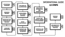
Steps for developing the Informational Guide. Porto Alegre, Rio Grande do Sul, Brazil, 2025
Step 1 - Situational diagnosis (October 2024)
The situational diagnosis was conducted through weekly collegiate meetings with multiprofessional participation, lasting approximately one hour, in which matters related to the management and operation of the DIC were discussed. Among these meetings, three specific sessions were held focused on the adequate preparation of patients for radiological and diagnostic imaging examinations.
The need to deepen this topic emerged from the analysis of management and care indicators, which showed examination reschedulings and cancellations, mainly due to inadequate patient preparation. These findings were corroborated by the experience of the researchers who have worked in the DIC for many years.
Additionally - in a meeting with the radiology nursing team at the DIC - frequent calls received from the healthcare teams of inpatient units requesting specific information on the preparation required for examinations were discussed. These demands consume a significant volume of nursing team working hours dedicated to passing on guidelines by telephone and email. The time wasted with patient transportation to the DIC was also discussed.
Although the DIC has institutional instructions on the necessary preparations, such guidelines are scattered across different documents and sources, without systematic organization in a single clear, objective, and easily accessible material. This fragmentation has compromised the standardization of conduct and the accuracy of guidelines on patient preparation.
Step 2 - Research project (October 2024 to November 2024)
The research project was executed according to the established schedule and included the following steps: elaboration of the summary, abstract, and abstract; introduction; justification; objective; literature review; method; schedule; budget; references; Informed Consent Form (ICF); appendices; research instrument; and submission to the Research Ethics Committee. The methodological framework adopted11) comprises 13 steps, as presented in Figure 1.
The construction and execution of the project followed the Guidelines and Regulatory Standards for Research Involving Human Beings, in accordance with Resolutions No. 466/2012 and No. 510/2016 of the National Health Council (CNS). All participants signed the Informed Consent Form (ICF), in compliance with Law No. 13,709/2018 (General Personal Data Protection Law). The study was approved by the Research Ethics Committee under opinion No. 7,167,846, with Certificate of Presentation for Ethics Assessment No. 83785824.8.0000.5327.
Step 3 - Definition and selection of content (December 2024 to January 2025)
For the definition and selection of content, the LILACS, BDENF, SciELO, PubMed/MEDLINE, CINAHL, Scopus, and CAPES Periódicos databases were consulted, in addition to considering the practical experience of the authors in the field of radiology and diagnostic imaging. The search focused on guidelines and scientific articles addressing patient preparation for radiological and imaging examinations.
The following descriptors were used, in Portuguese and English: “Diagnóstico por imagem/Diagnostic imaging”, “Serviço hospitalar de radiologia/Hospital radiology service”, “Enfermagem radiológica e de imagem/Radiological and imaging nursing”, and “Guia informativo/Information guide”, combined with the Boolean operator AND, to refine the results and ensure greater relevance to the identified studies.
The inclusion criteria considered complete articles published in the last five years - in Portuguese, English, or Spanish - that addressed guidelines and the adequacy of patient preparation for radiological and imaging examinations and ionizing radiation exposure. Editorials, reflective studies, duplicate articles, and those that did not present pertinence to the study objective were excluded.
Of the 1,375 publications initially identified, 17 studies were selected for detailed analysis and synthesis of the most relevant aspects. Of these, eight studies³⁻⁴·¹²⁻¹³·²¹⁻²⁴ addressed patient preparation for diagnostic imaging examinations specifically. Nine studies addressed management, performance, and multidisciplinary organization in DICs¹⁻²·⁵⁻¹¹. Despite the expanded database search, no higher-level evidence was identified, such as randomized clinical trials, quasi-experimental studies, or other research with greater methodological rigor. Thus, the literature review - combined with the clinical experience of the researchers - supported the development of the Pilot Informational Guide.
Step 4 - Language adaptation (February to October 2025)
The adaptation of the information identified in the literature to the institutional context formed the basis for the development of the informational guide, which was written in clear, objective, and concise language, with the purpose of facilitating understanding by healthcare teams and promoting the standardization and effectiveness of practices related to patient preparation for diagnostic imaging examinations. For language adaptation, the content was carefully structured to present relevant, accessible information aligned with the needs of the service, focusing on improving the guidelines provided to healthcare teams. The selection of information included in the guide considered the examinations currently performed at the DIC and, especially, current national and international recommendations²¹⁻²⁴, as well as the clinical experience of the researchers.
Step 5 - Inclusion of illustrations (February to October 2025)
With the aim of making the material more engaging and easy to understand, illustrations and photographs were included throughout the informational guide. The illustrations consist of images of the equipment and examination rooms, produced by the researchers themselves from the examinations performed at the DIC, contributing to greater visual lightness and better contextualization of the content presented. The size of the images was adjusted to fit the text formatting, with one illustration included in each chapter of the guide. It should be noted that the images do not feature patients or healthcare team members, being restricted exclusively to the equipment used in performing the examinations, in compliance with ethical and privacy requirements.
Step 6 - Development of the pilot material (December 2024 to January 2025)
The development of the Pilot Informational Guide was based on information and content related to the proposed theme, grounded in updated scientific literature and the clinical experience of the authors who work in the radiology and diagnostic imaging unit. The material was developed with the aim of meeting the needs and expectations of healthcare teams regarding patient preparation for imaging examinations, contributing to the standardization of guidelines, the improvement of care quality, and patient safety.
Step 7 - Study participants (February 2025)
Study participants were selected intentionally and informed in advance about the nature of the research, including the possibility of contributing to the content and appearance validation process of the informational guide on patient preparation for diagnostic imaging examinations. The invitation to participate was sent online, through the Google Forms® platform. In this process, each participant received the ICF, the research instrument, and the pilot informational guide.
Considering that the use of technologies based on the active participation of the multiprofessional team is essential for improving the quality of care and training of health professionals, referenced by Squire etal18, professionals working in the DIC, the inpatient units, the intensive care units, and the CRC of the institution were invited to participate in the study, as well as postgraduate students from the Professional Master’s Program.
The inclusion criteria covered participants’ expertise in the field of radiology, including professionals who request examinations, participate in patient preparation, or perform the examinations, as well as a minimum of two years of experience in their role. No exclusion criteria were established. Cases where participants, after accepting the invitation, did not respond to the research instrument within the stipulated period were considered losses; in these situations, other professionals were invited as substitutes.
In total, 45 participants were invited, of whom 12 were Professional Master’s students. The inclusion of master’s students was justified by the fact that one of the focuses of this professional training modality is to qualify professionals for transformative practice, aimed at meeting social, organizational, and labor market demands, as well as improving the efficiency and effectiveness of public and private organizations through innovation processes.
The sample size was defined based on previous methodological studies recommending the participation of specialists with expertise in the evaluated content, considering professional diversity and informational sufficiency as central criteria for the validation of educational materials, rather than a fixed number of participants²⁵⁻²⁷.
Step 8 - Content and appearance validation (March to July 2025)
The content and appearance validation process of the Informational Guide was carried out using a digital instrument developed by the researchers on the Google Forms® platform, whose content was based on instruments previously used in a study validating educational material28. The questionnaire consisted of seven statements assessed using a Likert scale, with the options: strongly agree, agree, neutral, disagree, and strongly disagree, as well as eight open-ended questions for collecting suggestions and qualitative observations.
The statements covered aspects related to the adequacy of the material for its target audience, clarity and objectivity of the information, ability to clarify doubts, language used, importance of the material, overall quality, and elements related to design. Participants had ten days to return the instruments (Pilot Informational Guide, ICF, and questionnaire) duly completed. In cases where there was no response within the stipulated period, new invitations were sent to other potential participants.
Content and appearance validation was performed by calculating the percentage of agreement among evaluators, considering valid those items that reached agreement equal to or greater than 90%, as recommended in the methodological literature²⁹. The Content Validity Index (CVI) was used to measure the proportion of judges who agreed with the different aspects assessed in the instrument and its items.
Step 9 - Analysis of information/suggestions (April to August 2025)
The comments and suggestions received during the validation process of the Pilot Informational Guide, recorded in the research instrument, were carefully analyzed by the researchers. All contributions were evaluated for their pertinence and applicability and, when considered relevant, were incorporated into the guide text, with the aim of improving the quality of the material and its adequacy for the target audience. The analysis covered different aspects of the guide, including the format and organization of items, the clarity and adequacy of titles, the instructions presented, the domains addressed, and the scores attributed to each domain. This process aimed to ensure the internal coherence of the material, achievement of the study objectives, and the effectiveness of the informational guide as a support instrument for healthcare teams.
Step 10 - Layout and graphic design (January to October 2025)
The layout and graphic design stage aimed to make the text clearer and the visual aspect more appropriate to the profile of the target audience. Based on the suggestions obtained during the validation phase, adjustments were made to the textual content, typography, color palette, font sizes, and graphic elements, aiming to improve the presentation, legibility, and usability of the informational guide.
Step 11 - Finalization and approval (October 2025)
The informational guide brings together updated recommendations on patient preparation and nursing care during the performance of radiological examinations, with the aim of strengthening care quality, promoting patient safety, and standardizing the practices of the multiprofessional team. It is organized into four main sections: patient preparation (fasting, contrast media, claustrophobia, sedation, and pain management), radiological protection (ionizing and non-ionizing radiation), radiology nursing care (venipuncture, monitoring, and transportation), and the guide itself (examinations and their respective preparations).
The final version of the guide was approved at a DIC collegiate meeting, with multiprofessional participation, whose members also participated in the content and appearance validation process of the pilot material. Furthermore, the informational guide was submitted to the qualification committee and the Professional Master’s thesis defense, as part of the academic requirements of the study.
Step 12 - Implementation in clinical practice (October 2025 to January 2026)
With the aim of facilitating the implementation of the informational guide in clinical practice, it was decided to actively involve the healthcare teams of different hospital areas in the material pilot validation process. This strategy favored the alignment of the guide with institutional routines and the real daily needs of healthcare professionals.
This step is part of the methodological framework adopted¹⁴⁻¹⁵, providing for the insertion of the Informational Guide into the daily institutional routine as a means of validating its applicability. Some actions already carried out include: presentation and dissemination of the material to healthcare teams, distribution in physical and digital format to all units, ensuring broad access and support to care practices. The phase still under implementation refers to the update of the examination report structure in the Health Management System, with the incorporation of the guide guidelines directly into the field for patient preparation for examinations. This action, already underway, aims to make the process more robust, standardized, and systematic, ensuring that relevant information is easily accessible to healthcare professionals at the appropriate time. This step combines completed actions with ongoing processes, ensuring coherence between the theoretical-methodological framework and practical application, strengthening the integration of the guide into institutional routines and multiprofessional clinical practice.
Step 13 - Periodic update (Whenever there is a need for changes)
The Informational Guide shall be updated whenever there are changes in institutional routines, inclusion of new examinations, or changes in conduct related to patient preparation for diagnostic imaging examinations. Updates shall be submitted to a careful review by professionals in the field, with the aim of ensuring that the information remains aligned with the most recent scientific evidence and best care practices.
It is also noteworthy the interest and commitment of the DIC healthcare teams in the continuous maintenance and updating of the guide, recognizing it as a relevant tool for the practice of the multiprofessional team and the professional training of students and residents.
RESULTS
A total of 45 individuals participated in the study, of whom 33 were healthcare professionals and 12 were postgraduate students. Five professionals accepted to participate but did not respond to the research instrument within the stipulated period; in these cases, new participants were invited as substitutes.
The mean professional experience among healthcare professionals was 14 years, ranging from four to 30 years. Among postgraduate students, professional experience ranged from two to 22 years. Of the healthcare professionals, 27 had expertise in the Radiology Service, while six worked in other hospital areas responsible for the preparation and referral of patients for examinations. The characterization of the sample, with the breakdown of professional categories, is presented in Table 1.
For the development of the guide, the 13 methodological steps proposed in the project execution were followed; however, these steps were not necessarily sequential throughout the study, due to the suggestions and contributions of the participants, especially regarding the content, language, and design of the material.The process began with a situational diagnosis, which revealed examination reschedulings and cancellations due to inadequate preparation, consolidating the need for the guide. Upon completion of the validation, 94 items were included in the final version of the Informational Guide on Preparing Patients for Radiological and Diagnostic Imaging Examinations.
In the guide validation, participants’ comments were - in general - predominantly positive, evidencing the relevance and quality of the material. There was no significant disparity between the perceptions of judges from different professional categories.
In the guide validation, participants’ comments were - in general - predominantly positive, evidencing the acceptance of the material and the relevance of the content for clinical practice. The importance of the guide as an updating and training instrument in the context of professional health education was highlighted. The main compliments referred to the clarity of the information, the accessible format, the logical organization of the content, and the applicability of the guidelines in the care context.
In the open-ended questions of the instrument, participants suggested adjustments related to font size, writing structure, organization, and layout of the material, as well as the arrangement and presentation of content. Corrections of definitions, suggestions for the inclusion and exclusion of items, and - mainly - the need for conceptual revision of some terms were also pointed out. Most suggestions were incorporated into the text; however, some were not accepted because they were inconsistent with the study objectives. Contributions related to design highlighted aspects such as the reorganization of content, the harmonization of the color palette, and the adjustment of font sizes.
The agreement rate was calculated from the sum of “agree” and “strongly agree” responses, divided by the total number of responses, expressed as a percentage. An agreement rate ≥80% was established as the minimum acceptance criterion.
After analyzing and incorporating participants’ suggestions, the Informational Guide on Preparing Patients for Radiological and Diagnostic Imaging Examinations now contains 71 pages, with guidelines for patient preparation for 60 types of radiological and diagnostic imaging examinations. The information is organized into the following topics: general considerations; important information; radiological examinations; contrast X-ray examinations; bone densitometry; computed tomography; magnetic resonance imaging; ultrasound or echography; invasive procedures guided by imaging examinations; and final considerations. This structure aimed to group the examinations by area of performance, distributing them into specific chapters, in order to facilitate consultation and application of the guidelines in care practice.
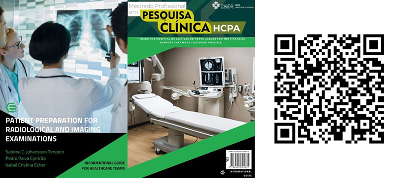
The Informational Guide was developed on the Canva® platform, usingTitillium Web font for titles, at size 43.7. In the body text, the Cooper Hewitt and Titillium Web fonts, both at size 18. For topics, the Titillium web font at size 18 was used, and for subtopics,Cooper Hewitt at size 28.4, following legibility and visual harmony criteria, as shown in Figure 2.
The material can be accessed free of charge via the hyperlink https://drive.google.com/file/d/1D8sT4bPVxvTzcL1bOTyzv4dbmhfOYUZq/view?usp=sharing
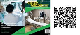
Cover, back cover, and QR Code of the Informational Guide: Preparing patients for radiological and diagnostic imaging examinations. Porto Alegre, Rio Grande do Sul, Brazil, 2025.
DISCUSSION
Based on the theoretical-methodological framework14-15, the Informational Guide was developed and validated with scientific rigor, achieving the proposed objectives. The process began with a situational diagnosis that revealed weaknesses in the care context, dispersion, and low accessibility of previously existing institutional guidelines - aspects described in the literature as factors that hinder professional practice, generate rework, and negatively impact the productivity of health services. In this regard, clear, accessible, and systematized educational materials prove essential for improving patient preparation and optimizing care flows30).
The growing use of these resources - especially in therapeutic and care contexts - reinforces the need to adopt quality criteria, such as coherence, cohesion, organization, and accessible language in educational materials intended for healthcare professionals. Objectivity and clarity play a central role in this process, since excessively lengthy or complex content can compromise the understanding and adherence of professionals26-27. The scarcity of publications specifically focused on nursing care and educational technologies in the context of diagnostic imaging highlights the contribution of this study to filling a relevant gap in the evidence available for professional practice in the area.
The active participation of a multiprofessional team in the process of developing and validating the guide constitutes a relevant aspect, since it broadens the scope of the material and favors its applicability in different care and educational contexts, consistent with the scientific literature on the construction of educational technologies for healthcare29-32.
In the present study, the validation process proved central to defining objective, relevant, and aligned information for the needs of clinical practice. The contributions of the professionals enhanced the clarity and applicability of the content, demonstrating the utility of the material. These findings converge with scientific evidence and guidelines21-24) that highlight adequate patient preparation as a strategic element for safety and quality of care in diagnostic imaging services. The guidelines of the American College of Radiology (ACR), the European Society of Urogenital Radiology (ESUR), and the Brazilian College of Radiology (CBR) on contrast media and radiological protection were considered references for the content developed...
The literature also indicates that scientifically grounded guides support clinical practice by functioning as reference instruments for clinical decision-making, assist in making decisions, and promote greater professional autonomy32. Furthermore, the critical analysis by experts contributes to ensuring that the information is up-to-date, consistent, and aligned with the proposed objectives.
The commitment to updating will be maintained, with periodic revisions whenever necessary, ensuring the relevance and reliability of the content. Making it digitally available on institutional and public platformscan be used as a reference tool and pedagogical resource in teaching-learning processes30,33, with potential
As study limitations, the scarcity of more robust evidence on patient preparation for diagnostic imaging examinations should be noted, as well as the unicentric nature of the study, which may limit the generalizability of the findings. The need to complete the implementation of the guide in clinical practice and future evaluations measuring its effectiveness is emphasized. Despite these limitations, the results indicate that the informational guide constitutes a consistent and promising resource for improving care quality, with the potential to contribute to safer, more standardized, and more efficient practices.
CONCLUSION
The study achieved its objective by developing and validating an informational guide for patient preparation in diagnostic imaging examinations, intended for healthcare teams. Content and appearance validation, performed by 45 evaluators from different health areas and postgraduate students, resulted in a mean agreement rate of 95.8%, demonstrating the clarity, relevance, and applicability of the material. Participants’ contributions were analyzed and incorporated when pertinent, qualifying the final version of the guide.
The Informational Guide meets a concrete demand of diagnostic imaging services for clear, standardized, evidence-based guidelines, with the potential to qualify care processes, favor the standardization of conduct, and contribute to patient safety and service efficiency.
Furthermore, the material presents potential for replication and adaptation in other institutional contexts, especially in hospital and academic settings, constituting an educational support strategy for the improvement of care quality in diagnostic imaging.
Acknowledgments
The authors thank Hospital de Clínicas de Porto Alegre for the scholarship grant and methodological support that contributed to the development of this study.
REFERENCES
-
1. Wachholz AC, Schacker LC. Centro de diagnóstico por imagem: papel do enfermeiro. Braz J Implantol Health Sci. 2025;7(1):2073-801. https://doi.org/10.36557/2674-8169.2025v7n1p2073-2801
» https://doi.org/10.36557/2674-8169.2025v7n1p2073-2801 -
2. Cordeiro CR, Souza SJP, Cordeiro P, Burci LM. O papel do profissional de enfermagem no centro de diagnóstico por imagem: revisão de literatura. Rev Gest Saúde [Internet]. 2021[cited 2026 Jan 27];23(1):136-45. Available from: https://www.herrero.com.br/files/revista/file80071263113ff6546896b61d9b2dced6.pdf
» https://www.herrero.com.br/files/revista/file80071263113ff6546896b61d9b2dced6.pdf -
3. Belo FBM, Reis LLL, Pereira JV. O papel do enfermeiro em exames de diagnóstico por imagem. Braz J Health Rev. 2023;6(6):30301-12. https://doi.org/10.34119/bjhrv6n6-286
» https://doi.org/10.34119/bjhrv6n6-286 -
4. Almaliky M, Alamri EA, Albishri FF, Algarni AF, Alsharif NM, Khabrani NH, et al. Interprofessional collaboration in diagnostic imaging services: roles, communication and impact on healthcare outcomes. Rev Diabet Stud [Internet]. 2024 [cited 2026 Jan 27];20(Suppl 7):45. Available from: https://diabeticstudies.org/index.php/RDS/article/view/1516
» https://diabeticstudies.org/index.php/RDS/article/view/1516 -
5. Silva FAF, Silva FAF Filho, Nitão FF, Medeiros EMM. Atuação do enfermeiro em centro de diagnóstico por imagem: uma abrangência multidisciplinar. Temas Saúde. 2020;20(6):205-17. https://doi.org/10.56083/RCV4N10-080
» https://doi.org/10.56083/RCV4N10-080 -
6. Faria AP, Lima EF, Peruzini G Filho, Almeida MV, Coelho MC, Silva RI, et al. Atuação da enfermagem na tomografia computadorizada. Enferm Foco. 2025;16:e-2025006. https://doi.org/10.21675/2357-707X.2025.v16.e-2025006
» https://doi.org/10.21675/2357-707X.2025.v16.e-2025006 -
7. Acauan LV, Seda JM, Silva SCSB, Paes GO, Trotte LAC, Stipp MAC. A atuação da equipe de enfermagem em serviços ambulatoriais de radiologia e diagnóstico por imagem. Rev Gaúcha Enferm. 2022;43: e20210079. https://doi.org/10.1590/1983-1447.2022.20210079.pt
» https://doi.org/10.1590/1983-1447.2022.20210079.pt -
8. Acauan LV, Seda JM, Silva SCSB, Paes GO, Trotte LAC, Stipp MAC. Quality management in imaging diagnosis and the nursing team: a case study. Rev Bras Enferm. 2021;74(6): e20210014. https://doi.org/10.1590/0034-7167-2020-0912
» https://doi.org/10.1590/0034-7167-2020-0912 -
9. Rockall AG, Justich C, Helbich T, Vilgrain V. Patient communication in radiology: Moving up the agenda. Eur J Radiol. 2022;155:110464. https://doi.org/10.1016/j.ejrad.2022.110464
» https://doi.org/10.1016/j.ejrad.2022.110464 -
10. Dias JVF, Sousa LR, Brito RL, Santana JCG, Oliveira IC, Moura FG, et al. Capacitação da equipe multidisciplinar em procedimentos de preparo para exames radiológicos: uma abordagem interdisciplinar. Cad Impacto Ext [Internet]. 2025 [cited 2026 Jan 29];5(1). Available from: https://revistas.editora.ufcg.edu.br/index.php/cite/article/view/6821
» https://revistas.editora.ufcg.edu.br/index.php/cite/article/view/6821 -
11. Mendes AMS, Nogueira LB, Fontainha CCP, Batista ASM, Santos TO. Relevância do conhecimento de protocolos de preparo de exames contrastados para a diminuição de exposições na radiografia médica. Braz J Develop. 2022;8:15667-74. https://doi.org/10.34117/bjdv8n3-012
» https://doi.org/10.34117/bjdv8n3-012 -
12. Lima GPSN. Implantação da Sistematização da Assistência de Enfermagem (SAE) em um serviço de diagnóstico por imagem. Braz J Health Rev . 2022;5(5):20898-906. https://doi.org/10.34119/bjhrv5n5-247
» https://doi.org/10.34119/bjhrv5n5-247 -
13. Jucá RF, Verçosa RCM, Maciel MPGS, Santos DAS. A importância do enfermeiro na gestão de riscos em exames de ressonância: um relato de experiência. Braz J Dev. 2023;9(1):256-72. https://doi.org/10.34117/bjdv9n1-019
» https://doi.org/10.34117/bjdv9n1-019 -
14. Echer IC, Lorenz Y, Severo IM. Tecnologias de educação em saúde: guia prático para o desenvolvimento de materiais educativos [Internet]. Porto Alegre: Moriá; 2024 [cited 2026 Jan 27]. Available from: http://hdl.handle.net/10183/271013
» http://hdl.handle.net/10183/271013 -
15. Echer IC. Elaboração e validação de materiais educativos em saúde: proposta atualizada. In: Levi - 75 anos da enfermagem da Universidade Federal do Rio Grande do Sul [Internet]. Porto Alegre: UFRGS; 2025 [cited 2026 Mar 05]. p. 87-89. Available from: http://hdl.handle.net/10183/65589
» http://hdl.handle.net/10183/65589 -
16. Nunes SM, Soares SS, Silva WCM, Brener CEdeS, Lodi D, Silva CS. Construção de material educativo: uma estratégia potencial para melhorar o cuidado em uma unidade de saúde. Rev Pesqui Cuid Fundam. 2025;17: e13599. https://doi.org/10.9789/2175-5361.rpcfo.v17.13599
» https://doi.org/10.9789/2175-5361.rpcfo.v17.13599 -
17. Cordeiro RB, Galina VSS, Primo CC, Cerqueira LB, Araújo DCSA, Rocha KSS. Desenvolvimento e validação de materiais educativos sobre HIV. Saúde Pesq. 2025;18: e13270. https://doi.org/10.17765/2176-9206.2025v18e13270
» https://doi.org/10.17765/2176-9206.2025v18e13270 -
18. Oliveira IKM, Marques MAPM, Teixeira LG, Salles DL. Construção e validação de cartilha para mitigar vulnerabilidades em saúde de cardiopatas. Rev Enferm Atual In Derme. 2025;99(Suppl 1): e025078. https://doi.org/10.31011/reaid-2025-v.99-n.supl.1-art.2431
» https://doi.org/10.31011/reaid-2025-v.99-n.supl.1-art.2431 - 19. Paranhos LR, Rodolpho PJ. Metodologia da pesquisa aplicada à tecnologia. São Paulo: SENAI-SP Editora; 2018. 160p.
-
20. Viana PLSC, Paiva DSBS. Construção de tecnologia educacional para gestantes acerca do trabalho de parto e seus direitos enquanto parturientes. Rev Eletrôn Acervo Saúde. 2025;25: e19012. https://doi.org/10.25248/reas.e19012.2025
» https://doi.org/10.25248/reas.e19012.2025 -
21. American College of Radiology. Manual on contrast media [Internet]. 2024 [cited 2026 Jan 27]. Available from: https://geiselmed.dartmouth.edu/radiology/wp-content/uploads/sites/47/2024/08/ACR-contrast-2024.pdf
» https://geiselmed.dartmouth.edu/radiology/wp-content/uploads/sites/47/2024/08/ACR-contrast-2024.pdf -
22. European Society of Urogenital Radiology (ESUR). Contrast Media Safety Committee Guidelines - Version 10.0 [Internet]. Vienna: ESUR; 2018[cited 2026 Jan 27]. Available from: https://www.esur.org/esur-contrast-media-safety-committee/
» https://www.esur.org/esur-contrast-media-safety-committee/ -
23. Colégio Brasileiro de Radiologia e Diagnóstico por Imagem(CBR). Diretrizes para o uso de meios de contraste intravenosos [Internet]. São Paulo: CBR; 2024[cited 2026 Jan 27]. Available from: https://cbr.org.br/wp-content/uploads/2024/01/Diretrizes-para-o-uso-de-meios-de-contrastes-intravenosos.pdf
» https://cbr.org.br/wp-content/uploads/2024/01/Diretrizes-para-o-uso-de-meios-de-contrastes-intravenosos.pdf -
24. Comissão de Proteção Radiológica do Colégio Brasileiro de Radiologia e Diagnóstico por Imagem (CPR-CBR). Informativos e materiais educativos para informar e preparar pacientes sobre exames radiológicos [Internet] São Paulo: CBR ; 2025[cited 2026 Jan 29]. Available from: https://cbr.org.br/comissao-de-protecao-radiologica/informativo-para-pacientes-cpr-cbr/
» https://cbr.org.br/comissao-de-protecao-radiologica/informativo-para-pacientes-cpr-cbr/ -
25. Squire CM, Giombi KC, Rupert DJ, Amoozegar J, Williams P. Determining an appropriate sample size for qualitative interviews to achieve true and near code saturation: secondary analysis of data. J Med Internet Res. 2024;26: e52998. https://doi.org/10.2196/52998
» https://doi.org/10.2196/52998 -
26. Salomé GM, Miranda FD. Validation of a brochure to guide health professionals in the dressing and undressing of personal protective equipment during the SARS-CoV-2 pandemic. J Coloproctol. 2021;42(1): 7-13. https://doi.org/10.1055/s-0041-1730424
» https://doi.org/10.1055/s-0041-1730424 -
27. Miranda LHD, Reis JS, Oliveira SR. Construção e validação de ferramenta educativa sobre insulinoterapia para adultos com diabetes mellitus. Ciênc Saúde Coletiva. 2023;28(5): 1513-24. https://doi.org/10.1590/1413-81232023285.09502022
» https://doi.org/10.1590/1413-81232023285.09502022 -
28. Zuchetti M, Severo IM, Echer IC, Borba DSM, Nectoux CLS, Azzolin KO. Validação de manual para complementar a transição de cuidados na alta da terapia intensiva. Rev Gaúcha Enferm [Internet]. 2022[cited 2026 Jan 27];43(esp): 20220142. Available from: https://seer.ufrgs.br/index.php/rgenf/article/view/127957
» https://seer.ufrgs.br/index.php/rgenf/article/view/127957 -
29. Alexandre NMC, Coluci MZO. Validade de conteúdo nos processos de construção e adaptação de instrumentos de medidas. Cienc Saúde Coletiva. 2011;16: 3061-8. https://doi.org/10.1590/S1413-81232011000800006
» https://doi.org/10.1590/S1413-81232011000800006 -
30. França T, Magnago C, Belisário SA. Diagnóstico situacional das Escolas de Saúde Pública da rede SUS. Gest Trab Educ Saúde. 2021;1(1): 170-88. https://doi.org/10.37885/210303433
» https://doi.org/10.37885/210303433 -
31. Manochio-Pina MG, Crivellenti LC, Sartorelli DS, Diez-Garcia RW. Educational instrument for intervention in the lifestyle of overweight pregnant women. Rev Bras Saúde Mater Infant. 2022;22(2): 385-98. https://doi.org/10.1590/1806-9304202200020011
» https://doi.org/10.1590/1806-9304202200020011 -
32. Andrade JC, Brito ASF, Santos KAV, Dini AP, Belasco AGS, Gasparino RC. Construção e validação de uma cartilha educativa multiprofissional para transplantados renais. Rev Bras Enferm . 2025;78(1). https://doi.org/10.1590/0034-7167-2024-0103pt
» https://doi.org/10.1590/0034-7167-2024-0103pt -
33. Almeida SLP, Primo CC, Almeida MVS, Freitas PSS, Lucena AF, Lima EFA, et al. Guide for systematization of care and nursing process: educational technology for professional practice. Rev Bras Enferm . 2023;76: e20210975. https://doi.org/10.1590/0034-7167-2021-0975pt
» https://doi.org/10.1590/0034-7167-2021-0975pt
Access to the dataset may be obtained upon request to the corresponding author.

 Source: developed by the authors
 Source: Developed by the authors, 2025.