
 Fabrication and characterization of rutin-loaded liquid crystalline nanoparticles with in vitro anti-inflammatory and antitumor activities
ABSTRACT
A rutin-loaded liquid crystalline nanoparticle (R-LCNP) formulation was developed using glyceryl monooleate and Poloxamer 407 by high-pressure homogenization to improve the solubility and biological performance of rutin. The optimized R-LCNP-2 dispersion showed a mean particle size of 176.1 ± 4.5 nm, a polydispersity index (PDI) of 0.211 ± 0.013, and a moderately negative zeta potential of –20.4 ± 1.1 mV, consistent with steric-electrostatic stabilization. The encapsulation efficiency and drug loading were 98.1 ± 1.5% and 2.45 ± 0.04%, respectively, while FTIR, XRD, and DSC analyses indicated molecular dispersion of rutin within the lipid matrix. The formulation showed biphasic diffusion-controlled release and suppressed nitric oxide, TNF-α, and IL-6 more effectively than free rutin in an in vitro LPS-stimulated RAW 264.7 model. R-LCNP-2 also reduced the IC50 values in MCF-7, HeLa, and A549 cells by 3.8–4.5 fold relative to free rutin. These findings support LCNP-based encapsulation as a promising strategy to broaden the functional performance of rutin and justify subsequent in vivo pharmacokinetic and efficacy studies.
Keywords:
Nanoencapsulation; Bioavailability; Macrophages; Cytokine inhibition; Cytotoxicity
INTRODUCTION
Rutin (quercetin-3-O-rutinoside) is a naturally occurring flavonoid glycoside ubiquitously found in a variety of dietary sources, including citrus fruits, buckwheat, apples, and tea [1]. As a prominent member of the polyphenol family, rutin has garnered significant scientific interest due to its broad spectrum of pharmacological activities. Extensive preclinical research has demonstrated its potent antioxidant, anti-inflammatory, anti-diabetic, neuroprotective, and cardioprotective properties [2]. These beneficial effects are largely attributed to its ability to scavenge free radicals, modulate key inflammatory signaling pathways, and induce apoptosis in aberrant cells. Consequently, rutin is considered a promising therapeutic agent for the prevention and treatment of various chronic and degenerative diseases, including cancer, cardiovascular disorders, and neurodegenerative conditions [3].
Despite its compelling therapeutic potential, the clinical translation of rutin is severely hampered by its challenging physicochemical properties. Rutin is classified as a Biopharmaceutics Classification System (BCS) Class II or IV compound, characterized by extremely low aqueous solubility (approximately 0.8 mg/mL) and poor membrane permeability This inherent hydrophobicity leads to low and erratic absorption from the gastrointestinal tract, resulting in poor oral bioavailability, which is estimated to be around 20% [4]. Furthermore, its polyhydroxy structure makes it susceptible to enzymatic degradation in the gut and rapid metabolism, further diminishing its systemic exposure and therapeutic efficacy [5]. These pharmacokinetic limitations necessitate the development of advanced formulation strategies to unlock the full therapeutic potential of this valuable natural compound.
Nanotechnology-based drug delivery systems have emerged as a transformative approach to overcome the bioavailability challenges associated with poorly soluble drugs like rutin [6]. By encapsulating active pharmaceutical ingredients within nanocarriers, it is possible to enhance their solubility, protect them from degradation, and facilitate their transport across biological barriers. Among the various nanoplatforms, lipid-based systems such as liposomes, solid lipid nanoparticles, and nanoemulsions are particularly attractive due to their excellent biocompatibility, biodegradability, and ability to encapsulate lipophilic molecules [7]. These systems can improve the dissolution rate of a drug by maintaining it in an amorphous or solubilized state, thereby increasing its effective concentration at the site of absorption.
Recent studies have shown that the efficacy of rutin can be improved through diverse nanocarriers, including oral nanoemulsions, nano-lipid complexes, nanovesicles, and coated nanoliposomes [8,9,10,11]. In addition, earlier rutin-loaded liquid-crystalline nanoparticle reports demonstrated that this class of carrier can modulate oxidative stress and inhibit non-small cell lung cancer behavior in cell-based models [10, 12, 13]. Accordingly, the present work is not positioned as the first rutin-LCNP platform; rather, it aims to provide a broader formulation-to-function dataset by coupling loading optimization with orthogonal physicochemical characterization, release modeling, anti-inflammatory assays, and cross-line antitumor screening.
Within the landscape of advanced lipid nanocarriers, lyotropic liquid crystalline nanoparticles (LCNPs), including cubosomes and hexosomes, represent a particularly sophisticated and promising platform [14]. LCNPs are thermodynamically stable, self-assembled nanostructures formed from amphiphilic lipids, such as glyceryl monooleate (GMO), in the presence of a stabilizer like Poloxamer 407 Their defining feature is a unique internal architecture composed of a continuous, curved lipid bilayer that partitions space into intertwined but non-intersecting aqueous channels [15]. This intricate, ordered internal matrix provides an exceptionally large interfacial surface area, making LCNPs highly effective carriers for both hydrophobic and hydrophilic drugs. The selection of LCNPs for rutin delivery is strategic; the rigid and tortuous internal environment is uniquely suited to sterically hinder the encapsulated drug molecules from aggregating and recrystallizing, a common failure point in less-ordered systems. This structural stabilization fundamentally alters the physical state of rutin from crystalline to amorphous, which is the direct mechanistic basis for enhancing its bioavailability and biological activity.
This study hypothesizes that the encapsulation of rutin into GMO-based LCNPs will overcome its inherent solubility limitations, leading to a nanomedicine formulation with significantly enhanced in vitro anti-inflammatory and antitumor activities compared to the free drug. The objectives of this research were threefold: (1) to fabricate and optimize stable rutin-loaded LCNPs (R-LCNPs) with varying drug payloads; (2) to conduct an exhaustive physicochemical characterization of the R-LCNPs using a suite of advanced analytical techniques to elucidate their morphology, structure, surface chemistry, and drug encapsulation state; and (3) to comparatively evaluate the biological efficacy of the optimized R-LCNP formulation against free rutin in validated in vitro models of inflammation and cancer.
2. Materials and Methods
2.1. Materials
Rutin, glyceryl monooleate (GMO, Myverol 18-99K), and Poloxamer 407 (Pluronic® F-127) were purchased from Aladdin Reagent Co., Ltd. (Shanghai, China). Ethanol, methanol, chloroform, and dimethyl sulfoxide (DMSO) were of HPLC grade and obtained from Macklin Biochemical Co., Ltd. (Shanghai, China). For cell culture, RAW 264.7 (murine macrophage), MCF-7 (human breast adenocarcinoma), HeLa (human cervical adenocarcinoma), and A549 (human lung carcinoma) cell lines were procured from the Cell Bank of the Chinese Academy of Sciences (Shanghai, China). Dulbecco’s Modified Eagle’s Medium (DMEM), fetal bovine serum (FBS), penicillin-streptomycin solution, and trypsin-EDTA were obtained from Gibco (Thermo Fisher Scientific, Shanghai, China). Lipopolysaccharide (LPS, from Escherichia coli O111:B4), 3-(4,5-dimethylthiazol-2-yl)-2,5-diphenyltetrazolium bromide (MTT), and Griess reagent were supplied by Solarbio Science & Technology Co., Ltd. (Beijing, China). Human Tumor Necrosis Factor-alpha (TNF-α) and Interleukin-6 (IL-6) ELISA kits were obtained from Beyotime Biotechnology (Shanghai, China).
2.2. Fabrication of rutin-loaded LCNPs (R-LCNPs)
R-LCNPs were prepared using a top-down emulsification and high-pressure homogenization technique adapted from previously reported glyceryl monooleate/Poloxamer 407 liquid-crystalline nanoparticle protocols [12, 15]. First, the lipid phase was prepared by melting a precisely weighed amount of GMO at 60 °C. Concurrently, rutin was dissolved in a minimal volume of ethanol (2 mL) and subsequently dispersed into the molten GMO under continuous magnetic stirring at 500 rpm until a homogeneous solution was formed. The aqueous phase was prepared by dissolving Poloxamer 407 (1% w/v) in deionized water, which was then heated to 60 °C. The hot lipid phase was then added dropwise to the aqueous phase under high-speed homogenization using an Ultra-Turrax T25 homogenizer at 10,000 rpm for 10 minutes to form a coarse pre-emulsion. This pre-emulsion was immediately passed through a high-pressure homogenizer (Avestin EmulsiFlex-C3, Ottawa, ON, Canada) for five cycles at a pressure of 1000 bar. The resulting nano-dispersion was cooled to room temperature and stored at 4 °C for further characterization. Three different formulations were prepared with varying theoretical drug loadings: R-LCNP-1 (1% w/w rutin relative to total lipid), R-LCNP-2 (2.5% w/w), and R-LCNP-3 (5% w/w). A blank LCNP formulation without rutin was also prepared using the same procedure to serve as a control. The formulation composition was defined on a 100 mg GMO lipid basis: R-LCNP-1, R-LCNP-2, and R-LCNP-3 contained 1.0, 2.5, and 5.0 mg rutin per 100 mg GMO, respectively, while Poloxamer 407 was maintained at 1% (w/v) in the aqueous phase.
The formulation workflow was arranged to mirror the analytical sequence later adopted in the Results and Discussion. After preparation of the dispersions, particle size, PDI, and zeta potential were determined by dynamic light scattering (DLS), while encapsulation efficiency (EE) and drug loading (DL) were quantified from the non-encapsulated fraction. The optimized batch was then advanced to morphological, structural, surface, release, and biological studies.
Formulation selection was based on the balance among loading capacity, dispersion uniformity, and surface charge. Three independent batches of the optimized R-LCNP-2 formulation were prepared under identical processing conditions to confirm reproducibility. Mean particle size, PDI, zeta potential, encapsulation efficiency, and drug loading were calculated from the replicate batches and compared before selecting the formulation for downstream experiments.
Scanning electron microscopy (SEM) and transmission electron microscopy (TEM) were used to examine particle morphology and internal organization. Fourier-transform infrared spectroscopy (FTIR), X-ray diffraction (XRD), and differential scanning calorimetry (DSC) were applied separately to evaluate molecular interactions, crystalline-state conversion, and thermal behavior, respectively. X-ray photoelectron spectroscopy (XPS) and Brunauer-Emmett-Teller (BET) surface-area analysis were performed to assess surface composition and porosity-related characteristics of the optimized formulation.
2.3. In vitro drug release study
The in vitro release of rutin from the optimized R-LCNP-2 formulation was evaluated using the dialysis bag method and compared with a suspension of free rutin. A sample volume equivalent to 2 mg of rutin was placed inside a dialysis bag (Spectra/Por, MWCO 12 kDa). The bag was sealed and immersed in 100 mL of release medium (phosphate-buffered saline, PBS, pH 7.4, containing 0.5% v/v Tween 80 to ensure sink conditions). The entire setup was maintained at 37 ± 0.5 °C with continuous stirring at 100 rpm. At predetermined time points (0.5, 1, 2, 4, 8, 12, 24, 48, and 72 hours), 2 mL aliquots of the release medium were withdrawn and replaced with an equal volume of fresh, pre-warmed medium. The concentration of rutin in the collected samples was determined by UV-Vis spectrophotometry at 354 nm.
2.4. In vitro anti-inflammatory assay
RAW 264.7 murine macrophages were cultured in DMEM supplemented with 10% FBS and 1% penicillin-streptomycin at 37 °C in a humidified 5% CO₂ atmosphere. Cell viability was first assessed using the MTT assay to determine non-toxic concentrations of the formulations. Cells were seeded in 96-well plates (1 × 10⁴ cells/well) and treated with various concentrations of Blank LCNPs, free rutin, and R-LCNP-2 for 24 hours. For the anti-inflammatory assays, cells were seeded at 5 × 10⁴ cells/well and pre-treated with non-toxic concentrations of the samples for 1 hour. Inflammation was then induced by adding LPS (1 µg/mL) to the wells, and the plates were incubated for an additional 24 hours. The concentration of nitric oxide (NO) in the culture supernatant was indirectly measured by quantifying its stable metabolite, nitrite, using the Griess reagent assay. The absorbance was measured at 540 nm [16]. The concentrations of the pro-inflammatory cytokines TNF-α and IL-6 in the supernatants were quantified using commercial ELISA kits, following the manufacturer’s protocols.
2.5. In vitro antitumor assay
The cytotoxic effects of free rutin and R-LCNP-2 were evaluated against MCF-7, HeLa, and A549 cancer cell lines using the MTT assay. Cells were seeded in 96-well plates at a density of 5 × 10³ cells/well and allowed to attach overnight. The medium was then replaced with fresh medium containing serial dilutions of free rutin or R-LCNP-2. After 48 hours of incubation, the medium was removed, and 100 µL of MTT solution (0.5 mg/mL in serum-free medium) was added to each well. The plates were incubated for another 4 hours at 37 °C. The resulting formazan crystals were dissolved by adding 100 µL of DMSO to each well. The absorbance was measured at 570 nm using a microplate reader (BioTek Instruments, Winooski, VT, USA). The percentage of cell viability was calculated relative to untreated control cells. The half-maximal inhibitory concentration (IC50) was determined from the dose-response curves [17].
3. Results and Discussion
3.1. Formulation, optimization, and physicochemical properties
Stable, homogenous, and milky-white colloidal dispersions of rutin-loaded liquid crystalline nanoparticles (R-LCNPs) were successfully fabricated using a high-pressure homogenization method. This top-down approach is highly effective for producing nanoparticles with uniform size distribution and is readily scalable [12]. The fundamental physicochemical properties of the blank LCNPs and the three R-LCNP formulations with increasing drug loads are summarized in Table 1. All formulations exhibited mean particle sizes within the desirable nanometer range of 160–190 nm. Specifically, the blank LCNPs had a mean diameter of 162.4 ± 4.1 nm, which slightly increased with the incorporation of rutin, reaching 185.7 ± 5.3 nm for R-LCNP-3 (5% theoretical loading). This modest increase suggests that the drug molecules are being accommodated within the internal lipid matrix without significantly disrupting the overall particle structure. The Polydispersity Index (PDI) for all formulations was below 0.25, indicating a narrow and monomodal particle size distribution, which is crucial for predictable in vivo behavior and stability. The zeta potential values were consistently negative, ranging from –18.5 mV to –22.1 mV. This moderately negative surface charge is attributed to the interfacial organization of GMO together with the adsorbed Poloxamer 407 layer, and the observed stability is more appropriately explained by combined electrostatic repulsion and steric hindrance rather than electrostatic effects alone.
Formulation composition, parameters, and physicochemical properties of LCNPs. Composition note: the lipid amount is expressed as a 100 mg GMO basis; the corresponding rutin amounts were 0, 1.0, 2.5, and 5.0 mg for Blank LCNP, R-LCNP-1, R-LCNP-2, and R-LCNP-3, respectively, with Poloxamer 407 fixed at 1% (w/v).
One of the most significant findings was the exceptionally high encapsulation efficiency (EE), which exceeded 97% for all drug-loaded formulations. Even at the highest theoretical loading of 5%, the EE remained at 97.2 ± 1.8%, corresponding to a drug loading (DL) of 4.86 ± 0.09%. This remarkable loading capacity is a hallmark of LCNP systems and is attributed to the high lipophilicity of rutin, which drives its partitioning into the extensive hydrophobic domains of the bicontinuous GMO lipid matrix. This efficient encapsulation is a prerequisite for overcoming rutin’s solubility issues, as it ensures that the vast majority of the drug is incorporated within the nanocarrier rather than remaining as free, insoluble crystals in the dispersion.
Among the three drug-loaded batches, R-LCNP-2 was selected as the lead formulation because it provided a favorable compromise between loading capacity and dispersion quality. Compared with R-LCNP-3, the optimized batch maintained a narrower size distribution while still preserving high entrapment. Reproducibility evaluation of three independently prepared R-LCNP-2 batches showed closely clustered values for mean particle size (176.4 ± 3.2 nm), PDI (0.210 ± 0.008), zeta potential (-20.3 ± 0.7 mV), encapsulation efficiency (98.0 ± 0.4%), and drug loading (2.44 ± 0.05%), supporting the robustness of the selected processing conditions.
The morphology of the nanoparticles was investigated by SEM and TEM. SEM images (Figure 1A) revealed that the R-LCNPs were predominantly spherical in shape with a relatively smooth surface, consistent with the DLS data showing a uniform population. TEM analysis provided deeper insight into the internal structure (Figure 1B). The negatively stained images of R-LCNP-2 showed nanoparticles with well-defined, ordered internal nanostructures. The observed periodic patterns of dark (lipid) and light (aqueous channel) regions are characteristic of a bicontinuous cubic or inverted hexagonal liquid crystalline phase, confirming the successful formation of LCNPs rather than simple lipid emulsions or liposomes. This ordered internal architecture is the key structural feature responsible for the high drug loading and controlled release properties of the system.

3.2. Structural and thermal analysis
To confirm the successful encapsulation of rutin and to determine its physical state within the LCNP matrix, a combination of FTIR, XRD, and DSC analyses was performed. This multi-technique approach provides a comprehensive picture, where bulk techniques like XRD and DSC probe the overall physical state of the drug, while spectroscopic methods like FTIR offer insights into molecular interactions. For clarity of interpretation, FTIR, XRD, and DSC were considered as complementary but distinct analytical outputs: FTIR was used to evaluate changes in the vibrational environment of rutin after incorporation into the lipid matrix, XRD to detect the presence or absence of crystalline drug domains, and DSC to confirm the thermal disappearance of the rutin melting endotherm.
The FTIR spectra of pure rutin, the physical mixture, and the lyophilized R-LCNPs are presented in Figure 2, with key peak assignments listed in Table 2. The spectrum of pure rutin displayed several characteristic sharp absorption bands, including a broad peak for O-H stretching around 3420 cm⁻1, a sharp peak for C=O stretching of the γ-pyrone ring at 1658 cm⁻1, and multiple peaks in the 1600–1450 cm⁻1 region corresponding to aromatic C=C stretching [18]. In the spectrum of the physical mixture, these characteristic peaks of rutin were still clearly distinguishable alongside the dominant peaks of GMO and Poloxamer 407. However, in the spectrum of the R-LCNPs, the sharp peaks corresponding to rutin were either completely absent or significantly broadened and masked by the intense signals from the lipid matrix. This disappearance indicates that the rutin molecules are molecularly dispersed within the nanoparticle core and that their vibrational modes are restricted, providing strong evidence of successful physical encapsulation without the formation of new covalent bonds [19].

FTIR spectra of pure rutin, physical mixture (rutin + GMO + Poloxamer 407), and lyophilized R-LCNPs.
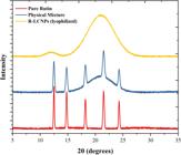
The physical state of the encapsulated rutin was definitively determined by XRD and DSC. The XRD pattern of pure rutin (Figure 3) exhibited numerous sharp, intense diffraction peaks at 2θ values of 12.5°, 14.8°, 18.2°, 21.5°, and 24.3°, confirming its highly crystalline nature [20]. In stark contrast, the XRD pattern of the lyophilized R-LCNPs showed only a broad, diffuse halo centered around 2θ = 21°, which is characteristic of amorphous materials. Crucially, all the sharp diffraction peaks corresponding to crystalline rutin were absent. This result provides unequivocal evidence that rutin was converted from its native crystalline state to an amorphous or molecularly dispersed state upon encapsulation within the LCNP matrix. This amorphization is the primary mechanism for overcoming the dissolution rate-limiting step of solubility, as amorphous forms possess higher free energy and thus greater apparent solubility than their crystalline counterparts.
The DSC thermograms (Figure 4) further corroborated the XRD findings. Pure rutin displayed a sharp, single endothermic peak at 196.5 °C, corresponding to its melting point. The thermogram for the physical mixture showed this characteristic melting peak of rutin, albeit at a slightly lower temperature due to the presence of other components. In the thermogram for the R-LCNPs, however, the melting endotherm of rutin was completely absent. This confirms the loss of crystallinity and indicates that rutin is homogenously dispersed within the lipid matrix at a molecular level, rather than existing as separate crystalline domains [20]. The combination of FTIR, XRD, and DSC results provides a cohesive and powerful confirmation of the successful encapsulation and amorphization of rutin within the LCNPs.

DSC thermograms of pure rutin, physical mixture, and R-LCNPs. The absence of the sharp endothermic peak of rutin at 196.5 °C in the R-LCNPs confirms loss of crystallinity and homogeneous molecular dispersion within the lipid matrix.
3.3. Surface and porosity characterization
XPS analysis was conducted to probe the surface chemistry of the R-LCNPs, providing critical information about the spatial distribution of the components within the nanoparticle. Since XPS is a surface-sensitive technique with a probing depth of only 5–10 nm, it can effectively distinguish between components on the surface versus those encapsulated in the core [21]. The survey spectrum of R-LCNP-2 (Figure 5A) revealed the presence of carbon (C 1s at ~285 eV) and oxygen (O 1s at ~532 eV) as the predominant elements, which is expected for a formulation composed of GMO (a lipid) and Poloxamer 407 (a polyether).

(A) XPS survey spectrum of R-LCNP-2 showing predominant carbon (C 1s at ~285 eV) and oxygen (O 1s at ~532 eV) peaks. (B) High-resolution C 1s spectrum deconvoluted into C–C/C–H (284.9 eV), C–O (286.8 eV), and C=O (288.2 eV) components.
The high-resolution C 1s spectrum (Figure 5B) was deconvoluted into three main components. The largest peak at 284.9 eV corresponds to C–C and C–H bonds, primarily from the long aliphatic chains of GMO. The peak at 286.8 eV is assigned to C–O bonds, characteristic of the ethylene glycol units of the Poloxamer 407 stabilizer. A smaller peak at 288.2 eV corresponds to C=O ester bonds from the GMO molecule. The key insight from this analysis is the chemical signature of the surface. The surface is dominated by signals from the lipid matrix (GMO) and, importantly, the hydrophilic PEG chains of the Poloxamer stabilizer (C–O peak). The absence of any unique spectral features attributable to rutin strongly indicates that the drug is successfully sequestered within the core of the nanoparticle, and the surface is effectively shielded by the stabilizer layer [22]. This core-shell-like architecture is ideal for protecting the drug from the external environment and facilitating a sustained release profile.
The UV–Vis spectrum of rutin in ethanol (Figure 6A) shows a characteristic absorption maximum at 354 nm, which was used for the quantification in the EE and drug release studies. BET analysis was performed on the lyophilized R-LCNPs to determine their specific surface area. The nitrogen adsorption-desorption isotherm (Figure 6B) was classified as a Type IV isotherm with a Type H3 hysteresis loop, typical for mesoporous materials. The calculated BET specific surface area was found to be 61.5 m2/g. This high surface area is a direct consequence of the small particle size and provides a large interface for interaction with biological media, which can contribute to an enhanced dissolution rate compared to bulk rutin powder, which has a surface area of less than 1 m2/g [23].

(A) UV–Vis absorption spectrum of rutin in ethanol showing a characteristic λₘₐₓ at 354 nm used for drug quantification. (B) Nitrogen adsorption–desorption isotherm of lyophilized R-LCNPs exhibiting a Type IV isotherm with a Type H3 hysteresis loop.
3.4. In vitro drug release
The in vitro release profiles of rutin from the optimized R-LCNP-2 formulation and a free rutin suspension were compared over 72 hours in PBS (pH 7.4) containing 0.5% Tween 80. As shown in Figure 7, the free rutin suspension exhibited very poor and incomplete dissolution, with only about 28.4 ± 3.1% of the drug released after 72 hours. This is a direct reflection of its low aqueous solubility, which is the primary barrier to its therapeutic use [19]. In contrast, the R-LCNPs demonstrated a significantly improved and controlled release profile. The release pattern was biphasic, characterized by an initial burst release of approximately 21.5 ± 2.5% within the first 4 hours, followed by a sustained and gradual release over the subsequent period. The cumulative release from R-LCNPs reached 89.7 ± 4.2% by 72 hours. The initial burst can be attributed to the release of rutin molecules located closer to the nanoparticle surface or within the outer aqueous channels. The subsequent sustained release phase is governed by the diffusion of the drug through the tortuous, complex network of lipid bilayers and water channels that form the core of the LCNP. This sustained release profile is highly desirable for therapeutic applications, as it can maintain drug concentrations within the therapeutic window for an extended period, potentially reducing dosing frequency and side effects.

In vitro cumulative release profiles of rutin from free drug suspension and R-LCNP-2 formulation in PBS (pH 7.4, 0.5% Tween 80) at 37 ± 0.5 °C over 72 hours. The R-LCNPs exhibit an initial burst release followed by a sustained release phase, reaching ~90% cumulative release by 72 h, whereas free rutin shows only ~28% release. Data expressed as mean ± SD (n = 3).
To elucidate the mechanism of drug release, the data were fitted to several kinetic models (Table 3). The release from R-LCNPs was best described by the Korsmeyer-Peppas model, with the highest correlation coefficient (R2 = 0.9912). The release exponent (n) value was calculated to be 0.41, which is less than 0.45, indicating that the primary mechanism of drug release is Fickian diffusion from the spherical matrix. This confirms that the liquid crystalline structure acts as a reservoir, controlling the diffusion rate of the encapsulated rutin into the surrounding medium.
Despite the favorable sustained-release profile observed in PBS (pH 7.4), the present system should not be interpreted as fully biorelevant. Under acidic gastric conditions, the apparent dissolved fraction of free rutin may remain limited, whereas intestinal surfactants and bile salts could enhance the dispersion and release of the encapsulated fraction. In a mildly acidic tumor-like microenvironment, partial changes in interfacial organization and diffusion resistance may alter the early release phase, which warrants dedicated follow-up testing under simulated gastrointestinal and disease-relevant conditions [24,25,26].
3.5. In vitro anti-inflammatory activity
The anti-inflammatory potential of the R-LCNPs was evaluated in an LPS-stimulated RAW 264.7 macrophage model. First, a preliminary MTT assay was conducted to ensure that the concentrations of the formulations used were non-toxic to the cells. As shown in Figure 8A, neither the Blank LCNPs nor the R-LCNP-2 formulation exhibited significant cytotoxicity at concentrations up to 50 µM (rutin equivalent), with cell viability remaining above 90%. This confirms that any observed reduction in inflammatory mediators is due to the pharmacological activity of rutin and not a result of cell death.
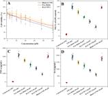
(A) Cell viability of RAW 264.7 macrophages treated with Blank LCNPs, free rutin, and R-LCNP-2 for 24 h, confirming absence of cytotoxicity at ≤50 µM rutin equivalent. (B–D) Inhibitory effects of formulations on LPS-induced (A) NO, (B) TNF-α, and (C) IL-6 production. R-LCNP-2 exhibits significantly greater anti-inflammatory efficacy compared to free rutin (*p < 0.01). Data presented as mean ± SD (n = 3).
LPS is a potent inducer of inflammation in macrophages, leading to the overproduction of pro-inflammatory mediators such as NO, TNF-α, and IL-6 [27]. As depicted in Figures 8B-8D and summarized in Table 4, stimulation of RAW 264.7 cells with LPS (1 µg/mL) caused a dramatic increase in the production of all three mediators compared to the untreated control group. Pre-treatment with free rutin at concentrations of 10 and 20 µM resulted in a modest, dose-dependent inhibition of NO, TNF-α, and IL-6 production. However, pre- treatment with R-LCNP-2 at equivalent rutin concentrations led to a significantly more potent anti-inflammatory effect. For instance, at a concentration of 20 µM, R-LCNP-2 inhibited NO production by 68.3 ± 4.5%, whereas free rutin only achieved an inhibition of 35.1 ± 3.8% (p < 0.01). Similarly, R-LCNP-2 treatment resulted in a much greater reduction in the secretion of TNF-α and IL-6 compared to free rutin.
This markedly superior efficacy of the nanoformulation can be directly attributed to the physicochemical advantages conferred by encapsulation. The LCNP system maintains rutin in a solubilized, bioavailable state within the aqueous cell culture medium, preventing its precipitation and thereby increasing the effective concentration of the drug that can interact with the cells [28]. Furthermore, nanoparticles are known to be internalized by macrophages more efficiently than soluble small molecules, often via endocytic pathways [29]. This enhanced cellular uptake delivers a higher payload of rutin to its intracellular targets, such as the NF-κB and MAPK signaling pathways, which are responsible for regulating the expression of iNOS, TNF-α, and IL-6 [30]. The Blank LCNPs showed no significant effect, confirming that the observed activity is solely due to the encapsulated rutin.
3.6. In vitro antitumor activity
The potential of the R-LCNP formulation to enhance the anticancer activity of rutin was evaluated against three human cancer cell lines: MCF-7 (breast cancer), HeLa (cervical cancer), and A549 (lung cancer). The cytotoxicity was assessed after 48 hours of treatment using the MTT assay, and the results are presented as dose-response curves in Figures 9A-C and summarized by the calculated IC50 values in Table 5.

Dose–response cytotoxicity curves of (A) MCF-7, (B) HeLa, and (C) A549 cancer cells treated with free rutin and R-LCNP-2 for 48 h, determined by the MTT assay. R-LCNP-2 exhibited significantly greater cytotoxic potency than free rutin across all cell lines (*p < 0.01). Data are expressed as mean ± SD (n = 3).
Across all three cell lines, R-LCNP-2 demonstrated significantly greater cytotoxic potency compared to free rutin. For example, in MCF-7 cells, the IC50 value for R-LCNP-2 was 28.6 ± 2.1 µM, which is approximately 4.2-fold lower than that of free rutin (119.5 ± 9.8 µM). Similar enhancements were observed for HeLa cells (3.8-fold decrease in IC50) and A549 cells (4.5-fold decrease in IC50). These results provide definitive quantitative evidence that encapsulating rutin in LCNPs dramatically enhances its ability to inhibit cancer cell proliferation.
The mechanism behind this enhanced antitumor activity is analogous to that observed in the anti-inflammatory assays. Free rutin, due to its poor solubility, likely precipitates in the cell culture medium, leading to a low actual concentration of dissolved drug available to the cancer cells [31]. The R-LCNP formulation effectively acts as a delivery vehicle that maintains a high local concentration of solubilized rutin, facilitating its passive diffusion or endocytic uptake into the cancer cells. By delivering a higher intracellular dose, the nanoformulation enables rutin to more effectively exert its known anticancer mechanisms, which include inducing cell cycle arrest at the G2/M phase and triggering apoptosis through the modulation of pathways involving p53, caspases, and Bcl-2 family proteins [2]. The superior performance of the R-LCNPs is therefore a direct and quantifiable consequence of overcoming the fundamental pharmacokinetic barrier of solubility, thereby unlocking the intrinsic cytotoxic potential of the rutin molecule. An important limitation is that the biological evidence presented here remains restricted to cell-based assays. While the marked improvements in anti-inflammatory and antitumor readouts are encouraging, in vivo pharmacokinetics, biodistribution, tolerability, and therapeutic efficacy must be determined before translational conclusions can be drawn. These studies are planned as the next step of the current formulation program.
4. CONCLUSION
In summary, the optimized R-LCNP-2 formulation achieved a favorable balance between loading capacity and dispersion quality, with reproducible particle size, narrow size distribution, moderately negative surface charge, and high encapsulation efficiency. The combined SEM/TEM, FTIR, XRD, DSC, XPS, and BET data supported successful incorporation of rutin within an ordered liquid-crystalline matrix and its conversion to a largely non-crystalline, molecularly dispersed state. The formulation further exhibited diffusion-governed sustained release and translated these physicochemical advantages into stronger in vitro bioactivity, including suppression of nitric oxide, TNF-α, and IL-6 in LPS-stimulated macrophages and enhanced cytotoxicity across MCF-7, HeLa, and A549 cells. Taken together, these results support LCNP encapsulation as a promising strategy to improve the functional performance of rutin, while also underscoring that in vivo pharmacokinetic and efficacy studies remain essential for future validation.
5. DATA AVAILABILITY
The data that support the findings of this study are available from the corresponding author upon reasonable request.
6. BIBLIOGRAPHY
-
[1] NOURI, Z., FAKHRI, S., NOURI, K., et al, “Targeting multiple signaling pathways in cancer: the rutin therapeutic approach”, Cancers (Basel), v. 12, n. 8, pp. 2276, Aug. 2020. doi: https://doi.org/10.3390/cancers12082276. PubMed PMID: 32823876.
» https://doi.org/10.3390/cancers12082276 -
[2] PRASAD, R., PRASAD, S.B., “A review on the chemistry and biological properties of Rutin, a promising nutraceutical agent”, Asian Journal of Pharmacy and Pharmacology, v. 5, n. 1, pp. 1–20, 2019. doi: https://doi.org/10.31024/ajpp.2019.5.s1.1.
» https://doi.org/10.31024/ajpp.2019.5.s1.1 -
[3] NAEEM, A., YU, C., ZANG, Z., et al, “Synthesis and evaluation of rutin–hydroxypropyl β-cyclodextrin inclusion complexes embedded in xanthan gum-based (HPMC-g-AMPS) hydrogels for oral controlled drug delivery”, Antioxidants, v. 12, n. 3, pp. 552, Feb. 2023. doi: https://doi.org/10.3390/antiox12030552. PubMed PMID: 36978800.
» https://doi.org/10.3390/antiox12030552 -
[4] PANDEY, P., KHAN, F., QARI, H.A., et al, “Rutin (bioflavonoid) as cell signaling pathway modulator: prospects in treatment and chemoprevention”, Pharmaceuticals (Basel, Switzerland), v. 14, n. 11, pp. 1069, Nov. 2021. doi: https://doi.org/10.3390/ph14111069. PubMed PMID: 34832851.
» https://doi.org/10.3390/ph14111069 -
[5] LEU, J.S.L., TEOH, J.J.X., LING, A.L.Q., et al, “Recent advances in the development of liquid crystalline nanoparticles as drug delivery systems”, Pharmaceutics, v. 15, n. 5, pp. 1421, May. 2023. doi: https://doi.org/10.3390/pharmaceutics15051421. PubMed PMID: 37242663.
» https://doi.org/10.3390/pharmaceutics15051421 -
[6] MALEKPOUR, M., EBRAHIMINEZHAD, A., KARIMI, Z., et al, “Current strategies for rutin nano-formulation, a promising bioactive compound with increased efficacy”, Bioprocess and Biosystems Engineering, v. 48, n. 6, pp. 877-898, 2025. doi: https://doi.org/10.1007/s00449-025-03156-y. PubMed PMID: 40148481.
» https://doi.org/10.1007/s00449-025-03156-y -
[7] SUBASH, P., KHUTE, S., “Recent advances in lyotropic liquid crystal nanoparticle formulations for drug delivery systems”, Frontiers in Soft Matter, v. 5, pp. 1658466, Sep. 2025. doi: https://doi.org/10.3389/frsfm.2025.1658466.
» https://doi.org/10.3389/frsfm.2025.1658466 -
[8] ALSHAHRANI, S.M., “Development and optimization of oral nanoemulsion of rutin for enhancing its dissolution rate, permeability, and oral bioavailability”, Pharmaceutical Development and Technology, v. 27, n. 5, pp. 588–597, May. 2022. doi: https://doi.org/10.1080/10837450.2022.2090957. PubMed PMID: 35703396.
» https://doi.org/10.1080/10837450.2022.2090957 -
[9] RAVI, G.S., CHARYULU, R.N., DUBEY, A., et al, “Nano-lipid complex of rutin: development, characterisation and in vivo investigation of hepatoprotective, antioxidant activity and bioavailability study in rats”, AAPS PharmSciTech, v. 19, n. 8, pp. 3631–3649, Nov. 2018. doi: https://doi.org/10.1208/s12249-018-1195-9. PubMed PMID: 30280357.
» https://doi.org/10.1208/s12249-018-1195-9 -
[10] MEHTA, M., PAUDEL, K.R., SHUKLA, S.D., et al, “Rutin-loaded liquid crystalline nanoparticles attenuate oxidative stress in bronchial epithelial cells: a PCR validation”, Future Medicinal Chemistry, v. 13, n. 6, pp. 543–549, Mar. 2021. doi: https://doi.org/10.4155/fmc-2020-0297. PubMed PMID: 33538615.
» https://doi.org/10.4155/fmc-2020-0297 -
[11] HAN, J., PANG, Y., SHEN, X., “Fucoidan and chitosan electrostatically coated nanoliposomes enhance physicochemical stability and bioavailability of rutin”, International Journal of Biological Macromolecules, v. 301, pp. 140450, Apr. 2025. doi: https://doi.org/10.1016/j.ijbiomac.2025.140450. PubMed PMID: 39884615.
» https://doi.org/10.1016/j.ijbiomac.2025.140450 -
[12] PAUDEL, K.R., WADHWA, R., TEW, X.N., et al, “Rutin loaded liquid crystalline nanoparticles inhibit non-small cell lung cancer proliferation and migration in vitro”, Life Sciences, v. 276, pp. 119436, Jul. 2021. doi: https://doi.org/10.1016/j.lfs.2021.119436. PubMed PMID: 33789146.
» https://doi.org/10.1016/j.lfs.2021.119436 -
[13] PAUDEL, K.R., WADHWA, R., MEHTA, M., et al, “Rutin loaded liquid crystalline nanoparticles inhibit lipopolysaccharide induced oxidative stress and apoptosis in bronchial epithelial cells in vitro”, Toxicology In Vitro : An International Journal Published in Association with BIBRA, v. 68, pp. 104961, Oct. 2020. doi: https://doi.org/10.1016/j.tiv.2020.104961. PubMed PMID: 32771431.
» https://doi.org/10.1016/j.tiv.2020.104961 -
[14] PUJITHA, R., CHELLAKUMARI, S.D., DAMAYANTHI, R.D., et al, “Engineered nanocrystals for poorly soluble drug delivery: a review”, Indian Journal of Pharmaceutical Sciences, v. 86, n. 3, pp. 742–754, Jun. 2024. doi: https://doi.org/10.36468/pharmaceutical-sciences.1332.
» https://doi.org/10.36468/pharmaceutical-sciences.1332 -
[15] AL-DHABI, N.A., ARASU, M.V., PARK, C.H., et al, “An up-to-date review of rutin and its biological and pharmacological activities”, EXCLI Journal, v. 14, pp. 59–63, Jan. 2015. doi: https://doi.org/10.17179/excli2014-663. PubMed PMID: 26535031.
» https://doi.org/10.17179/excli2014-663 -
[16] GARCÍA DÍAZ, J., GONZÁLEZ FERNÁNDEZ, R., ESCALONA ARRANZ, J.C., et al, “Inhibitory effect on nitric oxide release in LPS-stimulated macrophages and free radical scavenging activity of Croton linearis Jacq. Leaves”, Antioxidants, v. 11, n. 10, pp. 1915, Oct. 2022. doi: https://doi.org/10.3390/antiox11101915. PubMed PMID: 36290638.
» https://doi.org/10.3390/antiox11101915 -
[17] GUTIÉRREZ-RODRÍGUEZ, A.G., JUÁREZ-PORTILLA, C., OLIVARES-BAÑUELOS, T., et al, “Anticancer activity of seaweeds”, Drug Discovery Today, v. 23, n. 2, pp. 434–447, Feb. 2018. doi: https://doi.org/10.1016/j.drudis.2017.10.019. PubMed PMID: 29107095.
» https://doi.org/10.1016/j.drudis.2017.10.019 -
[18] MEENA, K.P., CHOUDHARY, P., KARRI, T., et al, “Preparation and characterization of rutin loaded microparticles for the treatment of diabetes”, Research Journal of Pharmacy and Technology, v. 16, n. 10, pp. 4867–4874, Oct. 2023. doi: https://doi.org/10.52711/0974-360X.2023.00789.
» https://doi.org/10.52711/0974-360X.2023.00789 -
[19] BAUDOT, C., TAN, C.M., KONG, J.C., “FTIR spectroscopy as a tool for nano-material characterization”, Infrared Physics & Technology, v. 53, n. 6, pp. 434–438, 2010. doi: https://doi.org/10.1016/j.infrared.2010.09.002.
» https://doi.org/10.1016/j.infrared.2010.09.002 -
[20] LIAN, R., LU, Y., QI, J., et al, “Silymarin glyceryl monooleate/poloxamer 407 liquid crystalline matrices: physical characterization and enhanced oral bioavailability”, AAPS PharmSciTech, v. 12, n. 4, pp. 1234–1240, Sep. 2011. doi: https://doi.org/10.1208/s12249-011-9666-2. PubMed PMID: 21948306.
» https://doi.org/10.1208/s12249-011-9666-2 -
[21] LOPINSKI, G.P., KODRA, O., KUNC, F., et al, “X-ray photoelectron spectroscopy of metal oxide nanoparticles: chemical composition, oxidation state and functional group content”, Nanoscale Advances, v. 7, n. 6, pp. 1671–1685, Jan. 2025. doi: https://doi.org/10.1039/D4NA00943F. PubMed PMID: 39898279.
» https://doi.org/10.1039/D4NA00943F -
[22] BAER, D.R., “Guide to making XPS measurements on nanoparticles”, Journal of Vacuum Science & Technology. A, Vacuum, Surfaces, and Films, v. 38, n. 3, pp. 031201, 2020. doi: https://doi.org/10.1116/1.5141419.
» https://doi.org/10.1116/1.5141419 -
[23] SHAJI, A., ZACHARIAH, A.K., “Surface area analysis of nanomaterials”, In: Thomas, S., Thomas, R., Zachariah, A.K., Mishra, R.K. (eds), Thermal and Rheological Measurement Techniques for Nanomaterials Characterization, USA, Elsevier, pp. 197–231, 2017. doi: https://doi.org/10.1016/B978-0-323-46139-9.00009-8.
» https://doi.org/10.1016/B978-0-323-46139-9.00009-8 -
[24] SENGUPTA, P., DAS, D., BHATTACHARYA, S., et al, “A pH-driven method for liposomal encapsulation of dietary flavonoid rutin: sustained release and enhanced bioefficacy”, Food Bioscience, v. 52, pp. 102392, Apr. 2023. doi: https://doi.org/10.1016/j.fbio.2023.102392.
» https://doi.org/10.1016/j.fbio.2023.102392 -
[25] ASFOUR, M.H., MOHSEN, A.M., “Formulation and evaluation of pH-sensitive rutin nanospheres against colon carcinoma using HCT-116 cell line”, Journal of Advanced Research, v. 9, pp. 17–26, Jan. 2018. doi: https://doi.org/10.1016/j.jare.2017.10.003. PubMed PMID: 30034879.
» https://doi.org/10.1016/j.jare.2017.10.003 -
[26] RAJESH, S., ZHAI, J., DRUMMOND, C.J., et al, “Synthetic ionizable aminolipids induce a pH dependent inverse hexagonal to bicontinuous cubic lyotropic liquid crystalline phase transition in monoolein nanoparticles”, Journal of Colloid and Interface Science, v. 589, pp. 85–95, May. 2021. doi: https://doi.org/10.1016/j.jcis.2020.12.060. PubMed PMID: 33450463.
» https://doi.org/10.1016/j.jcis.2020.12.060 -
[27] ALBAAYIT, S. F. A., AL-KHAFAJI, A. S. K., ALNAIMY, H. S., “In Vitro macrophage nitric oxide and interleukin-1 beta suppression by Moringa peregrina seed”, Turkish Journal of Pharmaceutical Sciences, vol. 16, no. 3, pp. 362–365, Jul. 2019, doi: https://doi.org/10.4274/tjps.galenos.2018.52244.
» https://doi.org/10.4274/tjps.galenos.2018.52244 - [28] HASSAN, A.S., SOLIMAN, G.M., “Rutin nanocrystals with enhanced anti-inflammatory activity: preparation and ex vivo/in vivo evaluation in an inflammatory rat model”, Pharmaceutics, v. 14, n. 12, pp. 2727, Dec. 2022. https://doi.org/10.3390/pharmaceutics14122727. PubMed PMID: 36559220.
- [29] CHEN, J., ZENG, GAO, et al, “Lipid-based liquid crystalline nanoparticles as oral drug delivery vehicles for poorly water-soluble drugs: cellular interaction and in vivo absorption”, International Journal of Nanomedicine, v. 7, pp. 3703–3718, 2012. https://doi.org/10.2147/IJN.S32599. PubMed PMID: 22888230.
-
[30] SOGASU, D., KUMAR, J.K., “Comparative anti-inflammatory activity of rutin-based mouthwash and diclofenac sodium: an in-vitro evaluation”, Journal of Pioneering Medical Sciences, v. 12, n. 3, pp. 61–64, Dec. 2023. doi: https://doi.org/10.61091/jpms202312312.
» https://doi.org/10.61091/jpms202312312 -
[31] CAPARICA, R., JÚLIO, A., ARAÚJO, M.E.M., et al, “Anticancer activity of rutin and its combination with ionic liquids on renal cells”, Biomolecules, v. 10, n. 2, pp. 233, Feb. 2020. doi: https://doi.org/10.3390/biom10020233. PubMed PMID: 32033222.
» https://doi.org/10.3390/biom10020233


