
Abstract
The use of micropropagation techniques is crucial for the conservation of endangered moss species and their reestablishment in nature. This study aimed to establish in vitro cultures of gametophyte fragments of ten species of Cerrado mosses. After disinfestation with alcohol and commercial bleach, moss explants were grown in Petri dishes containing Knop medium. The species Bryum argenteum, B. coronatum, Isopterygium tenerifolium, Leucobryum crispum, Pogonatum pensilvanicum, and Vitalia cuspidifera were successively established with efficiency rate ranging from 1 to 31.2%. However, no aseptic cultures were obtained for the species Barbula indica, Bryum densifolium, Fissidens flaccidus, and Sphagnum platyphylloides. Even though, a few contaminated explants of these species were able to develop and grow. Thus, all ten species were submitted to rescue techniques to establish cultures in aseptic conditions, from partially contaminated explants (indirect establishment). Consequently, the indirect establishment resulted in higher percentages of explant development, which enhanced the establishment of in vitro cultures for most of the species tested. This fact is especially important for conservation purposes, mainly for species whose material is sensitive or scarce. Therefore, indirect establishment as a new in vitro culture methodology was a viable form of propagating the bryophyte species listed in this research. This fact is essential for conservation purpose, especially for species whose material is sensitive or scarcer.
Key words:
bryophytes; conservation; cultivation; gametophyte; rescue
Resumo
O uso de técnicas de micropropagação é crucial para a conservação de espécies de musgos ameaçadas e para o reestabelecimento da natureza. Este estudo teve como objetivo estabelecer culturas in vitro de fragmentos de gametófitos de dez espécies de musgos do Cerrado. Após a desinfestação com álcool e solução de hipoclorito de sódio, explantes de musgo foram cultivados em meio Knop. As espécies Bryum argenteum, B. coronatum, Isopterygium tenerifolium, Leucobryum crispum, Pogonatum pensilvanicum e Vitalia cuspidifera foram sucessivamente estabelecidas com taxa de eficiência variando de 1 a 31,2%. No entanto, não foram obtidas culturas assépticas para as espécies Barbula indica, Bryum densifolium, Fissidens flaccidus e Sphagnum platyphylloides. Contudo, alguns explantes contaminados apresentaram desenvolvimento. Assim, todas as dez espécies foram submetidas as técnicas de resgate para estabelecer culturas em condições assépticas, a partir de explantes parcialmente contaminados (estabelecimento indireto). Consequentemente, o estabelecimento indireto resultou no aumento das chances de estabelecimento de culturas in vitro para a maioria das espécies. Portanto, o estabelecimento indireto como nova metodologia de cultivo in vitro foi bastante viável para a propagação das espécies de musgos listadas nesta pesquisa. Este fato é especialmente importante para fins de conservação, principalmente para espécies cujo material é sensível ou escasso.
Palavras-chave:
briófitas; conservação; cultivo; gametófito; resgate
Introduction
The Brazilian bryoflora comprises 1,524 species with 11 anthrocerus, 633 hepatic and 880 mosses. There are 478 cataloged bryophyte species in the Cerrado, from which 63 classified as endemic (Costa & Peralta 2015Costa DP & Peralta DF (2015) Bryophytes diversity in Brazil. Rodriguésia 66: 2015.). The Brazilian Cerrado is a tropical savanna and has been considered a hotspot for conservation because of its immense biodiversity and an enormous anthropic pressure (Myers et al. 2000Myers N, Mittermeier RA, Fonseca GAB & Kente J (2000) Biodiversity Hotspots for Conservation Priorities. Nature 403: 853-858.). It is important to register that this tropical savanna has been severely threatened by agriculture, disorderly urbanization and tourism, which have a negative impact on its flora, and, consequently, on native bryophytes (Söderström 2006Söderström L (2006) Conservation biology of bryophytes. Lindbergia 31: 24-32.). Thus, Cerrado bryophytes are losing their niches, which makes essential to identify the endangered species and work towards their conservation. Therefore, the improvement and implementation of specific strategies to ensure the long-term conservation of Cerrado bryophytes is of paramount importance (Costa et al. 2016Costa MLM, Bajgielman T, Pereira TS, Maurenza D, Amaro R, Dalcin EC & Maunder M (2016) Estratégia nacional para a conservação ex situ de espécies ameaçadas da flora brasileira. Centro Nacional de Conservação da Flora - CNC Flora, Instituto de Pesquisas Jardim Botânico do Rio de Janeiro, Andrea Jakobsson, Rio de Janeiro. 24p.; Söderström 2006Söderström L (2006) Conservation biology of bryophytes. Lindbergia 31: 24-32.).
It is important to note that bryophytes (mosses, liverworts, and hornworts) represent the second largest group of plants, with about 18 thousand species (Goffinet & Shaw 2009Goffinet B & Shaw AJ (2009) Bryophyte biology. 2nd ed. Cambridge University Press, New York. 565p.). In addition, these plants have demonstrated great commercial potential as landscape (Frahm 2004Frahm JP (2004) Recent developments of commercial products from bryophytes. The Bryologist 107: 277-283.; Anderson et al. 2010Anderson M, Lambrinos J & Schroll E (2010) The potential value of mosses for stormwater management in urban environments. Urban Ecosystems 13: 319-332.), and ecological importance (Bates 2000Bates JW (2000) Mineral nutrition, substratum ecology, and pollution. In: Shaw AJ & Goffinet B (eds.) Bryophyte biology. 2nd ed. Cambridge University Press, New York. Pp 299-356.; Tuba et al. 2011Tuba Z, Slack NG & Stark LR (2011) Bryophyte ecology and climate change. Cambridge University Press, New York. 506p.; González & Pokrovsky 2014González AG & Pokrovsky OS (2014). Metal adsorption on mosses: toward a universal adsorption model. Journal of colloid and interface science 415: 169-178.; Capozzi et al. 2017Capozzi F, Adamo P, Di Palma A, Aboal JR, Bargagli R, Fernandez JA, Lopez MP, Reski R, Tretiach M, Spagnuolo V & Giordano S (2017) Sphagnum palustre clone vs native Pseudoscleropodium purum: a first trial in the field to validate the future of the moss bag technique. Environmental Pollution 225: 323-328.) as well as broad biotechnological uses (Reski 1998Reski R (1998) Development, genetics and molecular biology of mosses. Botanica Acta 111: 1-15.; Decker & Reski 2004Decker EL & Reski R (2004) The moss bioreactor. Current Opinion in Plant Biology 7: 166-170.; Cuvertino-Santoni & Montenegro 2013Cuvertino-Santoni J & Montenegro G (2013) Bioprospecting a tool to conserve Chilean bryophytes. Gayana Botanica 70: 1.). Nevertheless, the bryophytes have been neglected in conservation initiatives, maybe because they are considered plants of little relevance and difficult to identify (Goffinet et al. 2009Goffinet B, Buck WR & Shaw AJ (2009) Morphology and classification of the Bryophyta. In: Shaw AJ & Goffinet B (eds.) Bryophyte biology. 2nd ed. Cambridge University Press, New York. Pp. 55-138.; Glime 2017Glime JM (2017) Introduction. In: Glime JM (ed) Bryophyte ecology. Vol 1. Physiological ecology. Michigan Technological University, International Association of Bryologists. Available at <http://digitalcommons.mtu.edu/bryophyte-ecology/>. Access on 5 November 2017.
http://digitalcommons.mtu.edu/bryophyte-...
).
Despite that, some conservation techniques have been used in bryophytes. For instance, in situ conservation is the most common strategy applied to this class of plants. However, the efficiency of this approach is not always suitable for bryophytes conservation (Söderström 2006Söderström L (2006) Conservation biology of bryophytes. Lindbergia 31: 24-32.). Nevertheless, ex situ techniques, through axenic cultures, can be successfully applied for long- and medium-term conservation. Hence, cryopreservation, tissue culture, as well as genetic and molecular studies are suitable techniques for ecological research and environmental reintroduction (Duckett et al. 2004Duckett JG, Burch J, Fletcher PW, Matcham HW, Read DJ, Russell AJ & Pressel S (2004) In vitro cultivation of bryophytes: a review of practicalities, problems, progress and promise. Journal of Bryology 26: 3-20.). For example, Ros et al. (2013)Ros RM, Werner O & Pérez-Álvarez JR (2013) Ex situ conservation of rare and threatened Mediterranean Bryophytes. Flora Mediterranea 23: 223-235. tested the in vitro culture for drought-tolerant moss species with subsequent cryopreservation, resulting in high survival rate.
In fact, the use of tissue culture techniques in mosses is not unusual. Several studies show the efficiency of these techniques mainly in the conservation of rare and endangered species (Reski 1998Reski R (1998) Development, genetics and molecular biology of mosses. Botanica Acta 111: 1-15.; Duckett et al. 2004Duckett JG, Burch J, Fletcher PW, Matcham HW, Read DJ, Russell AJ & Pressel S (2004) In vitro cultivation of bryophytes: a review of practicalities, problems, progress and promise. Journal of Bryology 26: 3-20.). Furthermore, tissue culture has also been applied to the development of more efficient methods for propagation, commercial and industrial exploitation of mosses (Duckett et al. 2004Duckett JG, Burch J, Fletcher PW, Matcham HW, Read DJ, Russell AJ & Pressel S (2004) In vitro cultivation of bryophytes: a review of practicalities, problems, progress and promise. Journal of Bryology 26: 3-20.; Cuvertino-Santoni & Montenegro 2013Cuvertino-Santoni J & Montenegro G (2013) Bioprospecting a tool to conserve Chilean bryophytes. Gayana Botanica 70: 1.). It is worth mentioning the large-scale cultivation of Sphagnum palustre L. in bioreactors, to produce standardized and sustainable material for biomonitoring air quality through a technique termed as “Moss Bag” (Beike et al. 2014Beike AK, Spagnuolo V, Lüth V, Steinhart F, Ramos-Gomez J, Krebs M, Adamo P, Rey-Asensio AI, Fernandez JA, Giordano S, Decker EL & Reski R (2014) Clonal in vitro propagation of peat mosses (Sphagnum L.) as novel green resources for basic and applied research. Plant Cell, Tissue and Organ Culture (PCTOC) 120: 1037-1049.; Capozzi et al. 2016Capozzi F, Giordano S, Aboal JR, Adamo P, Bargagli R, Boquete T, Di Palma A, Real C, Reski R, Spagnuolo V, Steinbauer K, Tretiach M, Varela Z, Zechmeister H & Fernandez JA (2016) Best options for the exposure of traditional and innovative moss bags: a systematic evaluation in three European countries. Environmental Pollution 214: 362-373.). This technique has also been applied with similar efficiency to other species of Sphagnum (Hu et al. 2018Hu R, Yan Y, Zhou X, Wang Y & Fang Y (2018) Monitoring heavy metal contents with Sphagnum junghuhnianum Moss bags in relation to traffic volume in Wuxi, China. International Journal of Environmental Research and Public Health 15: 374.), as well as to Hypnum cupressiforme Hedw. (Ares et al. 2012Ares A, Aboal JR, Carballeira A, Giordano S, Adamo P & Fernández JA (2012) Moss bag biomonitoring: a methodological review. Science of the Total Environment 432: 143-158.), and Pseudoscleropodium purum (Hedw.) Fleisch. (Capozzi et al. 2017Capozzi F, Adamo P, Di Palma A, Aboal JR, Bargagli R, Fernandez JA, Lopez MP, Reski R, Tretiach M, Spagnuolo V & Giordano S (2017) Sphagnum palustre clone vs native Pseudoscleropodium purum: a first trial in the field to validate the future of the moss bag technique. Environmental Pollution 225: 323-328.). Additionally, Bryum argenteum Hedw. has been micropropagated as a means of improving its production and commercial use to create greener environments (Liang et al. 2010Liang SF, Sun Y & Zhu RL (2010) In vitro micropropagation of Bryum argenteum Hedw. Cryptogamie. Bryologie 31: 233-239.).
In vitro culture of bryophytes has the advantage of being able to initiate cultures from either sexual (spores) or vegetative structures such as gametophyte fragments and gemmae (Duckett et al. 2004Duckett JG, Burch J, Fletcher PW, Matcham HW, Read DJ, Russell AJ & Pressel S (2004) In vitro cultivation of bryophytes: a review of practicalities, problems, progress and promise. Journal of Bryology 26: 3-20.). This broad application of tissue culture is essential for species that are not found in the fertile phase of their life cycle (Longton & Miles 1982Longton RE & Miles CJ (1982) Studies on the reproductive biology of mosses. Journal of the Hattori Botanical Laboratory 52: 219-239.; Löbel & Rydin 2010Löbel S & Rydin H (2010) Trade-offs and habitat constraints in the establishment of epiphytic bryophytes. Functional ecology 24: 887-897.). Besides, under specific conditions, the use asexual propagules for in vitro propagation of bryophytes may promote the formation of specialized resting propagules capable of withstanding extreme environmental conditions such as desiccation stress (Pressel et al. 2007Pressel S, Matcham HW & Duckett JG (2007) Studies of protonemal morphogenesis in mosses. XI. Bryum and allied genera: a plethora of propagules. Journal of Bryology 29: 241-258.; Rowntree et al. 2007Rowntree JK, Duckett JG, Mortimer CL, Ramsay MM & Pressel S (2007) Formation of specialized propagules resistant to desiccation and cryopreservation in the threatened moss Ditrichum plumbicola (Ditrichales, Bryopsida). Annals of Botany 100: 483-496.).
Concerning the establishment of axenic bryophyte cultures, sporophytes are usually more easily disinfested than gametophytes. This feature is very limiting for moss micropropagation because of the fragility of the tissues and the symbiotic interactions with diverse microorganisms, which is hard to replicate in vitro (Sabovljević et al. 2003Sabovljević M, Bijelovic A & Dragicevic IČ (2003) In vitro culture of Mosses: Aloina aloides (KF Schultz) Kindb., Brachythecium velutinum (Hedw.) BS & G., Ceratodon purpureus (Hedw.) Brid., Eurhynchium praelongum (Hedw.) BS & G. and Grimmia pulvinata (Hedw.) Sm. Turkish journal of botany 27: 441-446.; Duckett et al. 2004Duckett JG, Burch J, Fletcher PW, Matcham HW, Read DJ, Russell AJ & Pressel S (2004) In vitro cultivation of bryophytes: a review of practicalities, problems, progress and promise. Journal of Bryology 26: 3-20.). Despite that, once the in vitro culture has been established, the cultivation of these plants becomes relatively easy (Rowntree et al. 2011Rowntree JK, Pressel S, Ramsay MM, Sabovljević A & Sabovljević M (2011) In vitro conservation of European bryophytes. In vitro Cellular & Developmental Biology-Plant 47: 55-64.). A further limiting factor to initiate in vitro cultures is the low availability of gametophytic material from endangered species, especially when spores are not available. Likewise, the protocols for in vitro culture are often specific for each species, which further complicates the use of plant tissue culture techniques in this class of plants (Sarasan et al. 2006Sarasan V, Cripps R, Ramsay MM, Atherton C, McMichen M, Prendergast G & Rowntree JK (2006) Conservation in vitro of threatened plants-progress in the past decade. In vitro Cellular and Developmental Biology - Plant 42: 206-214.; Tacoronte et al. 2009Tacoronte BM, León Y, Olivo A & Vielma M (2009) Crecimiento in vitro de musgos del bosque nublado andino de Venezuela. Revista Forestal Latino americana 24: 69-89.).
In addition, researches on the propagation of non-endangered species are also important for rare and endangered species because they may provide essential information that could be applied to the conservation of bryophytes with limited material available (Rowntree 2006Rowntree JK (2006) Development of novel methods for the initiation of in vitro bryophyte cultures for conservation. Plant cell, tissue and organ culture 87: 191-201.; Sarasan et al. 2006Sarasan V, Cripps R, Ramsay MM, Atherton C, McMichen M, Prendergast G & Rowntree JK (2006) Conservation in vitro of threatened plants-progress in the past decade. In vitro Cellular and Developmental Biology - Plant 42: 206-214.). In this sense, the objective of current work was to establish in vitro cultures initiated from gametophyte fragments of ten mosses species occurring in the Cerrado.
Material and Methods
Plant collection and preparation
The plants with part of the substrate were collected in areas of Cerrado vegetation and urban areas of Brasília - DF, Brazil, between 2015 and 2017, and then, put in paper bags, according to Yano (1984)Yano O (1984) Briófitas. In: Fidalgo O & Bononi VLR (coords.) Técnicas de coleta, preservação e herborização de material botânico. Instituto de Botânica, São Paulo. Pp 27-30.. Ten species of mosses were collected: Barbula indica (Hook.) Spreng., Bryum argenteum Hedw., Bryum coronatum Schwägr., Bryum densifolium Brid., Fissidens flaccidus Mitt., Isopterygium tenerifolium Mitt., Leucobryum crispum Müll. Hal., Pogonatum pensilvanicum (Bartram ex Hedw.) P. Beauv., Sphagnum platyphylloides Warnst., Vitalia cuspidifera (Mitt.) P.E.A.S. Câmara, Carv.-Silva, and W.R. Buck, whose vouchers (Tab. 1) were deposited at UB Herbarium.
Disinfestation treatments with commercial bleach (2% active chlorine) – 1% concentration for 5 min (1% - 5'), 1% for 10 min (1% - 10'), and 2% for 2 min (2% - 2'), bryophyte species and respective collection data and number of explants per treatment.
The identification of the species was carried out in the Laboratory of Cryptogams of the University of Brasília - UnB. Gametophytes of each species were separated and washed in running water for 2 min, and then, in sterile water for 2 min. Preferably, the younger fragments of gametophytes, such as apices, leaves, parts of stems and branch leaves and new shoots were used as explants for micropropagation.
For Barbula indica, Bryum argenteum and Pogonatum pensilvanicum, due to their peculiarities, pre-cultures were carried out, aiming the production of gametophytes suitable for surface disinfestation. The precultivation procedure consisted of non-aseptic culture in closed containers, in which a portion of each sample, with part of the substrate, was packed in transparent pots of 250 mL containing either wet commercial substrate (Bioplant® plus, Nova Ponte, Brazil) or vermiculite. Afterward, the pots were closed and kept under natural light and room temperature (25 ºC) until the development of gametophytes. The pots were watered every 30 days. After the cultivation period, 0.5 to 1 cm gametophytes were collected and submitted to disinfestation and subsequent inoculation.
Culture medium and conditions
The inoculation of the explants, as well as the subcultures, were carried out in Knop medium (Knop 1865Knop W (1865) Quantitative Untersuchungen uber die Ernah rungsprozesse der Pflanze. Die Landwirtschaftlichen Versuchs-Stationen 7: 93-107.), with the addition of the following components: 0.8 g.L-1 of calcium nitrate [Ca (NO3)2 .4 H2O]; 0.2 g.L-1 potassium nitrate (KNO3); 0.2 g.L-1 of monopotassium phosphate (KH2PO4); 0.2 g.L-1 of magnesium sulfate (MgSO4 .7 H2O) and sterile water. The solidification of the solutions was carried out with the addition of 7.5 g.L-1 of agar, after adjusting the pH to 5.7 ± 0.1. Then, the media was sterilized at 121 ºC, 1.3 atm for 20 min, and 15 mL was poured into Petri dishes (90 × 15 mm).
The explants were previously disinfested in 70% alcohol for 5 s and solution of Sodium hypochlorite (commercial bleach containing 2% active chlorine, NaOCl) in three treatments: 1% for 5 min, 1% for 10 min, and 2% for 2 min (1% - 5’, 1% - 10’ and 2% - 2’), and then rinsed 3x in sterile water. The disinfestation treatments were applied to the species according to the availability and the sensitivity of the collected material. For the species with abundant material, all three disinfestation treatments were applied. For the species with limited material, only one treatment was performed. Additionally, eight explants per plate were inoculated. The number of inoculated explants was defined according to the quantity and quality of the material obtained in the collections. The total explants, as well as the treatments performed in each species are described in Table 1.
The experiment was completely randomized, with eight explants per plate, and each plate constituted a repetition. The number of repetitions per treatment was according to the quantity and quality of plant material obtained in the collections. The total number of explants, as well as the treatments performed in each species, are described in Table 1.
After inoculation, all plates were sealed with plastic PVC film and kept in a room, with the temperature set at 25 (± 2 ºC), photoperiod of 16 h and light intensity of approximately 41 µmol.m-2.s-1.
The explants were evaluated on the 5th and 10th day after inoculation and classified as follows: developed without contamination or developed with contamination. Explants that showed apical growth, budding or growth of protonemata were considered as developed. Explants that did not show any development of plant tissue in 10 days of cultivation were discarded.
In the first evaluation (5th day of cultivation), all the explants that showed some development, contaminated or not, were transferred to new plates (subculture plates). Then, the plates were again sealed and cultivated for another 5 days. In the second evaluation (10th day), the explants that presented development were transferred to subculture plates.
It was considered established those in vitro cultures that, after disinfestation, at least one explant had been developed without a visible presence of microorganisms. The subcultures were based on developed explants with no signs of contamination and formed colonies suitable for propagation in subsequent subcultures.
Results and Discussion
Figure 1 depicts the efficiency of disinfestation treatments with a solution of Sodium hypochlorite, which varied from 1 to 19% (1% for 5 min), 2.5 to 9% (1% for 10 min), and 3.5 to 31.2% (2% for 2 min). The use of hypochlorite in the disinfestation of fragments of bryophyte gametophytes has been shown to vary with the species, and with other parameters, according Sabovljević et al. (2003)Sabovljević M, Bijelovic A & Dragicevic IČ (2003) In vitro culture of Mosses: Aloina aloides (KF Schultz) Kindb., Brachythecium velutinum (Hedw.) BS & G., Ceratodon purpureus (Hedw.) Brid., Eurhynchium praelongum (Hedw.) BS & G. and Grimmia pulvinata (Hedw.) Sm. Turkish journal of botany 27: 441-446. as observed in Aloina aloides. This is consistent with the results obtained in this investigation. Note that almost 50% of the species tested were not successfully disinfested with lower concentrations of hypochlorite (Fig. 1; Tab. 2). Therefore, Liang et al. (2010)Liang SF, Sun Y & Zhu RL (2010) In vitro micropropagation of Bryum argenteum Hedw. Cryptogamie. Bryologie 31: 233-239. observed that the sterilization efficiency is dependent on the concentration and exposure time of explants. Furthermore, it was shown that disinfestation of Bryum argenteum explants achieved the highest efficiency when concentrations of 5% were used (Liang et al. 2010Liang SF, Sun Y & Zhu RL (2010) In vitro micropropagation of Bryum argenteum Hedw. Cryptogamie. Bryologie 31: 233-239.). Table 2 shows that the explants from 5 out of 6 species tested (80%) were better disinfested with 2% NaClO, which represented an average of 7.7% of all developed explants with no visible contamination. The only exception was Bryum densifolium that did not show decontamination with 2% NaClO (Fig. 1). Also, the highest percentage of decontamination was seen with Isopterygium tenerifolium, which had 19% of explants developed with no apparent contamination in treatment with NaClO 1% for 5 min (Fig. 1).
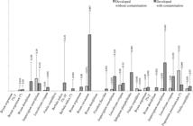
Explant development rate of ten Cerrado mosses species in different treatments (1% for 5 min; 1% for 10 min; and 2% for 2 min), after 10 days of in vitro cultivation. The bars represent the standard error. * Precultivated material.
Percentage of moss species developed with or without contamination after disinfestation treatments (solution of Sodium hypochlorite x exposure time) and cultivated in vitro for 10 days in Knop medium.
Additionally, Sabovljević et al. (2002)Sabovljević MS, Bijelović A & Dragićević IČ (2002) Effective and easy way of establishing in vitro culture of mosses, Bryum argenteum Hedw. and Bryum capillare Hedw. (Bryaceae). Archives of Biological Sciences 54: 7-8. also observed that variation in disinfestation efficiency depends on the morphology of the gametophyte, as well as on the position and density of the leaves. Some of the morphological characteristics could also be verified as limiting for disinfestation, as observed in Barbula indica that presents compact leaves, curved when dry and often with the presence of axillary gemmae (Sharp et al. 1994). Likewise, in Bryum argenteum leaves were ovate, concave, closely appressed and overlapped on the stem, which, usually, negatively interfered with the disinfestation (Sharp et al. 1994). Also, disinfestation difficulties were seen in Fissidens flaccidus that has a vaginant lamina in the leaves and in Sphagnum platyphylloides that presents empty cells that contain small openings named leucocysts (Costa et al. 2010Costa DP, Almeida JSS, Dias NS, Gradstein SR & Churchi SP (2010) Manual de briologia. Editora Interciência, Rio de Janeiro. 222p.). These morphological aspects, together with the fact that the species were collected directly from the soil, cemented pavement, and rocks, also contributed to difficult the disinfestation procedure as it was not possible to entirely remove these substrates from the samples.
Nevertheless, in six species out of 10 species, the in vitro establishment was obtained from the 1st subculture: Bryum argenteum, B. coronatum, I. tenerifolium, L. crispum, P. pensilvanicum, and V. cuspidifera. The percentage of explants established of the species mentioned above varied among them as well as treatments. Furthermore, the 2% solution for 2 min were more effective for five species (Fig. 1). Therefore, it has been considered established those in vitro cultures that at least one non-contaminated explant was obtained in the first subculture, which was termed as direct establishment (Fig. 2a-c).

a-g. Establishment and in vitro propagation of mosses. a-c. direct establishment (without contamination) - a. developed explants were transferred to new plates; b. at the end of the 1st subculture, the established explants were fragmented and transferred to new plates; c. propagation of the species. a, d-g and c. indirect establishment (with contamination) - a. developed explants were transferred to new plates; d-e. the explants were subcultivated until they had produced uncontaminated regions; f. sufficiently developed explants were fragmented and subcultivated; g. presence of established explants with no visible contamination in the plates; c. explants, free of contamination, were used to establish in vitro aseptic cultures and used for propagation of the species.
The other four species: Barbula indica, Bryum densifolium, F. flaccidus, and S. platyphylloides did not development without visible explant contamination (Fig. 1). However, despite contamination, it was observed that some explants had developed, whose efficiency was 3.1% (F. flaccidus) to 28.4% (S. platyphylloides). Also, it is important to point out that in all ten species studied showed the development of contaminated explants (Fig. 1). Compared with non-contaminated explants, the development of contaminated explants was quite variable. However, contaminated explants had higher development rates than non-contaminated when the disinfecting treatment was with NaClO 1% for 10 min (1.5 to 86.7%) and with NaClO 2% for 2 min (5.2 to 43.5%). Differently, the NaClO 1% for 5 min treatment showed a lower developmental rate of contaminated explants (2 to 15%) than those without contamination (19%). Moreover, the development of non-contaminated and contaminated explants in cultures of S. platyphylloides, I. tenerifolium, and Bryum coronatum was 0 and 28.4%, 3.5% and 43.5%, and 7.1% and 86.7%, respectively (Fig. 1).
Although most of the species studied are considered to be of widespread occurrence in Brazil, explant rescue techniques were applied to all. According to Rowntree et al. (2011)Rowntree JK, Pressel S, Ramsay MM, Sabovljević A & Sabovljević M (2011) In vitro conservation of European bryophytes. In vitro Cellular & Developmental Biology-Plant 47: 55-64., contaminated explants that maintain their capacity to develop can be propagated in subsequent subcultures. Therefore, contaminated explants of all ten species were used to recover and establish in vitro cultures to obtain at least one explant under aseptic conditions. The rescue technique was initially introduced by Rowntree (2006)Rowntree JK (2006) Development of novel methods for the initiation of in vitro bryophyte cultures for conservation. Plant cell, tissue and organ culture 87: 191-201., in which they described the recovery of plant material from partially contaminated cultures, using sodium dichloroisocyanurate (NaDCC) and PPMTM (Plant Cell Technology, Inc., Washington, USA) as sterilizing agents, increasing the success rate of 28 % to 52% in the in vitro establishment of rare plants. A similar method also appears to have been tested in other plants, such as Thamnobryum alopecurum and Hypnum cupressiform but was unsuccessful (Vujičić et al. 2011Vujičić M, Sabovljević A & Sabovljević M (2011) Axenically culturing the bryophytes: establishment and propagation of the moss Hypnum cupressiforme Hedw. (Bryophyta, Hypnaceae) in in vitro conditions. Botanica Serbica 35: 71-77.; Sabovljević et al. 2012Sabovljević A, Vujičić M, Skorić M, Bajić-Ljubičić J & Sabovljević M (2012) Axenically culturing the bryophytes: establishment and propagation of the pleurocarpous moss Thamnobryum alopecurum Nieuwland ex Gangulee (Bryophyta, Neckeraceae) in in vitro conditions. Pakistan Journal of Botany 44: 339-344.).
The protocol for the rescue techniques is outlined in Fig. 2 (in the following order a, d-g, and then back to c), which was called indirect establishment, as it does not follow the regular protocol used in in vitro establishment of bryophytes of aseptic cultures. Thus, the indirect establishment uses techniques to recover explants developed after disinfestation that were not adequately decontaminated. Therefore, this procedure was applied to all species in this study. This procedure was constituted by successive subcultures of the explants developed with contamination, wherein the during the first two subcultures, the contaminated explants were transferred to new plates every 5-10 days. In the following subcultures (30 to 40 days each), the explants formed colonies, and the regions of the cultures with no visible contamination were transferred to new plates. The level of explant contamination decreased after each subculture, which made possible to gradually achieve asepsis until the in vitro establishment of the species.
Figure 3 shows the indirect establishment of Bryum argenteum and Isopterygium tenerifolium cultures. These species were submitted to the disinfestation treatment, which did not result in adequate decontamination. However, despite the contamination, the explants showed capacity to develop and grow (Fig. 3a-f, i-j). Hence, the rescue techniques were applied, and after successive subcultures, protonemata (Fig. 3c,d), young gametophytes (Fig. 3e) or branches of elongated gametophytes (Fig. 3i) were carefully transferred to new plates and subcultivated until no contamination was observed. A similar procedure was repeated to reduce contamination (Fig. 3g,k) and establish an aseptic in vitro culture of the two species (Fig. 3h,l).

a-l. Indirect establishment of Bryum argenteum (a-h) and Isopterygium tenerifolium (i-l) - a. contaminated colony of explants in the 3rd subculture (arrow); b. partial contamination (arrow) in the 4th subculture; c. partial contamination (arrow) in the 5th subculture after 24 days of cultivation; d. partial contamination (arrow) in the 5th subculture after 35 days, showing the growth of protonemata external to the medium with contamination (asterisk); e. growth erect gametophytes, externally to the medium with contamination (arrow) in the 5th subculture after 24 days; f. growth of gametophytes near contaminated areas (arrows); g. apical fragment of gametophyte without contamination in the 6th subculture after 11 days of cultivation; h. established Bryum argenteum culture during the 6th subculture; i. Isopterygium tenerifolium with contamination (arrow) in the 3rd subculture with 40 days; j. growth of gametophyte near the contamination (arrows); k. apical fragment of gametophyte without contamination in the 4th subculture after 40 days; l. established Isopterygium tenerifolium culture free of contamination in the 5th subculture after 40 days. Bars: k. = 0.1 mm; f, g, j, l = 0.5 mm; c, d, e, h, i = 1 mm; a, b = 2 mm.
It was also observed that the time and number of subcultures required to obtain the indirect establishment of aseptic cultures varied among the species. Besides, none the studied species showed any correlation between the number and time of subcultures to establish in vitro cultures, as well as with factors such as plant growth habit, type of explant, and natural growth conditions of the species (Tab. 3).
Characterization of moss species and responses observed during in vitro establishment in Knop medium. (GA = gametophytes apex; FG = female gametangium; MG = male gametangium; LA = leaf axillary; SS = place of the stem section; ALd = axillary of the leaf detached; ABd = axillary of the branch detached; LL = leaf lamina; AG = apical growth; B = branching.
Furthermore, Barbula indica, Bryum argenteum, and P. pensilvanicum were submitted to a precultivation procedure, a stage before disinfestation with the purpose of reducing contamination and increasing the production of explants. For Barbula indica and Bryum argenteum, the precultivation treatment resulted in neither enhancement of the number of explants nor better decontamination (Fig. 1). Nevertheless, the precultivation of P. pensilvanicum and subsequent disinfestation (2% bleach for 2 min) yielded 31.2% of explants with no visible contamination (Fig. 1).
In precultivated Bryum argenteum with subsequent disinfestation disinfested with 1% bleach for 5 min, which yielded 1.5% of explants with no visible contamination. Moreover, in this condition was also observed the development of four additional contaminated explants. It appears that, compared with non-precultivated samples, there was no improvement in explant production and decontamination. Conversely, Rowntree (2006)Rowntree JK (2006) Development of novel methods for the initiation of in vitro bryophyte cultures for conservation. Plant cell, tissue and organ culture 87: 191-201. found that precultivated bryophytes can improve disinfestation, and, thereby, reduce the level of contamination, compared with bryophytes disinfested after field collection. Hence, the precultivation treatment must be tested individually because it may yield different results depending on the species.
Concerning Pogonatum pensilvanicum explants, which had limited biological material, and reduced gametophytes size (2-3 mm), rudimentary leaves and persistent protonemata (Sharp et al. 1994) were disinfested only after precultivation (Tab. 3). The explants used in the disinfestation treatment were at least 8 mm long, which was achieved after nine months of precultivation. The precultivation treatment was very efficient for this species and increased gametophyte production, as well as a higher rate of disinfestation. Thus, the percentage of explants developed without contamination for P. pensilvanicum was relatively high (31.2%). In fact, this was the highest number of explants developed without contamination among all species studied (Fig. 1). Additionally, the explants that development with contamination (9.4%) required fewer subcultures and less time to establish aseptic cultures (Tab. 3).
For Barbula indica, the precultivation treatment was not relevant because occurred a decrease from 12.5% to 3.4% in explant production submitted to non-precultivation and precultivation treatment, respectively (Fig. 1). Besides, only contaminated explants developed in both conditions. Even with low success rates for in vitro establishment of B. indica, it is crucial to improving the cultivation techniques for this species. Viet et al. (2010)Viet HN, Frontasyeva MV, Thi TMT, Gilbert D & Bernard N (2010) Atmospheric heavy metal deposition in Northern Vietnam: Hanoi and Thainguyen case study using the moss biomonitoring technique, INAA and AAS. Environmental Science and Pollution Research 17: 1045-1052. state that this species is reliable for atmospheric biomonitoring, such as heavy metals air contamination of subtropical and tropical regions. However, to use B. indica for air biomonitoring a few obstacles must be overcome as it is an acrocarpic plant, about 1 cm high with strong adhesion to the substrate, which can influence the analysis on the level metal contamination of the air. Thus, it is essential to use B. indica for that purpose is necessary plants with no contact with external environment, which makes in vitro propagation the best choice to provide plant free of contaminants for biomonitoring. Furthermore, a high multiplication rate can be achieved through micropropagation techniques to provide plant material and place in more suitable substrates such as house walls, roofs, and tree trunks, as suggested by Viet et al. (2010)Viet HN, Frontasyeva MV, Thi TMT, Gilbert D & Bernard N (2010) Atmospheric heavy metal deposition in Northern Vietnam: Hanoi and Thainguyen case study using the moss biomonitoring technique, INAA and AAS. Environmental Science and Pollution Research 17: 1045-1052..
All the species had protonema as the initial form of development. Besides, the species Bryum coronatum, Bryum coronatum, I. tenerifolium, V. cuspidifera, and S. platyphylloides also showed the development of shoots from inoculated explants (Fig. 4a). For most species, the initial development was from structures such as gametophyte apex (Fig. 4b), and, or, from the axil of the leaf, attached or not to the stem. Also, Bryum argenteum, Bryum coronatum and V. cuspidifera developed protonemata from stem sectioned regions (Fig. 4c). It was also observed the development of protonemata on sexual structures, such as in Bryum argenteum, in the female gametangium (archegonium) and the male gametangium (antheridium) of I. tenerifolium (Fig. 4d-e). Bryum argenteum, Bryum coronatum and S. platyphylloides showed apical growth (Fig. 4f). Only L. crispum developed protonemata directly from the base and apex of the leaf (Fig. 4g; Tab. 3).

a-p. Structures developed in Cerrado mosses grown in vitro - a. Bryum argenteum shoots (7); b. apical protonemata in Fissidens flaccidus (5); c. Protonemata at the stem section site (arrow) in Vitalia cuspidifera (20); d. Archegonium (arrow) of Bryum argenteum (10); e. Antheridium (arrow) of Isopterygium tenerifolium (13); f. apical growth from tip of the apex (arrow) in Bryum coronatum (13); g. Protonemata on the leaf lamina of Leucobryum crispum (20); h. Protonemata colony of Bryum argenteum with gametophytic buds (24); i. Protonemata colony of Pogonatum pensilvanicum with young gametophores (130); j. Gametophytes of Sphagnum platyphylloides (55); k. Tubers (arrow) on the axil of leaf of Bryum coronatum (100); l. Tubers (arrow) on the caulonemas of Bryum densifolium (150); m. Protonemal gemmae of Bryum argenteum in segmentation (arrow) (300); n. Protonemata colony formed from the germination of protonemal gemmae of Bryum argenteum (32); o. Gemmae on the apex of the protonema of Barbula indica (100); p. Regeneration of Leucobryum crispum from the apex of the leaf. (Numbers in parentheses = days of cultivation, without exchange of culture medium). Bars: d, e, m = 50 µm; a, b, c, k, l, o = 0.5 mm; f, g, h, p = 1 mm, i, j, n = 2 mm.
With few exceptions, the usual form of growth and colony formation occurred from protonemata (Fig. 4h). Moreover, among the species, there was a considerable variation in the development of gametophytic buds, which usually took place within 30 days of cultivation. After this period of cultivation, most species presented more quantity and developed gametophytes, as seen in P. pensilvanicum (Fig. 4i). As well as V. cuspidifera, where gametophytic buds emerged the gametophytic buds appeared after 1 month of cultivation, and in 3 months, gametophytes were 1.5 mm long, and in 6 months they developed reproductive structures (archegonia and antheridia). In Bryum argenteum, the formation of reproductive structures was also observed after the 4th month of cultivation. Only Barbula indica did not develop gametophytic buds at any moment.
Isopterygium tenerifolium and S. platyphylloides (Fig. 4j) showed the formation of colonies from gametophytes as a result of apical growth and free branching. The in vitro development of plants with similar morphological characteristics to that occurring in nature, with formation and predominance of their respective structures (leaves and stems) throughout the growing period, presents excellent advantages in studies related to the morphogenesis of mosses. The use of plant hormones can also be effective in inducing growth and differentiation of mosses. Sabovljević et al. (2010)Sabovljević A, Sabovljević M & Grubišić D (2010) Gibberellin influence on the morphogenesis of the moss Bryum argenteum Hedw. in in vitro conditions. Archives of Biological Sciences 62: 373-380., investigated the influence of gibberellins (GA3 and GA7) on in vitro growth and multiplication of B. argenteum protonemata and confirmed their positive effects on the morphogenesis of this species. However, they also raised a question regarding the origin of the increase in protonemata growth of the studied species, whether cell expansion or division is the main factor in the growth of protonemata.
Thus, mosses that maintain their morphology during in vitro cultivation can help to clarify problems in such morphogenetic events, and may be very useful in studies of interest to conservation or biomonitoring of air quality, as has already been done at an international level, through the “Moss Bag” technique, with the species Pseudoscleropodium purum (Hedw.) M. Fleisch. native (Capozzi et al. 2016Capozzi F, Giordano S, Aboal JR, Adamo P, Bargagli R, Boquete T, Di Palma A, Real C, Reski R, Spagnuolo V, Steinbauer K, Tretiach M, Varela Z, Zechmeister H & Fernandez JA (2016) Best options for the exposure of traditional and innovative moss bags: a systematic evaluation in three European countries. Environmental Pollution 214: 362-373.) and Sphagnum palustre L. cloned (Capozzi et al. 2017Capozzi F, Adamo P, Di Palma A, Aboal JR, Bargagli R, Fernandez JA, Lopez MP, Reski R, Tretiach M, Spagnuolo V & Giordano S (2017) Sphagnum palustre clone vs native Pseudoscleropodium purum: a first trial in the field to validate the future of the moss bag technique. Environmental Pollution 225: 323-328.).
It is noteworthy to point out that the protonema phase is considered the most important for micropropagation. Protonemata increase the chances of in vitro establishment of bryophytes, and it is the most useful form for environmental and economic applications, such as bioprospecting, bioremediation, and biomonitoring because it is easy to propagate vegetatively, favors biomass production, and has excellent stability over time (Cuker et al. 2004; Duckett et al. 2004Duckett JG, Burch J, Fletcher PW, Matcham HW, Read DJ, Russell AJ & Pressel S (2004) In vitro cultivation of bryophytes: a review of practicalities, problems, progress and promise. Journal of Bryology 26: 3-20.; Beike et al. 2014Beike AK, Spagnuolo V, Lüth V, Steinhart F, Ramos-Gomez J, Krebs M, Adamo P, Rey-Asensio AI, Fernandez JA, Giordano S, Decker EL & Reski R (2014) Clonal in vitro propagation of peat mosses (Sphagnum L.) as novel green resources for basic and applied research. Plant Cell, Tissue and Organ Culture (PCTOC) 120: 1037-1049.).
Concerning Bryaceae family, all species developed asexual propagules in cultures with approximately 120 days of age. In Bryum coronatum and B. densifolium the development of tubercle-like structures in the axil of the leaves, (Fig. 4k) and in the caulonemata was observed (Fig. 4l). The vegetative propagules formation has not been described in the literature for B. densifolium, and, therefore, to best of our knowledge, it is the first time that these structures are observed in this species. According to Spence (2014)Spence JR (2014) A guide to identification of Bryaceae with an emphasis on North American species. Version 3. Available at <https://www.researchgate.net/publication/268278609>. Access on 13 July 2017.
https://www.researchgate.net/publication...
, vegetative structures are especially crucial for the identification of Bryaceae species, and in vitro cultivation conditions may favor the formation of these propagules. The production of vegetative gemmae is a crucial mechanism for successful dispersal, especially for species that show limitations in the development of sporophytes, as well as for the dispersion and germination of spores in natural conditions, as demonstrated for B. coronatum (Egunyomi 1982Egunyomi A (1982) Dispersal mechanisms of Bryum coronatum in Nigeria. Lindbergia 8: 89-92.).
In Bryum argenteum, the formation of spherical cells from protonemata filaments, termed as protonemic gemmae as described by Pressel et al. (2007)Pressel S, Matcham HW & Duckett JG (2007) Studies of protonemal morphogenesis in mosses. XI. Bryum and allied genera: a plethora of propagules. Journal of Bryology 29: 241-258.. These structures can be produced in response to dehydration of the culture medium and constitute an important survival strategy to drought. Nevertheless, in plants grown under natural conditions, these structures are difficult to observe (Duckett et al. 2001Duckett JG, Goode JA & Matcham HW (2001) Studies of protonemal morphogenesis in mosses. VIII. The gemmiferous protonemata of Orthodontium and Dicranoweisia. Journal of bryology 23: 181-193.).
According to Ares et al. (2014)Ares A, Duckett JG & Pressel S (2014) Asexual reproduction and protonemal development in vitro in Fontinalis antipyretica Hedw. Journal of Bryology 36: 122-133., the formation of protonemic gemmae was also verified in liquid culture media (only in contaminated media). The cultivation in liquid media has the ecological advantage of reducing the time of cultivation in about 1 month (Ares et al. 2014Ares A, Duckett JG & Pressel S (2014) Asexual reproduction and protonemal development in vitro in Fontinalis antipyretica Hedw. Journal of Bryology 36: 122-133.). has the ecological advantage of reducing the time of cultivation to about 1 month (Ares et al. 2014Ares A, Duckett JG & Pressel S (2014) Asexual reproduction and protonemal development in vitro in Fontinalis antipyretica Hedw. Journal of Bryology 36: 122-133.). In fact, in our studies, these gemmae were also observed in liquid cultures of B. argenteum; however, only in 4-month-old aseptic cultures (unpublished results). Furthermore, in both solid and liquid cultures, protonemic gemmae were observed in segmentation (Fig. 4m), without Tmena formation (abscission cells). Also, when these structures were transferred to new culture media, the germination occurred with the formation of protonema filaments, and, consequently, the formation of colonies of protonemata (Fig. 4n). The regeneration of protonemic gemmae in several Bryum species has been reported by Pressel et al. (2007)Pressel S, Matcham HW & Duckett JG (2007) Studies of protonemal morphogenesis in mosses. XI. Bryum and allied genera: a plethora of propagules. Journal of Bryology 29: 241-258.; however, in B. argenteum, these results have not yet been reported.
Another species that also developed vegetative propagules was Barbula indica, where it was observed the formation of gemmae directly from the protonema apex (Fig. 4o). This species did not produce gametophytes under any circumstance or condition tested. It appears that in either natural or cultivation conditions, the production of gemmae is related to the availability of nutrients, whereas tubers-like structures are produced in response to the absence of nutrients (Duckett et al. 2004Duckett JG, Burch J, Fletcher PW, Matcham HW, Read DJ, Russell AJ & Pressel S (2004) In vitro cultivation of bryophytes: a review of practicalities, problems, progress and promise. Journal of Bryology 26: 3-20.).
Some species were able to maintain viability for more than a year without the need of a medium exchange. Although some species had a dry or dead aspect after months of cultivation in the same medium, the regeneration from shoots or protonemata occurred after the transplantation to a new medium. This phenomenon was observed in Leucobryum crispum, which developed protonemata from the apex of the leaves (Fig. 4p).
Conclusions
The indirect establishment was possible for all species tested and could be an alternative for species with low availability of material. Therefore, the indirect establishment is proposed as a viable methodology for the establishment of in vitro cultures of bryophytes, because it has higher rates of development and can be used to cultivate bryophytes that are more sensitive or scarce. This new method will help expand the commercial, conservational, and biotechnological uses of bryophytes of ecological interest, which includes endangered species.
Thus, this technique can also be applied to achieve the establishment of species of conservational interest, which may help to obtain micropropagation in large scale to reintroduce them to their natural environment.
The improvement and structuring of strategies that increase the success rate in the in vitro establishment of bryophyte species, such as those that need to be cultivated from the gametophyte, is a fundamental practice and of inestimable significance to stimulate the development of new research on this group of promising plants.
Acknowledgments
We would like to thank Capes (Coordination for the Improvement of Higher Education Personnel), for providing the scholarship for the first author; and the University of Brasília and the Foundation for Research Support from the Federal District (FAP-DF), for providing the financial support for this research. We also thank the colleagues of the Cryptogamae Laboratory at the University of Brasilia, for their contributions and support, especially Dr. Paulo Câmara, Dr. Graça Machado and Denise Costa for their help, expeditions as well as the identification of Sphagnum platyphylloides. Also, we are grateful to Caio Felipe S. Silva, for the help with the collection of Bryum densifolium.
References
- Anderson M, Lambrinos J & Schroll E (2010) The potential value of mosses for stormwater management in urban environments. Urban Ecosystems 13: 319-332.
- Ares A, Aboal JR, Carballeira A, Giordano S, Adamo P & Fernández JA (2012) Moss bag biomonitoring: a methodological review. Science of the Total Environment 432: 143-158.
- Ares A, Duckett JG & Pressel S (2014) Asexual reproduction and protonemal development in vitro in Fontinalis antipyretica Hedw. Journal of Bryology 36: 122-133.
- Bates JW (2000) Mineral nutrition, substratum ecology, and pollution. In: Shaw AJ & Goffinet B (eds.) Bryophyte biology. 2nd ed. Cambridge University Press, New York. Pp 299-356.
- Beike AK, Spagnuolo V, Lüth V, Steinhart F, Ramos-Gomez J, Krebs M, Adamo P, Rey-Asensio AI, Fernandez JA, Giordano S, Decker EL & Reski R (2014) Clonal in vitro propagation of peat mosses (Sphagnum L.) as novel green resources for basic and applied research. Plant Cell, Tissue and Organ Culture (PCTOC) 120: 1037-1049.
- Capozzi F, Giordano S, Aboal JR, Adamo P, Bargagli R, Boquete T, Di Palma A, Real C, Reski R, Spagnuolo V, Steinbauer K, Tretiach M, Varela Z, Zechmeister H & Fernandez JA (2016) Best options for the exposure of traditional and innovative moss bags: a systematic evaluation in three European countries. Environmental Pollution 214: 362-373.
- Capozzi F, Adamo P, Di Palma A, Aboal JR, Bargagli R, Fernandez JA, Lopez MP, Reski R, Tretiach M, Spagnuolo V & Giordano S (2017) Sphagnum palustre clone vs native Pseudoscleropodium purum: a first trial in the field to validate the future of the moss bag technique. Environmental Pollution 225: 323-328.
- Costa DP, Almeida JSS, Dias NS, Gradstein SR & Churchi SP (2010) Manual de briologia. Editora Interciência, Rio de Janeiro. 222p.
- Costa DP & Peralta DF (2015) Bryophytes diversity in Brazil. Rodriguésia 66: 2015.
- Costa MLM, Bajgielman T, Pereira TS, Maurenza D, Amaro R, Dalcin EC & Maunder M (2016) Estratégia nacional para a conservação ex situ de espécies ameaçadas da flora brasileira. Centro Nacional de Conservação da Flora - CNC Flora, Instituto de Pesquisas Jardim Botânico do Rio de Janeiro, Andrea Jakobsson, Rio de Janeiro. 24p.
- Cuvertino-Santoni J & Montenegro G (2013) Bioprospecting a tool to conserve Chilean bryophytes. Gayana Botanica 70: 1.
- Decker EL & Reski R (2004) The moss bioreactor. Current Opinion in Plant Biology 7: 166-170.
- Duckett JG, Goode JA & Matcham HW (2001) Studies of protonemal morphogenesis in mosses. VIII. The gemmiferous protonemata of Orthodontium and Dicranoweisia. Journal of bryology 23: 181-193.
- Duckett JG, Burch J, Fletcher PW, Matcham HW, Read DJ, Russell AJ & Pressel S (2004) In vitro cultivation of bryophytes: a review of practicalities, problems, progress and promise. Journal of Bryology 26: 3-20.
- Egunyomi A (1982) Dispersal mechanisms of Bryum coronatum in Nigeria. Lindbergia 8: 89-92.
- Frahm JP (2004) Recent developments of commercial products from bryophytes. The Bryologist 107: 277-283.
- Glime JM (2017) Introduction. In: Glime JM (ed) Bryophyte ecology. Vol 1. Physiological ecology. Michigan Technological University, International Association of Bryologists. Available at <http://digitalcommons.mtu.edu/bryophyte-ecology/>. Access on 5 November 2017.
» http://digitalcommons.mtu.edu/bryophyte-ecology/ - Goffinet B & Shaw AJ (2009) Bryophyte biology. 2nd ed. Cambridge University Press, New York. 565p.
- Goffinet B, Buck WR & Shaw AJ (2009) Morphology and classification of the Bryophyta. In: Shaw AJ & Goffinet B (eds.) Bryophyte biology. 2nd ed. Cambridge University Press, New York. Pp. 55-138.
- González AG & Pokrovsky OS (2014). Metal adsorption on mosses: toward a universal adsorption model. Journal of colloid and interface science 415: 169-178.
- Hu R, Yan Y, Zhou X, Wang Y & Fang Y (2018) Monitoring heavy metal contents with Sphagnum junghuhnianum Moss bags in relation to traffic volume in Wuxi, China. International Journal of Environmental Research and Public Health 15: 374.
- Knop W (1865) Quantitative Untersuchungen uber die Ernah rungsprozesse der Pflanze. Die Landwirtschaftlichen Versuchs-Stationen 7: 93-107.
- Liang SF, Sun Y & Zhu RL (2010) In vitro micropropagation of Bryum argenteum Hedw. Cryptogamie. Bryologie 31: 233-239.
- Löbel S & Rydin H (2010) Trade-offs and habitat constraints in the establishment of epiphytic bryophytes. Functional ecology 24: 887-897.
- Longton RE & Miles CJ (1982) Studies on the reproductive biology of mosses. Journal of the Hattori Botanical Laboratory 52: 219-239.
- Myers N, Mittermeier RA, Fonseca GAB & Kente J (2000) Biodiversity Hotspots for Conservation Priorities. Nature 403: 853-858.
- Pressel S, Matcham HW & Duckett JG (2007) Studies of protonemal morphogenesis in mosses. XI. Bryum and allied genera: a plethora of propagules. Journal of Bryology 29: 241-258.
- Reski R (1998) Development, genetics and molecular biology of mosses. Botanica Acta 111: 1-15.
- Ros RM, Werner O & Pérez-Álvarez JR (2013) Ex situ conservation of rare and threatened Mediterranean Bryophytes. Flora Mediterranea 23: 223-235.
- Rowntree JK (2006) Development of novel methods for the initiation of in vitro bryophyte cultures for conservation. Plant cell, tissue and organ culture 87: 191-201.
- Rowntree JK, Duckett JG, Mortimer CL, Ramsay MM & Pressel S (2007) Formation of specialized propagules resistant to desiccation and cryopreservation in the threatened moss Ditrichum plumbicola (Ditrichales, Bryopsida). Annals of Botany 100: 483-496.
- Rowntree JK, Pressel S, Ramsay MM, Sabovljević A & Sabovljević M (2011) In vitro conservation of European bryophytes. In vitro Cellular & Developmental Biology-Plant 47: 55-64.
- Sabovljević MS, Bijelović A & Dragićević IČ (2002) Effective and easy way of establishing in vitro culture of mosses, Bryum argenteum Hedw. and Bryum capillare Hedw. (Bryaceae). Archives of Biological Sciences 54: 7-8.
- Sabovljević M, Bijelovic A & Dragicevic IČ (2003) In vitro culture of Mosses: Aloina aloides (KF Schultz) Kindb., Brachythecium velutinum (Hedw.) BS & G., Ceratodon purpureus (Hedw.) Brid., Eurhynchium praelongum (Hedw.) BS & G. and Grimmia pulvinata (Hedw.) Sm. Turkish journal of botany 27: 441-446.
- Sabovljević A, Sabovljević M & Grubišić D (2010) Gibberellin influence on the morphogenesis of the moss Bryum argenteum Hedw. in in vitro conditions. Archives of Biological Sciences 62: 373-380.
- Sabovljević A, Vujičić M, Skorić M, Bajić-Ljubičić J & Sabovljević M (2012) Axenically culturing the bryophytes: establishment and propagation of the pleurocarpous moss Thamnobryum alopecurum Nieuwland ex Gangulee (Bryophyta, Neckeraceae) in in vitro conditions. Pakistan Journal of Botany 44: 339-344.
- Sarasan V, Cripps R, Ramsay MM, Atherton C, McMichen M, Prendergast G & Rowntree JK (2006) Conservation in vitro of threatened plants-progress in the past decade. In vitro Cellular and Developmental Biology - Plant 42: 206-214.
- Söderström L (2006) Conservation biology of bryophytes. Lindbergia 31: 24-32.
- Spence JR (2014) A guide to identification of Bryaceae with an emphasis on North American species. Version 3. Available at <https://www.researchgate.net/publication/268278609>. Access on 13 July 2017.
» https://www.researchgate.net/publication/268278609 - Tacoronte BM, León Y, Olivo A & Vielma M (2009) Crecimiento in vitro de musgos del bosque nublado andino de Venezuela. Revista Forestal Latino americana 24: 69-89.
- Tuba Z, Slack NG & Stark LR (2011) Bryophyte ecology and climate change. Cambridge University Press, New York. 506p.
- Viet HN, Frontasyeva MV, Thi TMT, Gilbert D & Bernard N (2010) Atmospheric heavy metal deposition in Northern Vietnam: Hanoi and Thainguyen case study using the moss biomonitoring technique, INAA and AAS. Environmental Science and Pollution Research 17: 1045-1052.
- Vujičić M, Sabovljević A & Sabovljević M (2011) Axenically culturing the bryophytes: establishment and propagation of the moss Hypnum cupressiforme Hedw. (Bryophyta, Hypnaceae) in in vitro conditions. Botanica Serbica 35: 71-77.
- Yano O (1984) Briófitas. In: Fidalgo O & Bononi VLR (coords.) Técnicas de coleta, preservação e herborização de material botânico. Instituto de Botânica, São Paulo. Pp 27-30.
Edited by
Publication Dates
-
Publication in this collection
08 Mar 2021 -
Date of issue
2021
History
-
Received
01 Apr 2019 -
Accepted
10 Feb 2020


