 New reports of species of Chaetomium and Humicola to Bahia state and Brazil
Abstract
The present study brings the first report of species of Chaetomium and Humicola from Bahia state. Chaetomium globosum was isolated from different substrates of cacao, jackfruit, papaya and soursop and C. pseudoglobosum from cow dung. Humicola cuyabenoensis was isolated from soil samples of cacao plantations in the southeast Bahia.
Key words:
Ascomycota; Chaetomiaceae; fungal diversity
Resumo
O presente estudo apresenta o primeiro relato de Chaetomium e Humicola para o estado da Bahia. Chaetomium globosum foi isolado de diferentes substratos de cacau, jaca, mamão e graviola e C. pseudoglobosum de fezes de vaca. Humicola cuyabenoensis foi isolado de amostras de solo de plantação de cacau no sudeste da Bahia.
Palavras-chave:
Ascomycota; Chaetomiaceae; diversidade fúngica
The Chaetomiaceae is a family with worldwide distribution, known to colonize different substrates, such as soil, dung, seeds, air, and indoor environments (Cooney & Emerson 1964; Betancourt et al. 2013; Wang et al. 2016). Members of this family are known as biological control agents and producers of industrially relevant enzymes (Berka et al. 2011; Harreither et al. 2011). Some species are known as well as causal agents of human mycoses, e.g., Canariomyces subthermophilus (Mouch.) X. Wei Wang & Houbraken, Chaetomium globosum Kunze, Humicola atrobrunnea X. Wei Wang, Houbraken, Y.L. Jiang & T.Y. Zhang, Subramaniula anamorphosa (S.A. Ahmed, Z.U. Khan, X. Wei Wang & de Hoog) X. Wei Wang & Samson (Ahmed et al. 2016; Wang et al. 2019). Despite the obvious importance, the Chaetomiaceae are still poorly comprehended in Brazil.
Chaetomium Kunze (Chaetomiaceae, Ascomycota) is a genus with more than 400 species known (Wang et al. 2016). Of worldwide occurrence, Chaetomium species colonize several substrates. Chaetomium is characterized by ostiolate ascomata, usually brownish to olivaceous buff, globose, ovate, obovate to ellipsoid, with sparse to dense straight, flexuous or coiled hair covering the ascoma. The asci are typically clavate or fusiform, bearing globose, subglobose to limoniform with a germ pore in one end. H. cuyabenoensis (Decock & Hennebert) X. Wei Wang & Houbraken (syn. C. cuyabenoensis Decock & Hennebert) was segregated from Chaetomium based on phylogenetic analysis (Wang et al. 2019). The species presents atypical morphology, even to other Humicola species: a typically long neck and prominently umbonate ends.
The aim of this study is to contribute with the knowledge of Chaetomium and Humicola, two diverse genera still poorly known in Brazil and Bahia. Thus, three isolation approaches were used according to the substrates from which the fungal species were obtained: isolation from cacao branches, cacao roots, and the cacao soil rhizosphere.
The isolation from cacao branches was carried out according to the protocol adapted from Bezerra et al. (2015), where the fragments of plant tissue were immersed in 70% ethanol for 1 minute, in 2% sodium hypochlorite for 4 minutes and again in 70% ethanol for 30 seconds, followed by triple washing in sterilized distilled water. Three fragments of each sample were placed equidistantly into Petri dishes containing PDA using a flamed forceps. As an asepsis control, 50 µL of the water from the last wash was pipetted into Petri dishes containing PDA to check for the growth of possible contaminants.
For isolation from cacao roots, samples of roots from productive cocoa trees were collected, washed in water and neutral soap, disinfected in 70% alcohol for 1 minute, sodium hypochlorite for 3 minutes, washed twice in sterilized distilled water, and, after quickly dried on sterile filter paper, cut into small fragments with the aid of a flamed scalpel and plated four root fragments per plate on water-agar medium. After the fungus grew, a portion of the colony was moved to a different Petri dish containing the PDA medium.
Samples of soil from rhizosphere of productive cocoa trees were collected and taken to the laboratory in order to obtain Chaetomium isolates using the filter paper disc method, according to the methodology described by Thiep & Soytong (2015). A layer of soil in a Petri dish was moistened with sterilized distilled water, and pieces of filter paper were placed on the surface to serve as bait. They were incubated for a few days. The fungal ascomata, formed on the paper baits, were transferred to water-agar medium and, after mycelial growth, the ends of the hyphae were cut and transferred to plates with PDA medium, to obtain pure culture.
For the identification of species, we followed the descriptions provided by Wang et al. (2016, 2019, 2022), observing the microscopic features of ascomata, ascomatal hairs, shape and dimensions of asci and ascospores. All samples were collected and determined by R.M.P. Gorguette and Jad. Pereira and deposited at the TFB Fungarium (Depto. Ciências Agrárias e Ambientais, Universidade Estadual de Santa Cruz), being available at <https://specieslink.net/col/TFB/>.
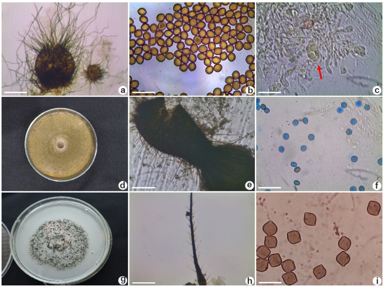
Chaetomium globosum Kunze, Mykologische Hefte (Leipzig) 1: 16 (1817).Fig. 1a-d
Ascomata superficial, dark olivaceous in reflected light, globose to subglobose, 138-140 µm diam., terminal hairs abundant, brown, tapering and fading towards the tips, undulate to loosely coiled with erect or flexuous lower part, up to 5 µm near the base; Asci fasciculate, fusiform to claviform; ascospores olivaceous brown when mature, limoniform, usually biapiculate, (8-)8.3-9.2(-10) × (6.3-)6.9-7.8(-8), with an apical germ pore. Asexual state absent. PDA colonies with sparse white aerial hyphae in the center, producing an orange luteal exudate that diffuses into the medium.
Material examined: Ilhéus, Universidade Estadual de Santa Cruz, branches and leaves, XI.2020, Theobroma cacao, IX.2022, roots TFB1750, leaves TFB1751, beans TFB1752; grocery store, Carica papaya, V.2022, seeds TFB1753. Una, Annona muricata, III.2021, leaves TFB1754. Itabuna, Artocarpus heterophyllus, XI.2022, leaves TFB1755.
Description of specimens studied herein corresponds to that of Wang et al. (2016), although the ascomata here were more globose than ellipsoid or obovate (this study × Wang et al. 2016). Chaetomium globosum was collected in Alagoas, Maranhão, Pernambuco and a single specimen from Bahia (URM 91169), collected back in 2014 in the Raso da Catarina Ecological Station (municipality of Paulo Afonso).
Chaetomium pseudoglobosum X. Wei Wang, Crous & L. Lombard, Persoonia36: 115 (2015) [2016].Fig. 1e-g
Ascomata superficial, olivaceous buff in reflected light, ovate, 200-350 µm in diam, terminal hairs abundant, forming a nearly globose head, lateral hairs sparse, flexuous to slightly undulate tapering and fading towards the tips. Asci fasciculate, claviform to fusiform, 10-27 µm. Ascospores olivaceous brown, when mature, limoniform, (5.8-)5.9-6.7(-7) × 5-6(-6.2) µm, with an apical pore. Asexual state absent. White mycelium, PDA colonies with sparse white aerial hyphae producing light color; posterior surface usually colorless.
Material examined: Ilhéus, Universidade Estadual de Santa Cruz, cow dung, VI.2018, TFB1756.
Chaetomium pseudoglobosum is part of the C. globosum-complex, being phylogenetically related to C. tenue X. Wei Wang, Crous & L. Lombard and C. afropilosum X. Wei Wang, Crous & L. Lombard. According to Wang et al. (2016), C. pseudoglobosum has ascospores bigger than the species mentioned above: 9-10 × 6.5-7.5 μm vs 8.5-9.5 × 6-7 μm (C. tenue) vs 7-8 × 5.5-6 μm (C. afropilosum), the specimen presented herein has even smaller ascospores, although the general morphological aspects are similar to that of C. pseudoglobosum.
Humicola cuyabenoensis (Decock & Hennebert) X. Wei Wang & Houbraken, Stud. Mycol.93: 80 (2018).Fig. 1i-h
Ascomata superficial, brown to blackish, ostiolate, elongate, pyriform to ampuliform, apically attenuated to a cylindrical thread-like neck with a truncate apex, 600-900 µm high, 100 µm at the widest part; terminal hairs arising from the adjacent ostiolar cells, smooth, 3 µm near the base, 75-80 µm long; lateral hairs sparse, scattered through the ascoma and the side of the neck, seta-like, fading towards the tips; Asci deliquesced, ascospores olivaceous brown, broad limoniform, umbonate at both ends, (6.9)7.4-8.1(8.3-) × (-5.9)6 -7(-7.1) µm, with an apical germ pore. Colonies in PDA with few aerial hyphae. Aerial mycelium burnt yellow, reverse orange.
Material examined: Ilhéus, Universidade Estadual de Santa Cruz, soil of cacao plantation, V.2022, TFB1757.
Humicola cuyabenoensis was unknown in Brazil until now. It was previously collected in Ecuador (Wang et al. 2019), which indicates that the species likely has a broader distribution, at least within the Neotropical region.. According to the study mentioned above, the striking characteristic of H. cuyabenoensis is a longneck covered with sparse lateral hairs and short terminal hairs. The ascospores with umbonate ends are also an important feature to confirm this species.
There are not enough studies on the Chaetomiaceae in Brazil. Out of the more than 50 known genera in this family, likely no more than 10 are known to occur in Brazil. According to Flora e Funga do Brasil (2015, continuously updated) and Specieslink (2023), only 7 to 16 Chaetomium species have been reported from Brazil so far, most of them deposited at Pe. Camille Torrend Herbarium (Depto. Micologia, Centro de Ciências Biológicas - UFPE). The second genus with more species reported from Brazil is Humicola, with only 3 species reported; from those, 2 were collected in Bahia.
a-d. Chaetomium globosum - a. ascomata; b. ascospores; c. asci; d. colony. e-g. Chaetomium pseudoglobosum - e. ascomata; f. ascospores; g. colony; h-i. Humicola cuyabenoensis - h. ascomata; i. ascospores. Scale bars: a,i = 100 µm; b-f = 20 µm; h = 200 µm.
Considering the impact of several species of Chaetomiaceae on Biotechnology, the indoor environment, and human health, more efforts should be put on this group, considering their worldwide distribution and diversity.
Acknowledgements
The first author thanks CAPES for granting financial support during the execution of this study and UESC for technical support.
Data availability statement
In accordance with Open Science communication practices, the authors inform that all samples collected and determined by R.M.P. Gorguette and Jad. Pereira are deposited at the TFB Fungarium (https://specieslink.net/col/TFB/
References
-
Ahmed SA, Khan Z, Wang XW, Moussa TAA, Alzahrani HS, Almaghrabi OA, Sutton DA, Ahmad S, Groenewald JZ, Izquierdo AA, Diepeningen AV, Menken SBJ, Najafzadeh MJ, Crous PW, Cornely O, Hamprecht A, Vehreschild MJGT, Kindo AJ & Hoog GS (2016) Chaetomium-like fungica using opportunistic infections in humans: a possible role for extreme tolerance. Fungal Diversity 76: 11-26. DOI: 10.1007/s13225-015-0338-5
» https://doi.org/10.1007/s13225-015-0338-5 - Betancourt O, Zaror L & Senn C (2013) Isolation of filamentous fungi from hair coat cats without skin lesions in Temuco, Chile. Revista Cientificade La Facultad de Ciencias Veterinárias de La Universidad del Zulia 23: 380-387.
-
Berka RM, Grigoriev IV, Otillar R, Salamov A, Grimwood J, Reid I, Ishmael N, John T, Darmond C, Moisan CM, Henrissat B, Coutinho PM, Lombard V, Natvig DO, Lindquist E, Schmutz J, Lucas S, Harris P, Powlowski J, Bellemare A, Taylor D, Butler G,Vries RP, Allijn IE, Brink JVD, Ushinsky S, Storms R, Powell A, Paulsen IT, Elbourne Ldh, Baker SE, Magnuson J, Laboissire S, Clutterbuck AJ, Martinez D, Wogulis M, Leon ALL, Rey MW &Tsang A (2011) Comparative genomic analysis of the thermophilic biomass-degrading fungi Myceliophthora thermophila and Thielavia terrestris Nature Biotechnology 29: 922-927. DOI: 10.1038/nbt.1976
» https://doi.org/10.1038/nbt.1976 -
Bezerra JD, Nascimento CC, Barbosa RDN, Silva DCV, Svedese VM, Silva-Nogueira EB, Gomes BS, Paiva LM, Souza-Mota CM (2015) Endophytic fungi from medicinal plant Bauhinia forficata: diversity and biotechnological potential. Brazilian Journal of Microbiology 46: 49-57. DOI: 10.1590/S1517-838246120130657
» https://doi.org/10.1590/S1517-838246120130657 - Cooney DG & Emerson R (1964) Thermophilic Fungi: an account of their biology, activities and classification. W.H. Freeman and Co., San Francisco, London. 188p.
-
Gusmão LFP & Melo RFR (2015) Sordariales inFlora e Funga do Brasil (continuously updated) Jardim Botânico do Rio de Janeiro. Available at <Available at https://floradobrasil.jbrj.gov.br/FB137449 >. Access on 10 November 2023.
» https://floradobrasil.jbrj.gov.br/FB137449 -
Harreither W, Sygmund C, Augustin M, Narciso M, Rabinovich ML, Gorton L, Haltrich D & Ludwig R (2011) Catalytic properties and classification of cellobiose dehydrogenases from ascomycetes. Applied and Environmental Microbiology 77: 1804-1815. DOI: 10.1128/AEM.02052-10
» https://doi.org/10.1128/AEM.02052-10 -
Specieslink (2023) Chaetomium and Humicola in species. Available at <Available at https://specieslink.net/search >. Access on 10 November 2023.
» https://specieslink.net/search - Thiep NV & Soytong K (2015) Chaetomium spp. as biocontrol potential to control tea and coffee pathogens in Vietnam. International Journal of Agricultural Technology 11: 1381-1392.
-
Wang XW, Han PJ, Bai FY, Luo A, Bensch K, Meijer M, Kraak B, Han DY, Sun BD, Crous PW & Houbraken J (2022) Taxonomy, phylogeny and identification of Chaetomiaceae with emphasis on thermophilic species. Studies in Mycology 101: 121-243. DOI: 10.3114/sim.2022.101.03
» https://doi.org/10.3114/sim.2022.101.03 -
Wang XW, Yang FY, Meijer M, Kraak B, Sun BD, Jiang YL, Wu YM, Bai FY, Seifert KA, Crous PW, Samson RA & Houbraken J (2019) Redefining Humicola sensu stricto and related genera in the Chaetomiaceae. Studies in Mycology 93: 65-153. DOI: 10.1016/j.simyco.2018.07.001
» https://doi.org/10.1016/j.simyco.2018.07.001 -
Wang XW, Lombard L, Groenewald JZ, Li J, Videira SIR, Samson RA, Liu XZ & Crous PW (2016) Phylogenetic reassessment of the Chaetomium globosum complex. Persoonia 36: 83-133. DOI: 10.3767/003158516X689657
» https://doi.org/10.3767/003158516X689657