Antimicrobial photodynamic therapy in the treatment of experimentally induced dental alveolitis in animals
Resumo
Introdução A reparação alveolar em áreas infectadas ainda carece de estudos adicionais, especialmente no que diz respeito a terapias adjuvantes ou alternativas.
Objetivo Este estudo teve como objetivo analisar histologicamente o processo de reparação em feridas infectadas após extração dentária tratadas com terapia de fotobiomodulação (FBM) isoladamente ou em combinação com um fotossensibilizador (FS) na terapia fotodinâmica antimicrobiana (aPDT).
Material e método Quarenta e oito ratos foram submetidos à extração do incisivo superior direito e após, receberam a indução da alveolite experimental. A seguir, foram aleatoriamente divididos em 4 grupos (n=12) que receberam os seguintes tratamentos: Grupo controle (C), sem tratamento intra ou extra-alveolar; Grupo FS, irrigação alveolar com azul de metileno (100 µg/mL); Grupo FBM, os alvéolos foram submetidos à irradiação com laser de baixa potência (685nm, 0,05W, 240s, 12J); Grupo aPDT: O tratamento foi semelhante ao do grupo FS, com um tempo de pré-irradiação de 60 segundos, seguido imediatamente da irradiação com laser, em condições semelhantes às do grupo FBM. Os animais foram eutanasiados no pós-operatório nos dias 7, 15, 21 e 28. Foram realizadas análises descritivas do processo do reparo alveolar em três locais.
Resultado As feridas do grupo C apresentaram atraso na cronologia da reparação alveolar. O grupo FS apresentou uma melhor resposta inflamatória inicial em comparação ao grupo controle. Os animais tratados com FBM e aPDT apresentaram reparação óssea mais acelerada do que os do grupo C.
Conclusão Conclui-se que houve atraso do processo de reparação alveolar; que o uso isolado do FS minimizou os efeitos deletérios da alveolite sobre o reparo ósseo; que as terapias fotônicas beneficiaram o reparo alveolar, principalmente quando a aPDT foi utilizada.
Descritores:
Alveolite seca; terapia por luz de baixa intensidade; fotoquimioterapia; regeneração óssea; ratos
Abstract
Introduction Alveolar repair in infected areas still lacks further studies, especially regarding adjuvant or alternative therapies.
Objective This study aimed to histologically analyze the repair process in infected wounds after tooth extraction treated with photobiomodulation (PBM) therapy alone or in combination with a photosensitizer (PS) in antimicrobial photodynamic therapy (aPDT).
Material and method Forty-eight rats underwent extraction of the right upper incisor and subsequently received experimental alveolitis induction. They were then randomly divided into 4 groups (n=12) that received the following treatments: Control group (C), no intra- or extra-alveolar treatment; PS group, alveolar irrigation with methylene blue photosensitizer (MB, 100 µg/mL); PBM group, the alveoli were subjected to PBM therapy mediated with a low-power laser (685nm, 0.05W, 240s, 12J); aPDT Group: The treatment was similar to that of the PS group, with a pre-irradiation time of 60 seconds, followed immediately by laser irradiation, under conditions similar to those of the PBM group. The animals were euthanized post-operatively on days 7, 15, 21, and 28.
Result The wounds in group C showed a delay in the chronology of alveolar repair, PS group showed an improved initial inflammatory response compared to the control group. The animals treated with PBM and aPDT showed faster bone repair than those in group C.
Conclusion It is concluded that there was a delay in the alveolar repair process; that the isolated use of the PS minimized the alveolitis deleterious effects on bone repair; those photonic therapies benefited alveolar repair, especially when aPDT was used.
Descriptors:
Dry socket; low-level light therapy; photochemotherapy; bone regeneration; rats
INTRODUCTION
Dental alveolitis is a clinical condition that requires rapid diagnosis and prompt professional intervention, occurring after tooth extractions, especially of third molars. It can also be referred to as dry or wet alveolitis. Diagnosis is based on the symptoms of intense pain after extraction in the dental socket region that radiates to the ear, eyes, and neck, peaking between 1 to 3 days, which may be accompanied by the presence of a totally or partially disintegrated blood clot1,2. Consequently, patients report a foul odor and bad taste, and lymph node swelling may also occur.
Various synonyms define this clinical condition, such as dry socket (absence of blood clot), fibrinolytic osteitis, alveolar osteitis, postoperative osteitis, alveolalgia, and purulent/suppurative alveolitis when infection and pus are present (wet alveolitis). Its incidence occurs in 10% of patients3, 1% to 5% of extractions, and 38% in lower third molar extractions1. With an etiology that is not yet fully clarified, many factors are associated, such as traumatic extractions, smoking, advanced age, oral contraceptives, infections at the extraction site, systemic diseases like diabetes mellitus, bone disorders, blood clotting problems, irradiation, and non-compliance with post-extraction instructions1,4,5.
Several treatment methods are reported in the literature6 and indicated according to the clinical presentation (dry or wet), such as irrigation with saline solution and 0.2% chlorhexidine gluconate to remove food debris and bacteria, debridement of the bone socket to reactivate new blood clot formation, blood products (fibrin-rich plasma, PRF), antibiotics, intra-alveolar medications (Alveogil, zinc oxide and eugenol, metronidazole, thermosensitive gel with lidocaine), and photobiomodulation (PBM) therapy with low-power laser7-14.
The benefits of PBM mediated by low-power laser have shown encouraging results in reducing pain, inflammation, and alveolar repair events in experimental animal studies15,16 and in humans17,18. However, publications attesting to the effectiveness of antimicrobial photodynamic therapy (aPDT) in the alveolar repair of infected dental extraction wounds are still scarce. Thus, the purpose of this study was to histologically evaluate in animals the repair of infected dental sockets treated with PBM and aPDT, with clinical similarity to wet alveolitis. The hypothesis of this study is that sockets treated with photonic therapies (PBM or aPDT) will present better histological patterns of bone repair in infected wounds compared to controls. The null hypothesis is that none of the treatments will benefit the biological response in the treatment of alveolitis.
MATERIAL AND METHOD
Animals
The research protocol was approved by the Ethics Committee on Animal Use (CEUA) of the School of Dentistry of Araçatuba, in accordance with the standards of the National Council for the Control of Animal Experimentation (CONCEA). Forty-eight male Wistar rats (Rattus norvegicus albinus), aged 90 to 120 days and weighing between 180 to 250 grams, were used. The animals were kept in appropriate plastic cages (4 animals per cage) in a temperature-controlled room (22 ± 2 °C) with a 12/12-hour light cycle and ad libitum access to balanced food and water.
Experimental Procedures
All procedures were in compliance with the ARRIVE guidelines for animal studies. The animals were anesthetized with general anesthesia via intramuscular injection of ketamine hydrochloride (0.7 mL/kg) combined with xylazine (0.3 mL/kg). To obtain a homogeneous suspension of purulent secretion, 10 animals were used following previously described methodology*: after extraction of the right upper incisor, a sterile absorbent paper point soaked in a millesimal adrenaline solution was inserted into the sockets for 60 seconds. After this period, animals were observed for 60 to 90 seconds to confirm the absence of bleeding and clot formation. Three days later, the sockets showed edge edema and purulent secretion, confirming the diagnosis. The secretion was collected by introducing sterile paper points into the socket for one minute. These were transferred to vials containing transport medium (MRT), agitated for 30 seconds, fractionated, and preserved in liquid nitrogen to ensure bacterial viability19.
Groups and Treatments
All animals underwent induction of clinical alveolitis. After confirmation, they were randomly distributed into: Control Group (C): Gentle cleaning of the coronal portion (soft tissue); no intra-alveolar treatment; PS Group: Gentle cleaning of the coronal portion followed by alveolar irrigation with methylene blue (MB, 100 µg/mL); PBM Group: Gentle surgical cleaning of the coronal portion followed by low-power laser irradiation; aPDT Group: Gentle surgical cleaning of the coronal portion, application of PS, and after a 60-second pre-irradiation time, low-power laser irradiation.
PBM and aPDT Therapy
In the PS and aPDT groups, 1 mL of methylene blue (Apothicário Farmácia de Manipulação, Araçatuba, SP, Brazil) at a concentration of 100 μg/mL was deposited into the alveoli and remained in the area for 60 seconds prior to irradiation. Subsequently, the alveoli were irradiated with a low-power laser.
In group PBM and aPDT a GaAlAs diode laser (Laser Beam®, Rio de Janeiro, RJ, Brazil) was used with the following protocol: 685 nm (visible red), single application, point contact on the vestibular soft tissue at the middle portion of the socket, 50 mW (0.05 W) power, 240 s exposure time, 12 J energy, and 600 J/cm2 energy density, 2 mm2 elliptical spot.
Euthanasia and Histologic Procedures
Animals were euthanized via anesthetic overdose at 7, 15, 21, and 28 days post-treatment. Specimens were removed, fixed in 10% formalin for at least 24 hours, and processed for histological analysis. After 48 hours in 10% formalin it was washed in running water, dehydrated, cleared, impregnated, embedded in paraffin and sectioned in a microtome with 4 µm thick. Semi-serial sections were performed and captured on histological slides. The histological sections were submitted to staining with hematoxylin-eosin (HE).
Histomorphological Analysis of the Samples
Sample analyses were performed by an expert examiner, who was blinded to the samples. Histological sections were analyzed under bright field illumination in an optical microscope (Axiolab, Carl Zeiss). A descriptive analysis of biological events was conducted across the cervical, middle, and apical thirds of the alveoli for all time periods. The evaluation included the presence of blood clots, inflammatory infiltrate, connective tissue organization (cells, fibers, and vessels), bone tissue formation, the alveolar bone crest, and the oral mucosal epithelium.
RESULT
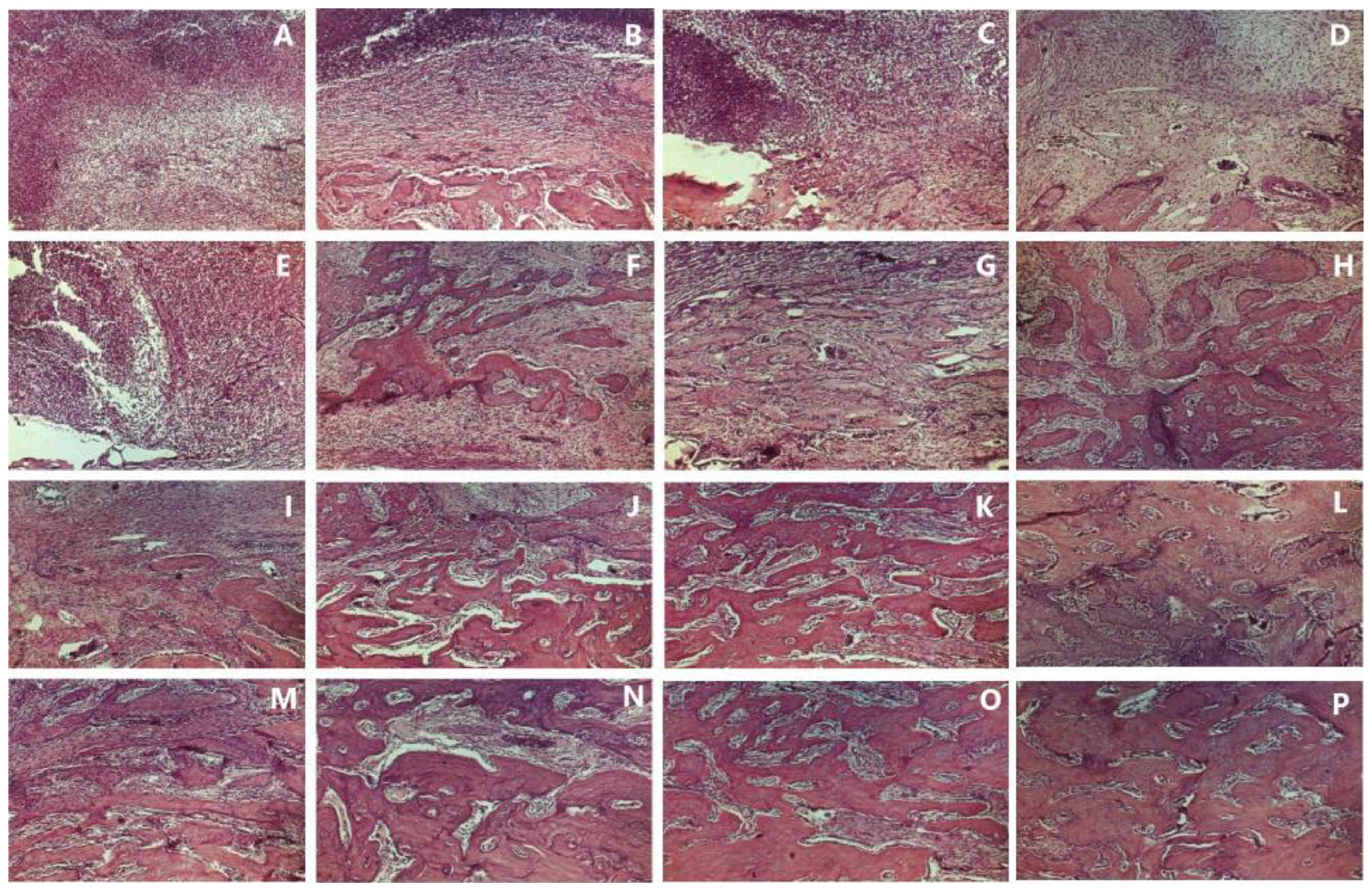
Histological events are detailed in Table 1 and documented in Figure 1.

Histological events across different groups, periods, and treatments: Control group (A, 7 days, cervical third; E, 15 days, cervical third; I, 21 days, cervical third; M, 28 days, cervical third); PS group (B, 7 days, cervical third; F, 15 days, cervical third; J, 21 days, cervical third; N, 28 days, cervical third); PBM group (C, 7 days, cervical third; G, 15 days, cervical third; K, 21 days, cervical third; O, 28 days, cervical third); aPDT group (D, 7 days, cervical third; H, 15 days, cervical third; L, 21 days, cervical third; P, 28 days, cervical third). Hematoxylin & Eosin. Original magnification: 63X.
DISCUSSION
The results obtained demonstrated that wounds treated with PS, PBM, and aPDT exhibited an accelerated repair process compared to those that received no treatment (Group C). Among the histological findings, greater organization and neoformation of connective tissue, increased fibroblastic proliferation, and earlier and more extensive bone remodeling and neoformation were observed. These characteristics were most prominent in wounds treated with aPDT, followed by those treated with PBM and PS, across all evaluation periods.
In the specimens from the PS group, the ability of the phenothiazine derivative (MB) to promote benefits in the repair of infected wounds when applied alone was demonstrated. Greater bone formation was noted in all periods, with well-vascularized connective tissue rich in fibroblasts and a lower degree of inflammation compared to control wounds. The observed reduction in the inflammatory process is likely due to the anti-inflammatory action of MB, which interferes with nitric oxide synthesis through an important signaling pathway (iNOS/NO), consequently promoting vasoconstriction in the area. It is known that MB is deeply related to its anti-inflammatory effect, which occurs through a variety of pathways and functions, decreasing the expression level of pro-inflammatory cytokines in several ways, including the inhibition of iNOS/NO signaling20,21. There is also evidence that the reduction of oxidative stress promoted by MB results in an increase in collagen synthesis22. These benefits of the PS are likely also due to the ability of phenothiazines to modulate the metabolism of macrophages, inducing the accumulation of intracellular reactive oxygen species (ROS), promoting the activation of autophagy and lysosomal activity, and thus increasing their bacterial destruction capacity. The increase in ROS contributes to the elimination of intracellular bacteria through direct microbial death, as well as by promoting lysosomal acidification and inducing autophagy23.
PBM therapy mediated by low-power laser has demonstrated established results in different conditions, both in dental extraction wounds under normal conditions16,18 and in infected extraction wounds24. Studies have shown that PBM effects may occur through the direct action of light on bacteria, acting on endogenous porphyrins which, when stimulated, release free radicals capable of damaging the cytoplasmic protein membrane and DNA25,26. Other studies have demonstrated the effects of PBM on the viability of microorganisms both in vitro** and in vivo27. An important factor to consider is the action of laser energy on the host response, promoting an indirect antimicrobial effect by increasing local blood flow and oxygenation28,29, which favors the presence of host defense cells in the area and, consequently, improves bone repair. Furthermore, PBM therapy is capable of inducing an increase in extracellular matrix synthesis and angiogenesis30.
The benefits of aPDT on biological events in infected areas proved to be superior to the other groups, becoming evident from the initial period of repair, with neoformation of connective tissue, bone formation, and reduction of the inflammatory process. These findings are likely due to three important hypotheses: first, the initial action of the PS on microorganisms, which remained in the area for sixty seconds (pre-irradiation time), sufficient for bacterial absorption and a primary action as previously described; second, the photodynamic action itself—the interaction of visible red light with MB capable of forming reactive oxygen species (ROS) resulting from Type I photosensitization (electron transfer leading to the production of superoxide anion [O2-], hydroxyl radical [OH-], and hydrogen peroxide [H2O2]) and Type II (energy transfer producing excited singlet oxygen [1O2]). Each of these ROS can combine with biomolecules, leading to subsequent oxidation and degradation, a reaction sufficient to cause cell/bacterial death31. The third hypothesis is the capacity of laser light to promote benefits in accelerating bone repair (photobiomodulation). Studies have demonstrated the benefits of aPDT in alveolar repair for the prevention of alveolitis in humans32 and as a preventive therapy for implant placement in animals33.
It is known that different conditions can interfere with laser effects on biological tissues, such as wavelength, power, number of applications, light interaction with the PS, physiological state of the cell, light absorption, emission and irradiation mode, exposure time, beam diameter, medium pH, tissue color, water content, thermal conductivity, and organic matrix.
The present study demonstrated the effective action of PBM and aPDT on alveolar repair in infected sockets. In the present study, a single session of PBM and aPDT therapy was used based on results from previous research developed in our research group24,33. However, future research may prove whether a greater number of sessions of these therapies could contribute to more favorable results in the repair of infected alveoli.
In addition, further studies are necessary, both in animals and clinical trials in humans, to build a more solid scientific basis for the development of treatment protocols for this condition, which can be difficult to manage. It should be noted that this animal study sought similarity with clinical conditions but presented limitations such as the sample size, laser clinical parameters, number of treatment sessions, and the need for a broader range of analyses.
CONCLUSION
Based on the results obtained, it can be concluded that there was a delay in the alveolar repair process in the control group; the isolated use of the PS minimized deleterious effects on bone repair; and photonic therapies benefited alveolar repair, with more favorable results achieved when aPDT was utilized.
-
*
Antonio GMD. Contaminação pós-exodôntica do alvéolo dental de ratos: estudo microbiológico e histológico [tese]. Araçatuba: Faculdade de Odontologia, Universidade Estadual Paulista; 1984. 52 p.
-
**
Ferreira JPR. Estudo in vitro da ação do laser em baixa intensidade, associado ou não a drogas fotossensibilizadoras, sobre a viabilidade de microorganismos bucal [dissertação]. Marília: Universidade de Marília; 2002. 58 p.
-
How to cite:
Garcia VG, Lopes Júnior W, Theodoro LH. Antimicrobial photodynamic therapy in the treatment of experimentally induced dental alveolitis in animals. Rev Odontol UNESP. 2026;55:e20260016. https://doi.org/10.1590/1807-2577.20260016
-
DATA AVAILABILITY
The datasets generated and/or analysed during the current study are not publicly available but are available from the corresponding author.
REFERENCES
-
1 Blum IR. Contemporary views on dry socket (alveolar osteitis): a clinical appraisal of standardization, etiopathogenesis and management: a critical review. Int J Oral Maxillofac Surg. 2002 Jun;31(3):309-17. https://doi.org/10.1054/ijom.2002.0263 PMid:12190139.
» https://doi.org/10.1054/ijom.2002.0263 -
2 Kolokythas A, Olech E, Miloro M. Alveolar osteitis: a comprehensive review of concepts and controversies. Int J Dent. 2010;2010:249073. https://doi.org/10.1155/2010/249073 PMid:20652078.
» https://doi.org/10.1155/2010/249073 -
3 Rosa A, Pujia AM, Arcuri C. Investigation of alveolar osteitis and the effectiveness of laser treatment: a unified Meta-analysis and review of the literature. BMC Oral Health. 2024;24(1):700. https://doi.org/10.1186/s12903-024-04461-w PMid:38886713.
» https://doi.org/10.1186/s12903-024-04461-w - 4 Bowe DC, Rogers S, Stassen LF. The management of dry socket/alveolar osteitis. J Ir Dent Assoc. 2011;57(6):305-10. PMid:22338284.
-
5 Eshghpour M, Rezaei NM, Nejat A. Effect of menstrual cycle on frequency of alveolar osteitis in women undergoing surgical removal of mandibular third molar: a single-blind randomized clinical trial. J Oral Maxillofac Surg. 2013 Sep;71(9):1484-9. https://doi.org/10.1016/j.joms.2013.05.004 PMid:23866782.
» https://doi.org/10.1016/j.joms.2013.05.004 -
6 Daly B, Sharif MO, Newton T, Jones K, Worthington HV. Local interventions for the management of alveolar osteitis (dry socket). Cochrane Database Syst Rev. 2012 Dec;12:CD006968. https://doi.org/10.1002/14651858.CD006968.pub2 PMid:23235637.
» https://doi.org/10.1002/14651858.CD006968.pub2 -
7 Eshghpour M, Ahrari F, Najjarkar NT, Khajavi MA. Comparison of the effect of low level laser therapy with alvogyl on the management of alveolar osteitis. Med Oral Patol Oral Cir Bucal. 2015 May;20(3):e386-92. https://doi.org/10.4317/medoral.20375 PMid:25662557.
» https://doi.org/10.4317/medoral.20375 -
8 Taberner-Vallverdú M, Nazir M, Sanchez-Garces MA, Gay-Escoda C. Efficacy of different methods used for dry socket management: a systematic review. Med Oral Patol Oral Cir Bucal. 2015 Sep;20(5):e633-9. https://doi.org/10.4317/medoral.20589 PMid:26116842.
» https://doi.org/10.4317/medoral.20589 -
9 Ramos E, Santamaria J, Santamaria G, Barbier L, Arteagoitia I. Do systemic antibiotics prevent dry socket and infection after third molar extraction? A systematic review and meta-analysis. Oral Surg Oral Med Oral Pathol Oral Radiol. 2016 Oct;122(4):403-25. https://doi.org/10.1016/j.oooo.2016.04.016 PMid:27499028.
» https://doi.org/10.1016/j.oooo.2016.04.016 -
10 Daly BJ, Sharif MO, Jones K, Worthington HV, Beattie A. Local interventions for the management of alveolar osteitis (dry socket). Cochrane Database Syst Rev. 2022 Sep;9(9):CD006968. https://doi.org/10.1002/14651858.CD006968.pub3 PMid:36156769.
» https://doi.org/10.1002/14651858.CD006968.pub3 -
11 Laforgia A, Inchingolo AD, Riccaldo L, Avantario P, Buongiorno S, Malcangi G, et al. The use of Platelet-Rich Fibrin (PRF) in the management of dry socket: a systematic review. Int J Mol Sci. 2024 Sep;25(18):10069. https://doi.org/10.3390/ijms251810069 PMid:39337554.
» https://doi.org/10.3390/ijms251810069 -
12 Camps-Font O, Sábado-Bundó H, Toledano-Serrabona J, Valmaseda-de-la-Rosa N, Figueiredo R, Valmaseda-Castellón E. Antibiotic prophylaxis in the prevention of dry socket and surgical site infection after lower third molar extraction: a network meta-analysis. Int J Oral Maxillofac Implants. 2024 Jan;53(1):57-67. https://doi.org/10.1016/j.ijom.2023.08.001 PMid:37612199.
» https://doi.org/10.1016/j.ijom.2023.08.001 -
13 Minervini G, Franco R, Martelli M, Hafedh S, Maddalena Marrapodi M, Di Blasio M, et al. Low-level laser treatment’s ability to reduce dry socket pain. Acta Odontol Scand. 2024 Nov;83:631-41. https://doi.org/10.2340/aos.v83.42261 PMid:39530606.
» https://doi.org/10.2340/aos.v83.42261 -
14 Supachawaroj N, Kerdmanee K, Limsitthichaikoon S. Lidocaine-loaded thermoresponsive gel for accelerated wound healing in dry socket and oral wounds. Gels. 2024 Nov;10(11):739. https://doi.org/10.3390/gels10110739 PMid:39590095.
» https://doi.org/10.3390/gels10110739 -
15 Çırak E, Özyurt A, Peker T, Ömeroğlu S, Güngör MN. Comparative evaluation of various low-level laser therapies on bone healing following tooth extraction: An experimental animal study. J Craniomaxillofac Surg. 2018 Jul;46(7):1147-52. https://doi.org/10.1016/j.jcms.2018.05.012 PMid:29805068.
» https://doi.org/10.1016/j.jcms.2018.05.012 -
16 Ribeiro LNS, Figueiredo FAT, Silva Mira PC, Arnez MFM, Matsumoto MAN, Menezes LM, et al. Low-level laser therapy (LLLT) improves alveolar bone healing in rats. Lasers Med Sci. 2022 Mar;37(2):961-9. https://doi.org/10.1007/s10103-021-03340-y PMid:34002343.
» https://doi.org/10.1007/s10103-021-03340-y -
17 Gururaj SB, Shankar SM, Parveen F, Chidambar CK, Bhushan K, Prabhudev CM. Assessment of healing and pain response at mandibular third molar extraction sites with and without pre- and postoperative photobiomodulation at red and near-infrared wavelengths: a clinical study. J Pharm Bioallied Sci. 2022 Jul;14(Suppl 1):S470-4. https://doi.org/10.4103/jpbs.jpbs_675_21 PMid:36110661.
» https://doi.org/10.4103/jpbs.jpbs_675_21 -
18 Hadad H, Santos AFP, Jesus LK, Poli PP, Mariano RC, Theodoro LH, et al. Photobiomodulation therapy improves postoperative pain and edema in third molar surgeries: a randomized, comparative, double-blind, and prospective clinical trial. J Oral Maxillofac Surg. 2022 Jan;80(1):37.e1-12. https://doi.org/10.1016/j.joms.2021.08.267 PMid:34656515.
» https://doi.org/10.1016/j.joms.2021.08.267 - 19 Sampaio Camargo P, Stoppelaar JD. The preservation of dental plaque samples in liquid nitrogen. Caries Res. 1976;(10):148.
-
20 Ahn H, Kang SG, Yoon SI, Ko HJ, Kim PH, Hong EJ, et al. Methylene blue inhibits NLRP3, NLRC4, AIM2, and non-canonical inflammasome activation. Sci Rep. 2017 Sep;7(1):12409. https://doi.org/10.1038/s41598-017-12635-6 PMid:28963531.
» https://doi.org/10.1038/s41598-017-12635-6 -
21 Zheng J, Li Q. Methylene blue regulates inflammatory response in osteoarthritis by noncoding long chain RNA CILinc02. J Cell Biochem. 2019 Mar;120(3):3331-8. https://doi.org/10.1002/jcb.27602 PMid:30548658.
» https://doi.org/10.1002/jcb.27602 -
22 Xiong ZM, O’Donovan M, Sun L, Choi JY, Ren M, Cao K. Anti-aging potentials of methylene blue for human skin longevity. Sci Rep. 2017;7(1):2475. https://doi.org/10.1038/s41598-017-02419-3 PMid:28559565.
» https://doi.org/10.1038/s41598-017-02419-3 -
23 Qiu L, Chen W, Wang J, Deng X, Liu H, Qiu J. Phenothiazines enhance antibacterial activity of macrophage by inducing ROS and autophagy. Front Immunol. 2025 Nov;16:1712724. https://doi.org/10.3389/fimmu.2025.1712724 PMid:41394842.
» https://doi.org/10.3389/fimmu.2025.1712724 - 24 Garcia VG, Carvalho PSP, Oliveira JAGP. Ação da radiação Laser na reparação de feridas de extração dental infectadas: estudo histológico em ratos. RGO. 1995;43(4):191-4.
-
25 Lubart R, Eichler M, Lavi R, Friedman H, Shainberg A. Low-energy laser irradiation promotes cellular redox activity. Photomed Laser Surg. 2005 Feb;23(1):3-9. https://doi.org/10.1089/pho.2005.23.3 PMid:15782024.
» https://doi.org/10.1089/pho.2005.23.3 -
26 Xu Y, Lin Y, Gao S, Shen J. Study on mechanism of release oxygen by photo-excited hemoglobin in low-level laser therapy. Lasers Med Sci. 2018;33(1):135-9. https://doi.org/10.1007/s10103-017-2363-y PMid:29067617.
» https://doi.org/10.1007/s10103-017-2363-y -
27 Rico-Holgado S, Ortiz-Díez G, Martín-Espada MC, Fernández-Pérez C, Baquero-Artigao MR, Suárez-Redondo M. Effect of low-level laser therapy on bacterial counts of contaminated traumatic wounds in dogs. J Lasers Med Sci. 2021 Dec;12(1):e78. https://doi.org/10.34172/jlms.2021.78 PMid:35155163.
» https://doi.org/10.34172/jlms.2021.78 -
28 Kubota J. Effects of diode laser therapy on blood flow in axial pattern flaps in the rat model. Lasers Med Sci. 2002;17(3):146-53. https://doi.org/10.1007/s101030200024 PMid:12181629.
» https://doi.org/10.1007/s101030200024 -
29 Colombo F, Valença A No, Sousa AP, Marchionni AM, Pinheiro AL, Reis SR. Effect of low-level laser therapy (lambda660 nm) on angiogenesis in wound healing: a immunohistochemical study in a rodent model. Braz Dent J. 2013;24(4):308-12. https://doi.org/10.1590/0103-6440201301867 PMid:24173246.
» https://doi.org/10.1590/0103-6440201301867 -
30 Usumez A, Cengiz B, Oztuzcu S, Demir T, Aras MH, Gutknecht N. Effects of laser irradiation at different wavelengths (660, 810, 980, and 1064 nm) on mucositis in an animal model of wound healing. Lasers Med Sci. 2014 Nov;29(6):1807-13. https://doi.org/10.1007/s10103-013-1336-z PMid:23636299.
» https://doi.org/10.1007/s10103-013-1336-z - 31 Wainwright M. Princípios básicos da terapia fotodinâmica na infecção. In: Garcia VG, Theodoro LH, editors. Lasers em odontologia: uma visão clínica baseada em evidências científicas. São Paulo: Santos Publicações Ltda; 2021.
-
32 Neugebauer J, Jozsa M, Kübler A. Antimicrobial photodynamic therapy for prevention of alveolar ostitis and post-extraction pain. Mund Kiefer Gesichtschir. 2004 Nov;8(6):350-5. https://doi.org/10.1007/s10006-004-0572-6 PMid:15583924.
» https://doi.org/10.1007/s10006-004-0572-6 -
33 Theodoro LH, Pires JR, Fernandes LA, Gualberto EC Jr, Longo M, Almeida JM, et al. Effect of antimicrobial photodynamic therapy on periodontally infected tooth sockets in rats. Lasers Med Sci. 2015 Feb;30(2):677-83. https://doi.org/10.1007/s10103-013-1400-8 PMid:23912780.
» https://doi.org/10.1007/s10103-013-1400-8
Edited by
-
Edited by
Editor: Rosemary Adriana Chierici Marcantonio
The datasets generated and/or analysed during the current study are not publicly available but are available from the corresponding author.