Neurological complications of Mycoplasma pneumoniae infection
Mycoplasma pneumoniae is a common cause of respiratory tract infections1, and may also lead to various extrapulmonary manifestations, including neurological complications2. Herein, we present three cases of neurological involvement associated with M. pneumoniae infection.
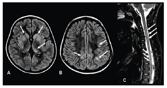
Case 1. A 7-year-old girl presented with seizures and reduced consciousness 3 weeks after completing amoxicillin treatment for tonsillitis. Brain and spinal magnetic resonance imaging (MRI) revealed lesions consistent with acute disseminated encephalomyelitis (Figure 1). Serum testing was positive for anti-myelin oligodendrocyte glycoprotein antibodies and for both immunoglobulin M (IgM) and IgG against M. pneumoniae. Cerebrospinal fluid (CSF) analysis demonstrated mild pleocytosis (77 lymphocytes/mm3) with normal protein and glucose levels. The patient was treated with immunoglobulin and methylprednisolone for 5 days and intravenous clarithromycin for 14 days, resulting in full recovery.
Brain magnetic resonance imaging (MRI) reveals multiple bilateral and asymmetrical lesions with hyperintense signal on FLAIR, involving the basal ganglia (arrows in A) and subcortical white matter (arrows in B), consistent with acute disseminated encephalomyelitis. Spine MRI demonstrates longitudinally extensive myelitis with hyperintense signal on T2-weighted imaging (arrows in C).
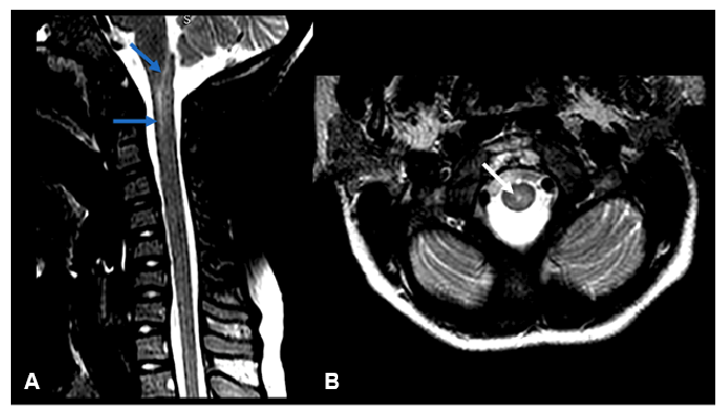
Case 2. A 10-year-old boy presented with a 3-day history of lower limb paresis and neck stiffness, without preceding respiratory symptoms. Spinal MRI revealed myelitis extending from the obex to C2 (Figure 2). Serum testing was positive for M. pneumoniae IgM and IgG. CSF analysis showed pleocytosis (206 lymphocytes/mm3, 12 monocytes/mm3, and 7 neutrophils/mm3), elevated protein (110 mg/dl), and normal glucose. Treatment with clarithromycin for 14 days and methylprednisolone for 5 days, followed by prednisolone for 6 months, led to complete recovery.
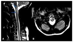
Spine magnetic resonance imaging (MRI) shows a hyperintense signal within the central spinal cord extending from the obex to the C2 level on T2-weighted imaging, visible in both sagittal (arrows in A) and axial (arrow in B) planes, consistent with myelitis. No gadolinium enhancement was observed. Brain MRI findings were normal.
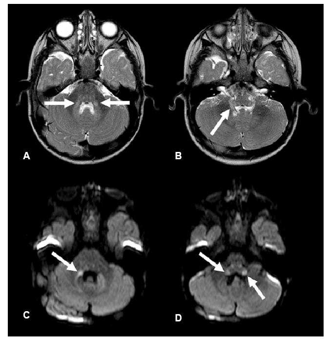
Case 3. A 19-month-old boy presented with a 2-day history of fever and ataxic gait. Brain MRI revealed lesions consistent with rhombencephalitis (Figure 3). Serum testing was positive for M. pneumoniae IgM. The patient was treated with levofloxacin for 10 days, with complete resolution of symptoms.
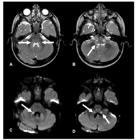
Brain magnetic resonance imaging demonstrates hyperintense lesions on T2-weighted imaging (A and B), with restricted diffusion (C and D), involving the pons and middle cerebellar peduncles (arrows in A and C), as well as the medulla and inferior cerebellar peduncles (arrows in B and D), consistent with rhombencephalitis.
The pathophysiology of M. pneumoniae-associated neurological complications remains incompletely understood. the following three mechanisms have been proposed, which may occur even in the absence of respiratory symptoms: (1) direct invasion, with bacterial presence at the site of inflammation (typically within 7 days of respiratory symptoms onset); (2) indirect immune-mediated processes involving autoimmunity or immune complexes (after 8 days of respiratory symptom onset, if present); and (3) vascular occlusion2,3. Encephalitis and myelitis may result from either direct invasion or immune-mediated mechanisms, whereas stroke is more commonly associated with vascular occlusion4.
REFERENCES
-
1 Narita M. Pathogenesis of extrapulmonary manifestations of Mycoplasma pneumoniae infection with special reference to pneumonia. J Infect Chemother. 2010;16(3):162-9. Available from:10.1007/s10156-010-0044-x.
» https://doi.org/10.1007/s10156-010-0044-x -
2 Tsiodras S, Kelesidis I, Kelesidis T, Stamboulis E, Giamarellou H. Central nervous system manifestations of Mycoplasma pneumoniae infections. J Infect. 2005;51(5):343-54. Available from:10.1016/j.jinf.2005.07.005.
» https://doi.org/10.1016/j.jinf.2005.07.005 -
3 Corrêa DG, Pacheco FT, da Cruz LCH Jr, Nunes RH, Maia ACM Jr, de Souza Godoy LF, et al. Intracranial vessel wall magnetic resonance imaging features of infectious vasculitis. Clin Imaging. 2023;98:26-35. Available from: 10.1016/j.clinimag.2023.03.014.
» https://doi.org/10.1016/j.clinimag.2023.03.014 -
4 Guleria R, Nisar N, Chawla TC, Biswas NR. Mycoplasma pneumoniae and central nervous system complications: a review. J Lab Clin Med. 2005;146(2):55-63. Available from: 10.1016/j.lab.2005.04.006.
» https://doi.org/10.1016/j.lab.2005.04.006
Edited by
-
Associated Editor:
Manoel O. da Costa Rocha Orcid: https://orcid.org/0000-0002-4721-1767