Tuberculous Spondylodiscitis with Sacroiliac Joint Involvement
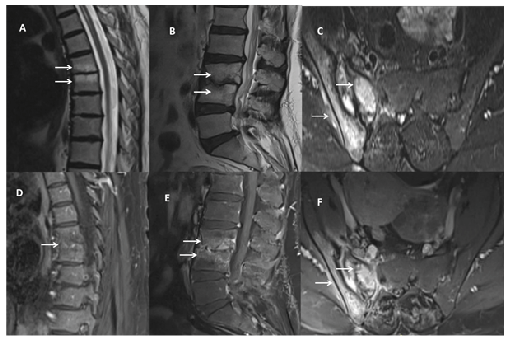
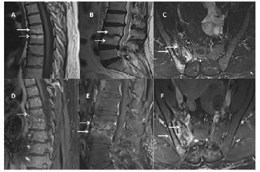
A 50-year-old woman presented with a 1 -year history of progressively worsening low back and hip pain, accompanied by a 10 kg weight loss. Laboratory evaluation revealed elevated inflammatory markers (erythrocyte sedimentation rate: 27 mm/h; C-reactive protein: 22 mg/L). Contrast-enhanced magnetic resonance imaging (MRI) revealed endplate irregularities, edema, disc space narrowing, and vertebral enhancement at T6-7, consistent with spondylitis. At L3-L4, MRI showed endplate destruction, edema, and intervertebral disc damage with marked enhancement, indicative of spondylodiscitis. T2-weighted images revealed hyperintense signals in the right sacroiliac joint, with postcontrast enhancement of the joint, adjacent bone, and surrounding soft tissues, consistent with sacroiliitis (Figure 1). A Quantiferon test performed for suspected tuberculosis was positive (TB2 antigen: 0.98). The patient was treated with a 6-month antituberculosis regimen, leading to clinical improvement and marked radiologic regression on follow-up MRI (Figure 2).
Magnetic resonance imaging of the thoracic and lumbar spine and right sacroiliac joint. T2-weighted images (A-C) demonstrate areas of high signal intensity (arrows), while postcontrast images (D-F) show corresponding contrast enhancement (arrows).
Follow-up magnetic resonance imaging after 6 months of antituberculosis treatment reveals decreased signal intensity on T2-weighted images (A-C, arrows) and reduced contrast enhancement on postcontrast images (D-F, arrows).
Bone and joint tuberculosis constitutes a small proportion of all tuberculosis cases but can cause substantial morbidity. Spinal tuberculosis is the most common form, with an incidence of 2.4-7.2 per 100,000 in developed countries1. Sacroiliac joint involvement is uncommon, accounting for 3-9.7% of musculoskeletal tuberculosis2. Coexisting sacroiliac and spinal involvement complicates diagnosis.
ACKNOWLEDGMENTS
No.
REFERENCES
- 1 Naselli N, Facchini G, Lima GM, Evangelisti G, Ponti F, Miceli M, et al. MRI in differential diagnosis between tuberculous and pyogenic spondylodiscitis. Eur Spine J. 2022;31(2):431-41.
- 2 Eren B, Guzey FK, Aktas OY, Gulec I, Bas NS. Lumbar tuberculous spondylodiscitis, sacroiliitis and paraspinal abscess with Staphylococcus aureus superinfection. J Clin Exp Orthop. 2018;4.
Edited by
-
Associated Editor:
Manoel O. da Costa Rocha Orcid: https://orcid.org/0000-0002-4721-1767