 Intermediate ultimate pH compromises color stability in aged beef from Bos indicus cattle
ABSTRACT:
Beef with high and intermediate pHu (ultimate pH) levels can contribute to the occurrence of dark-cutting beef, resulting in economic losses for the industry. This study aimed to assess beef color and its stability by examining color parameters, including lightness (L*), redness (a*), yellowness (b*), Chroma (C*), hue angle (h*), global variations in color (∆E), metmyoglobin percentage (MMb %), deoxymyoglobin percentage (DMb %), oxymyoglobin percentage (OMb %), ratio of light reflectance (%) (R630/580), relative oxygen consumption rate (OCR %), relative metmyoglobin reducing activity (MRA %), and myoglobin concentration of fresh beef across three pHu ranges: normal (pH < 5.8), intermediate (5.8 ≤ pH < 6.20), and high (pH ≥ 6.20). Longissimus lumborum (LL) muscles from Nellore cattle were vacuum-packed, and analyses were conducted after refrigerated storage at 2 ± 2 °C for 3, 14, and 21 days post-mortem. The blooming effect of normal and intermediate pHu steaks during simulated retail exposure (vacuum-packaging) at 3, 14, and 21 days was higher (> OMb %) compared to high pHu steaks. However, wet aging (vacuum-packaging) did not prevent discoloration in intermediate pHu steaks after 14 days; these displayed higher MMb levels (%) than normal pHu steaks, similar to those observed in high pHu steaks. Notably, higher MRA values in both intermediate and high pHu samples across all aging periods evaluated aligned with the increased myoglobin concentrations in these samples. These findings suggest that the dark color of steaks with pH > 5.8 may be associated with an increase in oxidative metabolism and greater oxygen consumption (OC) by the mitochondria over time.
Keywords:
Nellore; meat quality; instrumental color; dark-cutting
Introduction
Brazil is responsible for approximately 14 % of the global beef production, making it the largest beef exporter and accounting for 28.53 % of worldwide beef exports in 2023 (ABIEC, 2024). Among the various quality standards, the color of fresh beef is a primary focus of research and discussion, as it is the first attribute consumers assess and directly influences their purchasing decisions (Djenane and Roncalés, 2018; Patinho et al., 2024; Wang et al., 2020).
Dark-cutting meat is a phenomenon that negatively affects beef quality, characterized by an abnormal darkening of the meat surface, which is often associated with a high ultimate pH (pHu > 6.0) (Gagaoua et al., 2021; Terlouw et al., 2021). While the incidence of high pH in meat is relatively uncommon in Brazil (Ribeiro et al., 2022), a study that assessed 399 carcasses reported a 5.5 % occurrence of meat with high pHu and 47.6 % with intermediate pHu (5.8 ≤ pH < 6.20) 24 h post-mortem (Contreras-Barón et al., 2021). Although dark-cutting beef is typically associated with high pHu, there is limited information on the extent to which these undesirable characteristics appear in beef with intermediate pHu for Bos indicus L. animals. This gap underscores the need for further investigation into the color behavior of intermediate pHu beef.
The Brazilian cattle herd is predominantly comprised of Zebu breeds (B. indicus), with the Nellore breed representing approximately 78 % of all cattle produced in the country (Mueller et al., 2019; Ribeiro et al., 2022). The reactive behavior typical of this breed also influences the occurrence of pHu ≥ 5.8. The unstable temperament observed, particularly among dominant males, causes physiological changes in their metabolism that negatively affect meat quality attributes, including color. This results in the occurrence of dark-cutting beef, resulting in economic losses for the meat industry (Contreras-Barón et al., 2021; Ribeiro et al., 2022; Silveira et al., 2006). This study aimed to assess the behavior and color stability in the Longissimus lumborum (LL) muscle of Nellore cattle across different vacuum packaging aging times, assessing both physical and chemical characteristics at various pHu levels.
Materials and Methods
This study was conducted at the Laboratório de Qualidade e Processamento de Carnes of the Departamento de Ciência e Tecnologia de Alimentos at the Escola Superior de Agricultura Luiz de Queiroz (ESALQ) of Universidade de São Paulo (USP) (22°42’30" S, 47°38’00" W, altitude 546 m). It received approval from the Ethics Committee for the Use of Animals at the Escola Superior de Agricultura Luiz de Queiroz of the Universidade de São Paulo (USP/ESALQ), Piracicaba, São Paulo State, Brazil (Protocol No. 2019/22).
Animals and sample selection
Initially, a total of 200 carcasses from Nellore (B. indicus) bulls, raised on pasture and aged between 18 and 36 months (exhibiting two to six permanent incisor teeth), were selected at a commercial slaughterhouse in São Paulo State, Brazil, during the first semester of 2021. The selection process for the animals and samples followed procedures like those outlined by Gonza et al. (2023). At approximately 44 h post-mortem, the Longissimus lumborum (LL) muscles, sectioned between the first and sixth lumbar vertebrae, were deboned, vacuum-packed, and transported under refrigeration to the Meat Quality and Processing Laboratory. A 1 g sample of the Longissimus thoracis (LT) muscle, taken between the tenth and eleventh rib, was collected three days post-mortem for pHu determination. For this study, muscle samples were selected from 50 non-castrated male Nellore cattle. These carcasses were classified into three distinct pHu ranges: normal pHu (pH ≤ 5.79; n = 21), intermediate pHu (5.8 ≤ pH < 6.20; n = 16), and high pHu (pH ≥ 6.2; n = 13). Three steaks from each animal were used to analyze the instrumental color.
Sample preparation
Three days post-mortem, the muscles were removed from the vacuum-packaging, cut into 2.5 cm thick steaks, and individually vacuum-packed (VSA 211; Cryovac, Sealed Air, Nylon-polyethylene with an oxygen permeability of less than 25 cm3 m–2 per 24 h) using a packaging machine (Selovac 300-B). These steaks were then aged for 14 to 21 days post-mortem at a temperature of 2 ± 2 °C in darkness. Prior to the first instrumental color analysis, the portioned steaks underwent a blooming (oxygenation) process for 30 min in a refrigeration chamber, also in the absence of light. After the vacuum aging periods of 14- and 21-days post-mortem, the remaining steaks were subjected to the same blooming procedure before the instrumental color assessment.
Ultimate pH (pHu) analysis
The pHu was measured following the methodology established by England et al. (2017). One gram of beef sample was homogenized in 10 mL of buffer solution containing 5 mM sodium iodoacetate and 150 mM KCl (pH 7.0) using an Ultra Turrax (IKA, model T18 basic). The homogenized samples were then centrifuged at 13,000 × g for 5 min at room temperature using an Eppendorf centrifuge (model 5810R). The pH of the supernatant was measured using a digital pH meter (Lucadema, model LUCA-210) that includes automatic temperature compensation and a glass penetration electrode (SC-09, Sensoglass). Calibration of the pH meter was performed using standard buffer solutions with pH values of 4, 7, and 10 at 20 °C.
Drip loss
The drip loss analysis was conducted following the methodology outlined by Honikel (1998), with modifications from Torres Filho et al. (2017). Muscle samples, weighing between 30 and 50 g, were taken three days post-mortem and cut in the direction of the muscle fibers into a rectangular shape (30 mm thick × 60 mm long × 25 mm wide). These samples were then placed in a net and suspended in a cold room kept at 0 ± 4.0 °C for 48 h. After this period, the samples were weighed again, and the percentage of drip loss was calculated using Eq. (1):
Instrumental color evaluation
For the instrumental color analysis, a Hunter portable spectrophotometer, model MiniScan® XE Plus (Hunter Associates Laboratory Inc.), was utilized. The device was calibrated using a black and white ceramic plate (Y = 93.7, x = 0.3160, y = 0.3323), with a measuring area of 8 mm in diameter, an observation angle of 10°, and illuminant A. The CIELAB parameters – lightness (L*), redness (a*), yellowness (b*), Chroma (C*), and hue angle (h*) were determined by taking five readings from different points on the steak surface, avoiding connective tissue, after a 30 min oxygenation period (blooming time) at a temperature of 2 ± 2 °C.
To evaluate beef color stability, the ratio of light reflectance (%) at wavelengths of 630 nm and 580 nm (R630/580) was calculated. This ratio indicates the degree of beef discoloration, with a higher value indicating greater color stability (King et al., 2023). Surface reflectance was determined by averaging the light reflectance from the beef surface at wavelengths ranging from 400 nm to 700 nm (Ramanathan et al., 2022). Equations (2, 3, and 4) were used to calculate C*, h*, and ∆E, respectively:
The MMb %, DMb % and OMb % pigments were determined using the wavelengths recommended by King et al. (2023). These values were converted into absorbance (A) using the equation: A = log (1/R), which facilitated the calculation of myoglobin redox pigments. The specific percentages for MMb %, DMb %, and OMb % pigments were calculated using Eq. (5, 6, and 7) as outlined by King et al. (2023):
Relative oxygen consumption rate (OCR %)
The evaluation of the relative oxygen consumption rate (OCR %) was conducted following the methodology outlined by McKeith et al. (2016) and adapted by Ramanathan et al. (2019a). Steaks, aged 3, 14, and 21 days post-mortem, were exposed to oxygen for a duration of 2 h under refrigeration at ± 2 °C. Samples of meat cubes (4 cm × 4 cm × 2.5 cm), free of connective tissue and apparent fat, were prepared for the determination of OCR % and MRA %. These meat cubes were vacuum-packed and incubated in a water bath at 25 °C for 30 min. After incubation, two to three readings were taken for each sample using a Hunter Lab portable spectrophotometer (MiniScan® XE Plus - Hunter Associates Laboratory Inc.). The DMb % was quantified using the K/S474 ÷ K/S525 ratio (King et al., 2023). A lower K/S474 ÷ K/S525 ratio indicates a higher concentration of DMb or oxygen consumption (OC). The ratio was transformed using the Eq. (8):
which resulted in a higher number representing higher OC (King et al., 2023). The transformed values were then converted into percentages for visualization. To convert the ratio K/S474 ÷ K/S525 to a relative percentage, the higher numerical ratio of OC was taken as 100 %.
Relative metmyoglobin reducing activity (MRA)
The MRA % was determined according to the methodology outlined by McKeith et al. (2016) and modified by Ramanathan et al. (2019a). Meat cubes were placed in 200 mL beakers and immersed in 50 mL of 0.3 % sodium nitrite solution. The samples were then incubated in a BOD incubator at 30 °C for 20 min in the darkness. After the soak time, excess solution was removed from the samples using a paper towel, and they were subsequently vacuum-packed. Two to three readings of each sample were taken using a Hunter Lab portable spectrophotometer (MiniScan® XE Plus - Hunter Associates Laboratory Inc.) to determine the initial MRA % values. Myoglobin oxidation resistance was assessed using the formula K/S572 ÷ K/S525. A higher K/S572 ÷ K/S525 ratio indicates lower formation of MMb % and therefore a higher MRA %. For a better visualization of the MRA %, the ratio K/S572 ÷ K/S525 was converted into a relative percentage, with the highest numerical MRA % ratio serving as the baseline at 100 %.
Myoglobin concentration in fresh beef
To assess the concentration of myoglobin (Mb concentration in mg g–1) in fresh beef, the methodologies of Faustman and Phillips (2001) and Warris (1979) were adopted. Initially, five grams of beef were homogenized with 25 mL of 40 mM cold sodium phosphate buffer solution at pH 6.8, using an Ultra Turrax (IKA, model T18 basic) for 30-40 s. After homogenization, the samples were placed in an ice bath at ± 4 °C for 1 h. Subsequently, the samples were centrifuged at 4,500 × g for 30 min at 4 °C using an Eppendorf centrifuge (model 5810R). The supernatant was then filtered through Whatman No. 1 filter paper. The total myoglobin concentration was determined by measuring the absorbance at 525 nm with a spectrophotometer (Shimadzu, UV-vis mini 1240). The concentration of total myoglobin was calculated using Equation (9):
where: Millimolar extinction coefficient for Mb at 525 nm = 7.6; Average molecular weight of myoglobin in KiloDalton (KDa) = 17; Dilution factor = 6.
Statistical analysis
The experiment was conducted using a completely randomized design with varying numbers of replicates (animals). It followed a factorial experiment with repeated measurements over time. Specifically, the experiment employed a 3 × 3 factorial design, with the first factor represented by pH levels (high, intermediate, and normal) and the second factor indicated by aging times (3, 14, and 21 days). Analyses for drip loss, ∆E14, and ∆E21 were performed only once in the experiment, utilizing a completely randomized design with a focus on a single factor (pHu ranges). Mean differences were assessed using Tukey's test (p < 0.05). To identify patterns in the data, the variables were analyzed through principal component analysis (PCA). All statistical analyses were carried out using the R Studio software, specifically employing the FactoMineR and factoextra packages.
Results
The interactions between different pHu ranges (normal, intermediate, and high) and aging times (3, 14, and 21 days) significantly affected parameters related to meat color stability. Normal pHu meat exhibited greater stability throughout the aging period, while intermediate and high pHu meat showed progressive discoloration. The results from analysis of variance (Table 1) indicated significant interactions between the pHu ranges and aging times for the variables b* (p = 0.0454), MMb % (p = 0.0043), DMb % (p = 0.0041), OMb % (p = 0.0010), and MRA % (p = 0.0009). Normal pHu meat exhibited lower MMb % at all aging times (p < 0.05), indicating less myoglobin oxidation and reduced discoloration. The balance of high OMb % and low MMb % contributed to greater overall color stability. Although the MRA % in normal pHu samples was lower than in high pHu beef, it was sufficient to maintain color stability and prevent excessive accumulation of MMb %. Additionally, the relatively lower oxygen consumption rate (OCR) in normal pHu beef contributed to its superior color stability, as reduced oxygen depletion allowed for prolonged maintenance of the red oxymyoglobin state. Beef with intermediate and high pHu levels showed no effect for b* values at 3 and 14 days of aging. However, after 21 days, high pHu beef had a lower b* value (p < 0.05) compared to the other pHu ranges (Table 1). MMb is associated with beef darkening. At 14 days of aging, both intermediate and high pHu beef samples exhibited higher MMb % (p < 0.05) than normal pHu beef, indicating that pH values above 5.8 lead to reduced color stability due to the oxidation of DMb to MMb (Table 1). A bright cherry-red color on the meat surface characterizes the OMb. During the aging period, samples with high pHu showed lower proportions (p < 0.05) of OMb % compared to the other pHu ranges, suggesting a reduced ability to maintain a bright red color over time. Intermediate pHu beef also displayed lower OMb % (p < 0.05) compared to normal pHu beef at 3 and 14 days post-mortem. Moreover, high pHu beef did not exhibit a gradual increase in OMb % over the aging period, with the lowest mean value observed at 14 days of aging (Table 1).
Mean values between ultimate pH (pHu) ranges (normal, intermediate, and high) and aging times (3, 14 and 21 days) for the variables b*, MMb %, DMb %, OMb % and MRA %.
DMb is characterized by a purple color on the meat surface. The highest DMb % value was observed in beef with high pHu levels at 14 days of aging, which then showed a significant reduction (p < 0.05) by 21 days, although the averages remained higher than those of the other pHu ranges (Table 1). In contrast, intermediate pHu beef exhibited a decrease in DMb % values at 21 days of aging. Notably, high pHu beef samples demonstrated no variation in MRA % throughout the aging period (Table 1). Nevertheless, they displayed significantly higher values (p < 0.05) compared to normal and intermediate pHu beef at both 14 and 21 days (Table 1). Despite presenting the highest MRA % values (p < 0.05), which typically contribute to maintaining a red color, high pHu beef was unable to prevent MMb accumulation, likely due to elevated oxidative metabolism. Furthermore, the lower b* values at 21 days of aging indicate a reduction in yellow tones, which underscores the darker appearance of high pHu meat.
Although some variables did not show interaction, they still played a significant role in influencing the quality attributes of meat color across different pHu ranges and aging times (Table 2). The pHu significantly impacted several variables, including L* (p = 0.0012), a* (p = 0.0015), C* (p = 0.0027), h* (p = 0.0189), Mb concentration (mg g–1) (p = 0.0001) in fresh beef, and ∆E at 21 days (p = 0.0114). Aging times (3, 14, and 21 days) significantly affected the variables a* (p = 0.0001), C* (p = 0.0001), h* (p = 0.0001), R630/580 (p = 0.0004), and OCR % (p = 0.0003). Normal pHu beef exhibited the highest values for L*, a*, and C*, indicating greater lightness, redness intensity, and color saturation (p < 0.05) (Table 2). Higher h* values suggest color change, which is often associated with discoloration over time. Throughout the aging period, the h* values increased in all pHu ranges, indicating a gradual change in meat tone, which may be linked to a gradual discoloration process. Furthermore, this pHu range maintained higher OMb % values during the aging period, promoting a bright red color and enhanced color stability. Throughout aging, normal pHu beef exhibited a higher R630/580 reflectance ratio, indicative of lower MMb % accumulation and improved preservation of appealing color for consumers. OCR % values decreased throughout the maturation period, which may have contributed to improved color stability (p < 0.05). The ∆E at 21 days showcased observable changes yet remained within the expected range for aged beef.
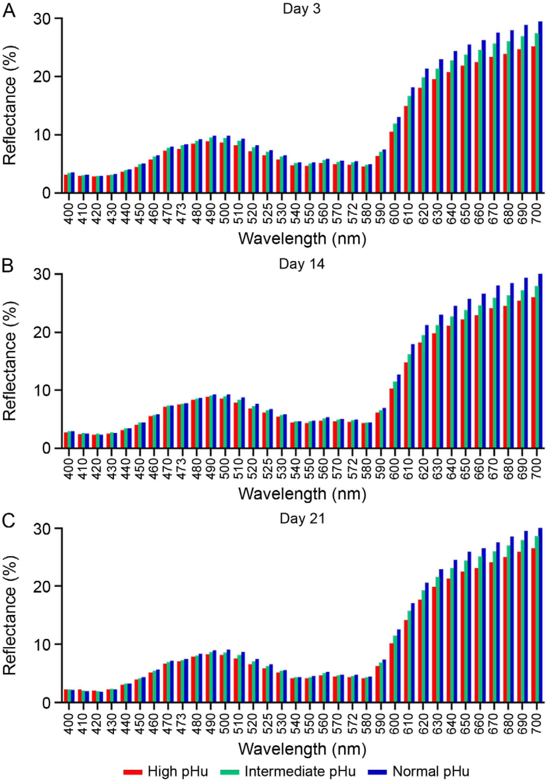
At three days post-mortem, normal pHu beef samples exhibited a more intense bright red color than intermediate pHu samples, demonstrating superior oxygenation capacity and greater stability throughout aging (Figure 1A-C). Initially, intermediate pHu beef exhibited characteristics like those of normal pHu beef, with high L*, a*, and C* values (p < 0.05). However, as the aging process continued, this stability diminished, particularly after 14 days, when an increase in MMb % levels was observed, indicating greater susceptibility to oxidation. In terms of h* values, intermediate pHu beef showed lower values than those of normal pHu beef but higher than those of high pHu beef. Although the R630/580 values were higher than those observed for high pHu beef, they remained lower than those recorded for normal pHu beef, suggesting reduced resistance to discoloration. Furthermore, the ∆E at 21 days reached the highest level among all pHu ranges, indicating that this type of meat experienced the most noticeable color changes over time.
Wavelengths (nm) of light reflected by the surface of the samples in the three pHu ranges (normal, intermediate, and high) illuminated by Illuminant A, showing the effect of pHu on the reflectance curve during the period of (A) 3, (B) 14, and (C) 21 days post-mortem.
High pHu beef displayed the lowest L*, a*, C*, and h* values, resulting in a darker and less saturated color (p < 0.05) (Table 2). The high Mb content contributed to this increased darkening, ultimately reducing its commercial appeal. During the aging process, this meat showed lower levels of OMb % and higher levels of DMb % and MMb %, indicating reduced oxygenation and greater discoloration. The lowest R630/580 reflectance ratio among all pHu ranges confirmed a faster MMb accumulation. Higher OCR % during early storage resulted in increased oxygen consumption, which hindered color stabilization. This suggests that the high OCR % counteracted the advantages of increased MRA %, leading to rapid OMb deoxygenation and subsequent MMb formation. High pHu beef exhibited the lowest color stability, further reinforcing its association with the dark-cutting beef phenomenon and reduced consumer acceptance (Figure 1A-C).
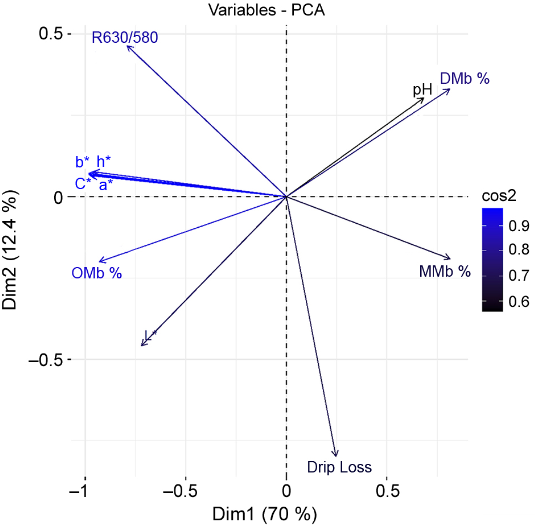
The PCA analysis indicates that the first two principal dimensions (Dim1 and Dim2) account for 82.4 % of the total variation in the data, with contributions of 70 % and 12.4 %, respectively (Figure 2). The angles cos < 90° amongst the parameters L*, a*, b*, C*, h*, OMb %, and R630/580 were highly positively correlated with each other and negatively correlated with pHu, MMb %, and DMb % (Dim1). Additionally, the drip loss variable demonstrated a strong correlation with the principal dimension Dim2. However, this relationship contributes minimally to the overall variation, as Dim2 represents only 12.4 % of the total variation in the dataset.
Principal component analysis (PCA) for dimension 1 (Dim1) (70 %) and dimension 2 (Dim2) (12.4 %) and squared cosine (cos2) for pH, lightness (L*), redness (a*), yellowness (b*), Chroma (C*), hue angle (h*), ratio of light reflectance (R630/580), metmyoglobin percentage (MMb %), deoxymyoglobin percentage (DMb %), oxymyoglobin percentage (OMb %) and drip loss of fresh beef from Nellore cattle.
Discussion
Higher b* values are linked to reduced oxidative stability during the aging period, primarily due to increased accumulation of MMb, similar to results reported by Aroeira et al. (2017). As the aging period progressed, higher b* values in the LL muscle were observed (Mckenna et al., 2005). In Nellore cattle, a rise in b* was also recorded at 14 days of aging (Krauskopf et al., 2024; Patinho et al., 2024).
High pHu samples exhibit a darker appearance after blooming and contain a higher proportion of MMb compared to other pHu ranges (Abril et al., 2001; Mancini and Ramanathan et al., 2020b). This discoloration is caused by the oxidation of the OMb molecule into MMb, a process known as auto-oxidation. This occurs as OMb deoxygenates to form DMb, a highly unstable compound that rapidly oxidizes to MMb (King et al., 2023; Mancini and Ramanathan, 2020b). Metabolic enzyme systems that facilitate oxygen consumption (OC) and the electron transport chain are associated with higher mitochondrial content, which accelerates the oxidative fiber discoloration process (Listrat et al., 2016; Mitacek et al., 2019; McKeith et al., 2014). Consequently, higher OC on the meat surface leads to reduced light scattering, contributing to a darker color on the meat surface (Picard and Gagaoua, 2020). The darkening observed in high pHu meat is attributed to a higher respiratory rate of the mitochondria compared to normal pHu beef, which decreases the availability of oxygen for binding with myoglobin (Wu et al., 2020a). Notably, a significant accumulation of MMb in high pHu beef was recorded at five and seven days of aging (Ijaz et al., 2020).
The lower intensity of the red color in high pHu LL is associated with a greater proportion of DMb and elevated OC levels, which reduce the bright red color of the meat (Holman and Hopkins, 2019; Mancini and Ramanathan, 2020b; Wu et al., 2020a). The rise in DMb and the reduction in OMb observed in high pHu meat are attributed to increased mitochondrial activity and OC (Gagaoua et al., 2021; Ijaz et al., 2020). High respiratory and metabolic activity likely occurred in intermediate and high pHu beef samples, resulting in higher OC and subsequently heightened mitochondrial activity, which contributes to meat darkening. Consequently, the bright red surface typically caused by the blooming effect, facilitated by OMb, is reduced, allowing the purple coloration of the meat to predominate and promoting the enhanced formation of DMb (England et al., 2017).
The higher values of MRA % at three days of aging (p < 0.05) compared to normal and intermediate pHu beef at 14 and 21 days (Table 1) indicate that high pHu beef may have a high concentration of lactic acid after slaughter. This factor may contribute to a reduction in MMb, promoting the reducing activity in high pHu beef samples compared to those with normal and intermediate pHu levels. The increase in MRA % with rising pHu is attributed to both enzymatic and non-enzymatic reductive pathways, as well as the influence of pH levels above 6.5, which tend to favor dark-cutting beef that exhibits higher MRA % compared to other pHu ranges. Similar results have been reported by several authors (Kiyimba et al., 2021; Wu et al., 2020a). Consequently, MRA % plays a crucial role in the stability of meat color, as evidenced by the DMb levels found in this study, where high pHu beef showed a relatively higher proportion of DMb % compared to normal and intermediate pHu beef across all aging times (Table 1). The enhanced MRA % can be attributed to the superior color stability observed in Nellore crossbred animals (Venturini et al., 2010).
High pHu beef showed higher MRA % compared to normal pHu samples when vacuum-packed and aged for 14 days. A study conducted by Krauskopf et al. (2024) reported a similar behavior. A decrease in MRA % and OCR % was observed over the aging period in normal pHu samples. During the aging process, various enzymatic and biochemical processes may occur due to the increased activity of proteolytic enzymes, especially in the degradation of muscle proteins, leading to a reduction in MRA % and OCR %. A reduction in MRA % and OCR % during the aging process has also been reported by English et al. (2016) and Mitacek et al. (2019).
The L* parameter exhibited a significant effect of pHu. Samples with intermediate and normal pHu demonstrated increased lightness compared to those with high pHu (p < 0.05) (Table 2). As pHu rises, muscle proteins enhance their capacity to retain water, leading to fiber swelling and a reduction in the spaces between muscle fibrils. This phenomenon decreases dispersion and increases light absorption, resulting in lower reflectance by myoglobin and promoting a darker appearance of the meat (Ramanathan et al., 2020a; Ribeiro et al., 2022; Wu et al., 2020a).
Meat darkening may be influenced by various factors, including breed characteristics that exhibit a reactive behavior (Silveira et al., 2006), transport conditions (Gallo et al., 2003), and pre-slaughter stress. The latter leads to a reduced concentration of muscle glycogen and an increase in lactic acid production (Ponnampalam et al., 2017). These factors can contribute to meat darkening in these animals (Apaoblaza et al., 2020; Ribeiro et al., 2022; Silva et al., 2019), ultimately resulting in meat with low lightness levels (Krauskopf et al., 2024; Patinho et al., 2024; Ramanathan et al., 2019b).
The bright cherry-red color, indicated by the a* parameter, is a key factor influencing consumers’ purchasing decisions regarding meat freshness and serves as a meat quality indicator (Djenane and Roncalés, 2018; Wang et al., 2020). Consequently, beef with normal and intermediate pHu levels exhibited a lighter reddish appearance compared to the high pHu samples, likely due to the blooming effect and the oxidative state of heme iron during the post-mortem period (Ramanathan et al., 2019b). Throughout the aging period, higher a* values were observed at 21 days compared to days 3 and 14 (Table 2), indicating a progressive increase in the red color intensity on the meat surface over time. This increase in a* values during aging may be attributed to the enhanced color stability of the Longissimus muscle, which has greater color stability and a higher concentration of glycolytic enzymes (Della Malva et al., 2022; Suman et al., 2023). The vacuum-packaging process employed during the aging period in this study may have contributed to a reduction in oxygen consumption (OC), resulting in a more intense red color and C* on the meat surface (Table 2). Vacuum-packed steaks demonstrated an increase in both a* and C* values throughout the aging period (Frank et al., 2017; Krauskopf et al., 2024).
The results indicate that an increase in C* is associated with enhanced color stability and intensity. This stability correlates with higher C* and R630/580 values in normal pHu beef (Ijaz et al., 2020). In contrast, high pHu beef exhibited lower color saturation, leading to a darker and less bright color. Several studies have documented the reduction in C* values as pH increases (Contreras-Barón et al., 2021; Krauskopf et al., 2024; Patinho et al., 2024). Throughout the aging period, significantly higher C* values (p < 0.05) were observed at 21 days compared to days 3 and 14 (Table 2). It is possible that the vacuum-packaging procedure contributed to a reduction in OC during the aging period, resulting in greater color saturation on the meat surface, a characteristic of aged meats. The increase in C* values during the aging period may be attributed to the depletion of substrates involved in mitochondrial activity, which promotes the concentration of OMb on the meat surface, leading to enhanced color saturation, as reported by several authors (Krauskopf et al., 2024; Patinho et al., 2024).
Our findings suggest that meat samples with high and intermediate pHu exhibit less intense coloration and reduced color stability. Higher h* values indicate a lower shade of red and correlate with reduced meat color stability, which is indicative of discoloration typical in high pHu meat (King et al., 2023). The post-mortem glycolysis process, followed by a decline in pH, can facilitate the phosphorylation of the myoglobin structure, making it more susceptible to oxidation and contributing to changes in meat color stability (Li et al., 2018). Throughout the aging period, h* values were notably higher at 21 days compared to days 3 and 14 (Table 2). The discoloration process in meat is characterized by an increase in h* during the aging period (Table 2). This phenomenon has also been observed by Lee et al. (2005) and Patinho et al. (2024). It is possible that during aging, myoglobin oxidizes, altering the h* of the meat, resulting in higher h* values and changes in beef color stability.
A higher R630/580 ratio indicates greater color stability in beef, reflecting a higher proportion of OMb and a lower accumulation of MMb on the meat surface (King et al., 2023). As the vacuum-aged steaks matured, their color stability increased, attributed to the higher proportion of OMb, lower accumulation of MMb on the meat surface, and increased C* values during the aging period (Table 2). The enhanced color stability noted in Longissimus steaks can be attributed to the antioxidant properties of sarcoplasmic proteins, which help mitigate lipid and myoglobin oxidation (Suman et al., 2023). Additionally, this study reported increased color stability during the vacuum-packaging aging period as observed by Wu et al. (2020b). Greater color stability associated with a higher R630/580 ratio in the LL muscles from Nellore cattle has also been documented (Patinho et al., 2024).
A high rate of post-mortem oxygen consumption (OC) can negatively impact the development of a bright red color on the surface of fresh beef. The findings of this study indicated a significant reduction (p < 0.05) in oxygen consumption rate (OCR) during the aging period, which in turn enhanced color development in vacuum-packed beef. This improvement is associated with the depletion of substrates for OC, such as lactate, succinate, and the reduced state of nicotinamide adenine dinucleotide (NADH). During storage, oxygen diffuses more quickly through the tissue, facilitating its binding to myoglobin, and thereby improving color development and stability (Mancini and Ramanathan, 2014; Ribeiro et al., 2022).
The concentration of myoglobin (Mb) is a significant factor in the development of dark color in meat. This concentration can vary due to several factors, including genetics, age, testosterone levels, muscle type, the types of oxidative fibers present in the muscle, and environmental influences (Fink et al., 2018; Ponnampalam et al., 2017; Ribeiro et al., 2022). Elevated Mb concentrations (mg g–1) in beef with high and intermediate pHu values above 6.0 from B. indicus cattle may be attributed to the rearing of uncastrated male animals that are finished on pasture and slaughtered at an age of approximately 36 to 42 months (Contreras-Castillo et al., 2016; Cardoso et al., 2016). The reactive behavior of Nellore males also induces physiological changes in their metabolism (Miguel et al., 2014). Research indicated that Mb concentration in steaks was 7.9 mg g–1 for high pHu meat and 5.7 mg g–1for normal pHu meat (English et al., 2016). Myoglobin (Mb) concentrations of 4.8 mg g–1, 5.2 mg g–1, and 6.6 mg g–1 were found in three pHu ranges, namely pH = 5.41, pH = 5.42, and pH = 5.84 in LT muscles, respectively (Hughes et al., 2017). For the Nellore breed of B. indicus, a Mb concentration of 4.96 mg g–1was observed in meat with normal pHu (Canto et al., 2016). Thus, it is evident that Mb concentration in mg g–1 of fresh beef varies across different pHu ranges.
Global variations in color (∆E) are assessed by the differences in color coordinates between the last and initial days of aging, reflecting the color changes perceivable to the human eye (King et al., 2023). Utilizing the scale proposed by Monteiro et al. (2022), where ∆E values ranging from 1.5 to 3.0 indicate noticeable visual differences and values from 3.0 to 6.0 denote pronounced visual differences, the findings of this study reveal that meat with normal and intermediate pHu exhibited more noticeable color changes. This indicates that these pHu levels influence beef color to a greater extent due to changes in the myoglobin structure of the meat, thereby enhancing the visual color perception. Similar ∆E values were reported by the same authors for Nellore cattle meat, with a mean ∆E value of 4.13 after 21 days for normal pHu.
As the aging time of the samples progressed across different pHu ranges, there was a noticeable increase in the intensity of the reflectance peak near 600 nm, accompanied by a decrease in the intensity of the reflectance peak around 540 to 550 nm (Figure 1A-C). The observed reflectance dips at approximately 550 nm and 580 nm indicated the presence of OMb on the meat surface, which is influenced by the oxygenation process. This process results in a decrease in reflectance in the blue region and an increase in reflectance in the red region, leading to a meat color change from purple-red to bright red throughout the aging period. This effect was particularly pronounced in samples with normal pHu, as observed by Hernández et al. (2015) and Salueña et al. (2019).
The beef samples with high and intermediate pHu levels, evaluated at various aging times, exhibited lower reflectance peaks (ranging from 400 to 700 nm) compared to normal pHu samples. This finding indicates reduced reflectance and, consequently, higher absorbance in the wavelengths associated with the red color spectrum (ranging from 650 to 700 nm) (Figure 1A-C). As a result, the red color appears less intense and brighter to the human eye. The presence of MMb displays characteristic features, including a depression near 630 nm and an increase in reflectance within the central region of the visible spectrum. This phenomenon leads to a diminished difference in reflectance values between 630 nm and 580 nm, producing an opaque, brownish appearance. As oxidation progresses, the difference between the reflectance values at 630 nm and 580 nm continues to decrease (Canto et al., 2016; Gatellier et al., 2001).
The darker surface color observed in high pHu beef (Figure 1A-C) may be attributed to the enhanced water-holding capacity of muscle proteins, which causes the fibers to swell. This swelling reduces the space between muscle fibers, leading to decreased light scattering and increased light absorption by myoglobin, ultimately resulting in a darker muscle surface color (English et al., 2016; Hughes et al., 2017; Ponnampalam et al., 2017). In contrast, normal and intermediate pHu samples exhibited greater numerical reflectance in the wavelengths of red coloring. At 14 and 21 days of aging, both sample types showed similar reflectance peaks (Figure 1B-C). Thus, the reflectance spectra corroborate the color parameter data already discussed in this study.
The PCA indicates that as pHu increases, there is a corresponding rise in DMb % and MMb % (Figure 2), which contributes to the beef surface darkening observed in this study (Table 1). As noted earlier, beef with a high pHu value is darker and subsequently exhibits a higher proportion of MMb % at the surface (Abril et al., 2001; Mancini and Ramanathan, 2020b). The increased OCR % observed in this study (Table 2) may be linked to heightened mitochondrial activity, which facilitates the formation of DMb and the oxidation of OMb (Table 1). This process leads to a reduction in the red color and an increase in the purple color on the meat surface. Consequently, the findings illustrate that the negative correlation between a* with the DMb % and MMb % parameters can be attributed to these factors (Figure 2). Furthermore, the strong positive correlation observed between the parameters L*, a*, b*, C*, h*, OMb %, and R630/580 (Figure 2) suggests that greater lightness and redness (a*) are positively correlated with OMb % and color stability, resulting in reduced surface discoloration of the meat. This relationship is evident with the increase in C*, R630/580, and h* when the pHu is not elevated. Similar correlations have also been reported by Ijaz et al. (2020) and Patinho et al. (2024).
Conversely, the parameters L*, a*, b*, C*, h*, OMb %, and R630/580 (Figure 2) exhibited negative correlations with the parameters pHu, DMb %, and MMb %. As pHu rises, the variables a*, b*, and C* decrease, leading to darker meat cuts and reduced L* values on the surface. Previous studies on meat quality in Nellore bulls have also established a link between decreased values of L*, a*, b*, and C* and the occurrence of dark meat cuts associated with high pHu levels (Contreras-Barón et al., 2021; Krauskopf et al., 2024; Patinho et al., 2024).
The accumulation of MMb on the surface of steaks throughout the vacuum-packaging aging process was found to be pHu-dependent. The findings of this study indicate that the high OCR observed throughout the aging period promoted the deoxygenation of OMb and rapid oxidation to MMb, particularly in steaks with intermediate and high pHu levels. This had an adverse effect on the beef color from B. indicus animals. Notably, steaks from Nellore cattle with intermediate pHu levels, despite exhibiting more pronounced blooming in the initial days of aging, were similarly susceptible to discoloration as those with high pHu levels after 14 days of aging at 2 °C. These results underscore the urgent need to reassess handling practices and production processes within the meat industry to mitigate oxidative metabolism and minimize events that lead to ultimate pH > 5.8. Such measures are essential for optimizing meat color quality, ensuring consumer satisfaction, and protecting brand reputation.
Data availability statement
Data may be made available upon reasonable request.
Acknowledgments
The Fundação de Amparo à Pesquisa do Estado de São Paulo (FAPESP, process number 2017/26667-2) funded this research. The authors thank the Coordenação de Aperfeiçoamento de Pessoal de Nível Superior (CAPES), which awarded the doctoral scholarship (Scholarship number 88887.595866/2020-00).
References
-
Abril M, Campo MM, Önenç A, Sańudo C, Albertí P, Negueruela AI. 2001. Beef colour evolution as a function of ultimate pH. Meat Science 58: 69-78. https://doi.org/10.1016/S0309-1740(00)00133-9
» https://doi.org/10.1016/S0309-1740(00)00133-9 -
Apaoblaza A, Gerrard D, Matarneh SK, Wicks JC, Kirkpatrick L, Sheffler TL, et al. 2020. Muscle from grass and grain fed cattle differs energetically. Meat Science 161: 107996. https://doi.org/10.1016/j.meatsci.2019.107996
» https://doi.org/10.1016/j.meatsci.2019.107996 -
Aroeira CN, Torres Filho RA, Fontes PR, Ramos ALS, Gomide LAM, Ladeira MM, et al. 2017. Effect of freezing before aging on myoglobin redox forms and CIE color of beef from Nellore and Aberdeen Angus cattle. Meat Science 125: 16-21. https://doi.org/10.1016/j.meatsci.2016.11.010
» https://doi.org/10.1016/j.meatsci.2016.11.010 -
Associação Brasileira das Indústrias Exportadoras de Carnes [ABIEC]. 2024. Beef report 2024. Available at: https://abiec.com.br/publicacoes/beef-report-2024-perfil-da-pecuaria-no-brasil/ [Accessed Feb 27, 2025] (in Portuguese)
» https://abiec.com.br/publicacoes/beef-report-2024-perfil-da-pecuaria-no-brasil/ -
Canto ACVCS, Costa-Lima BRC, Suman SP, Monteiro MLG, Viana FM, Salim APAA, et al. 2016. Color attributes and oxidative stability of longissimus lumborum and psoas major muscles from Nellore bulls. Meat Science 121: 19-26. https://doi.org/10.1016/j.meatsci.2016.05.015
» https://doi.org/10.1016/j.meatsci.2016.05.015 -
Cardoso AS, Berndt A, Leytem A, Alves BJR, Carvalho INO, Soares LHB, et al. 2016. Impact of the intensification of beef production in Brazil on greenhouse gas emissions and land use. Agricultural Systems 143: 86-96. https://doi.org/10.1016/j.agsy.2015.12.007
» https://doi.org/10.1016/j.agsy.2015.12.007 -
Contreras-Barón LC, Santos-Donado PR, Ramos PM, Donado-Pestana CM, Delgado EF, Contreras-Castillo CJ. 2021. Influence of ultimate pH on biochemistry and quality of Longissimus lumborum steaks from Nellore bulls during aging. International Journal of Food Science & Technology 56: 3333-3343. https://doi.org/10.1111/ijfs.14955
» https://doi.org/10.1111/ijfs.14955 -
Contreras-Castillo CJ, Lomiwes D, Wu G, Frost D, Farouk MM. 2016. The effect of electrical stimulation on post mortem myofibrillar protein degradation and small heat shock protein kinetics in bull beef. Meat Science 113: 65-72. https://doi.org/10.1016/j.meatsci.2015.11.012
» https://doi.org/10.1016/j.meatsci.2015.11.012 -
Della Malva A, Gagaoua M, Santillo A, De Palo P, Sevi A, Albenzio M. 2022. First insights about the underlying mechanisms of Martina Franca donkey meat tenderization during aging: a proteomic approach. Meat Science 193: 108925. https://doi.org/10.1016/j.meatsci.2022.108925
» https://doi.org/10.1016/j.meatsci.2022.108925 -
Djenane D, Roncalés P. 2018. Carbon monoxide in meat and fish packaging: advantages and limits. Foods 7: 12. https://doi.org/10.3390/foods7020012
» https://doi.org/10.3390/foods7020012 -
England EM, Matarneh SK, Scheffler TL, Gerrard DE. 2017. Perimortal muscle metabolism and its effects on meat quality. p. 63-89. In: Purslow PP, ed. New aspects of meat quality. Woodhead Publishing, Cambridge, England, UK https://doi.org/10.1016/B978-0-08-100593-4.00004-7
» https://doi.org/10.1016/B978-0-08-100593-4.00004-7 -
English AR, Wills KM, Harsh BN, Mafi GG, VanOverbeke DL, Ramanathan R. 2016. Effects of aging on the fundamental color chemistry of dark-cutting beef. Journal of Animal Science 94: 4040-4048. https://doi.org/10.2527/jas.2016-0561
» https://doi.org/10.2527/jas.2016-0561 -
Faustman C, Phillips A. 2001. Measurement of Discoloration in Fresh Meat. John Wiley, New York, NY, USA. https://doi.org/10.1002/0471142913.faf0303s00
» https://doi.org/10.1002/0471142913.faf0303s00 -
Fink J, Schoenfeld BJ, Nakazato K. 2018. The role of hormones in muscle hypertrophy. The Physician and Sportsmedicine 46: 129-134. https://doi.org/10.1080/00913847.2018.1406778
» https://doi.org/10.1080/00913847.2018.1406778 -
Frank DC, Geesink G, Alvarenga TIRC, Polkinghorne R, Stark J, Lee M, et al. 2017. Impact of high oxygen and vacuum retail ready packaging formats on lamb loin and topside eating quality. Meat Science 123: 126-133. https://doi.org/10.1016/j.meatsci.2016.09.010
» https://doi.org/10.1016/j.meatsci.2016.09.010 -
Gagaoua M, Warner RD, Purslow P, Ramanathan R, Mullen AM, López-Pedrouso M, et al. 2021. Dark-cutting beef: a brief review and an integromics meta-analysis at the proteome level to decipher the underlying pathways. Meat Science 181: 108611. https://doi.org/10.1016/j.meatsci.2021.108611
» https://doi.org/10.1016/j.meatsci.2021.108611 -
Gallo C, Lizondo G, Knowles TG. 2003. Effects of journey and lairage time on steers transported to slaughter in Chile. The Veterinary Record 152: 361-364. https://doi.org/10.1136/vr.152.12.361
» https://doi.org/10.1136/vr.152.12.361 -
Gatellier P, Hamelin C, Durand Y, Renerre M. 2001. Effect of a dietary vitamin E supplementation on colour stability and lipid oxidation of air- and modified atmosphere-packaged beef. Meat Science 59: 133-140. https://doi.org/10.1016/S0309-1740(01)00063-8
» https://doi.org/10.1016/S0309-1740(01)00063-8 -
Gonza MMC, Cavalcante CL, Saldaña E, Sartori AG, Contreras-Castillo CJ. 2023. Effect of ultimate pH on quality of aged Longissimus dorsi muscle of Zebu Nellore (Bos indicus during long-term frozen storage. Food Research International 174: 113675. https://doi.org/10.1016/j.foodres.2023.113675
» https://doi.org/10.1016/j.foodres.2023.113675 -
Hernández B, Sáenz C, Alberdi C, Diñeiro JM. 2015. Comparison between two different methods to obtain the proportions of myoglobin redox forms on fresh meat from reflectance measurements. Journal of Food Science and Technology 52: 8212-8219. https://doi.org/10.1007/s13197-015-1917-x
» https://doi.org/10.1007/s13197-015-1917-x -
Holman BWB, Hopkins DL. 2019. Contrasting the quality traits of aged bolar blade, topside and striploin cuts sourced from dark cutting and control Australian beef carcasses. Meat Science 149: 24-30. https://doi.org/10.1016/j.meatsci.2018.10.014
» https://doi.org/10.1016/j.meatsci.2018.10.014 -
Honikel KO. 1998. Reference methods for the assessment of physical characteristics of meat. Meat Science 49: 447-457. https://doi.org/10.1016/S0309-1740(98)00034-5
» https://doi.org/10.1016/S0309-1740(98)00034-5 -
Hughes J, Clarke F, Purslow P, Warner R. 2017. High pH in beef longissimus thoracis reduces muscle fibre transverse shrinkage and light scattering which contributes to the dark colour. Food Research International 101: 228-238. https://doi.org/10.1016/j.foodres.2017.09.003
» https://doi.org/10.1016/j.foodres.2017.09.003 -
Ijaz M, Li X, Zhang D, Hussain Z, Ren C, Bai Y, et al. 2020. Association between meat color of DFD beef and other quality attributes. Meat Science 161: 107654. http://doi.org/10.1016/j.meatsci.2019.107954
» http://doi.org/10.1016/j.meatsci.2019.107954 -
King DA, Hunt MC, Barbut S, Claus JR, Cornforth DP, Joseph P, et al. 2023. American meat science association guidelines for meat color measurement. Meat and Muscle Biology 6: 12473. https://doi.org/10.22175/mmb.12473
» https://doi.org/10.22175/mmb.12473 -
Kiyimba F, Hartson SD, Rogers J, VanOverbeke DL, Mafi GG, Ramanathan R. 2021. Changes in glycolytic and mitochondrial protein profiles regulates postmortem muscle acidification and oxygen consumption in dark-cutting beef. Journal of Proteomics 232: 104016. https://doi.org/10.1016/j.jprot.2020.104016
» https://doi.org/10.1016/j.jprot.2020.104016 -
Krauskopf MM, Araújo CDL, Santos-Donado PR, Dargelio MDB, Manzi JAS, Venturini ACJ, et al. 2024. The effect of succinate on color stability of Bos indicus bull meat: pH-dependent effects during the 14-day aging period. Food Research International 175: 113688. https://doi.org/10.1016/j.foodres.2023.113688
» https://doi.org/10.1016/j.foodres.2023.113688 -
Lee S, Decker EA, Faustman C, Mancini RA. 2005. The effects of antioxidant combinations on color and lipid oxidation in n-3 oil fortified ground beef patties. Meat Science 70: 683-689. https://doi.org/10.1016/j.meatsci.2005.02.017
» https://doi.org/10.1016/j.meatsci.2005.02.017 -
Li M, Li Z, Li X, Xin J, Wang Y, Li G, et al. 2018. Comparative profiling of sarcoplasmic phosphoproteins in ovine muscle with different color stability. Food Chemistry 240: 104-111. https://doi.org/10.1016/j.foodchem.2017.07.097
» https://doi.org/10.1016/j.foodchem.2017.07.097 -
Listrat A, Lebret B, Louveau I, Astruc T, Bonnet M, Lefaucheur L, et al. 2016. How muscle structure and composition determine meat quality. The Scientific World Journal 2016: 3182746. https://doi.org/10.1155/2016/3182746
» https://doi.org/10.1155/2016/3182746 -
Mancini RA, Ramanathan R. 2014. Effects of postmortem storage time on color and mitochondria in beef. Meat Science 98: 65-70. https://doi.org/10.1016/j.meatsci.2014.04.007
» https://doi.org/10.1016/j.meatsci.2014.04.007 -
Mancini RA, Ramanathan R. 2020. Molecular basis of meat color. p. 117-129. In: Biswas AK, Mandal PK, eds. Meat quality analysis. Elsevier, Amsterdam, The Netherlands. https://doi.org/10.1016/B978-0-12-819233-7.00008-2
» https://doi.org/10.1016/B978-0-12-819233-7.00008-2 -
McKeith RO, King DA, Grayson AL, Shackelford SD, Savell JW, Wheeler TL. 2014. Influence of mitochondrial efficiency on beef lean color stability. Meat Science 96: 476. https://doi.org/10.1016/j.meatsci.2013.07.116
» https://doi.org/10.1016/j.meatsci.2013.07.116 -
McKeith RO, King DA, Grayson AL, Shackelford SD, Gehring KB, Savell JW, et al. 2016. Mitochondrial abundance and efficiency contribute to lean color of dark cutting beef. Meat Science 116: 165-173. https://doi.org/10.1016/j.meatsci.2016.01.016
» https://doi.org/10.1016/j.meatsci.2016.01.016 -
McKenna DR, Mies PD, Baird BE, Pfeiffer KD, Ellebracht JW, Savell JW. 2005. Biochemical and physical factors affecting discoloration characteristics of 19 bovines muscles. Meat science 70: 665-682. https://doi.org/10.1016/j.meatsci.2005.02.016
» https://doi.org/10.1016/j.meatsci.2005.02.016 -
Miguel GZ, Faria MH, Roça RO, Santos CT, Suman SP, Faitarone ABG, et al. 2014. Immunocastration improves carcass traits and beef color attributes in Nellore and Nellore×Aberdeen Angus crossbred animals finished in feedlot. Meat Science 96: 884-891. https://doi.org/10.1016/j.meatsci.2013.08.030
» https://doi.org/10.1016/j.meatsci.2013.08.030 -
Mitacek RM, Ke Y, Prenni JE, Jadeja R, VanOverbeke DL, Mafi GG, et al. 2019. Mitochondrial degeneration, depletion of NADH, and oxidative stress decrease color stability of wet-aged beef longissimus steaks. Journal of Food Science 84: 38–50. https://doi.org/10.1111/1750-3841.14396
» https://doi.org/10.1111/1750-3841.14396 -
Monteiro PAM, Maciel ICF, Alvarenga RC, Oliveira AL, Barbosa FA, Guimaraes ST, et al. 2022. Carcass traits, fatty acid profile of beef, and beef quality of Nellore and Angus x Nellore crossbred young bulls finished in a feedlot. Livestock Science 256: 104829. https://doi.org/10.1016/j.livsci.2022.104829
» https://doi.org/10.1016/j.livsci.2022.104829 -
Mueller LF, Balieiro JCC, Ferrinho AM, Martins TS, Corte RRPS, Amorim TR, et al. 2019. Gender status effect on carcass and meat quality traits of feedlot Angus × Nellore cattle. Journal of Animal Science 90: 1078-1089. https://doi.org/10.1111/asj.13250
» https://doi.org/10.1111/asj.13250 -
Patinho I, Cavalcante CL, Saldaña E, Gagaoua M, Behrens JH, Contreras-Castillo CJ. 2024. Assessment of beef sensory attributes and physicochemical characteristics: a comparative study of intermediate versus normal ultimate pH striploin cuts. Food Research International 175: 113778. https://doi.org/10.1016/j.foodres.2023.113778
» https://doi.org/10.1016/j.foodres.2023.113778 -
Picard B, Gagaoua M. 2020. Muscle fiber properties in cattle and their relationships with meat qualities: an overview. Journal of Agricultural and Food Chemistry 68: 6021-6039. https://doi.org/10.1021/acs.jafc.0c02086
» https://doi.org/10.1021/acs.jafc.0c02086 -
Ponnampalam EN, Hopkins DL, Bruce H, Li D, Baldi G, Bekhit AE. 2017. Causes and contributing factors to "dark cutting" meat: current trends and future directions; a review. Comprehensive Reviews in Food Science and Food Safety 16: 400-430. https://doi.org/10.1111/1541-4337.12258
» https://doi.org/10.1111/1541-4337.12258 -
Ramanathan R, Hunt MC, English AR, Mafi GG, VanOverbeke DL. 2019a. Effects of aging, modified atmospheric packaging, and display time on metmyoglobin reducing activity and oxygen consumption of high-pH beef. Meat and Muscle Biology 3: 276-288. https://doi.org/10.22175/mmb2019.05.0017
» https://doi.org/10.22175/mmb2019.05.0017 -
Ramanathan R, Nair MN, Hunt MC, Suman SP. 2019b. Mitochondrial functionality and beef colour: a review of recent research. South African Journal of Animal Science 49: 9-19. http://dx.doi.org/10.4314/sajas.v49i1.2
» http://dx.doi.org/10.4314/sajas.v49i1.2 -
Ramanathan R, Hunt MC, Mancini RA, Nair MN, Denzer ML, Suman SP, et al. 2020a. Recent updates in meat color research: integrating traditional and high-throughput approaches. Meat and Muscle Biology 4: 1-24. https://doi.org/10.22175/mmb.9598
» https://doi.org/10.22175/mmb.9598 -
Ramanathan R, Mafi GG, Yoder L, Perry M, Pfeiffer M, VanOverbeke DL, et al. 2020b. Biochemical changes of postmortem meat during the aging process and strategies to improve the meat quality analysis p. 67-80. In: Biswas AK, Mandal PK, eds. Meat quality analysis. Elsevier, Amsterdam, The Netherlands. https://doi.org/10.1016/B978-0-12-819233-7.00005-7
» https://doi.org/10.1016/B978-0-12-819233-7.00005-7 -
Ramanathan R, Mancini RA, Konda MR, Bailey K, More S, Mafi GG. 2022. Evaluating the failure to bloom in dark-cutting and lactate-enhanced beef longissimus steaks. Meat Science 184: 108684. https://doi.org/10.1016/j.meatsci.2021.108684
» https://doi.org/10.1016/j.meatsci.2021.108684 -
Ribeiro CCS, Contreras-Castillo CJ, Santos-Donado PR, Venturini AC. 2022. New alternatives for improving and assessing the color of dark-cutting beef: a review. Scientia Agricola 79: e20200079. https://doi.org/10.1590/1678-992X-2020-0079
» https://doi.org/10.1590/1678-992X-2020-0079 -
Salueña BH, Gamasa CS, Rubial JMD, Odriozola CA. 2019. CIELAB color paths during meat shelf life. Meat Science 157: 107889. https://doi.org/10.1016/j.meatsci.2019.107889
» https://doi.org/10.1016/j.meatsci.2019.107889 -
Silva LHP, Assis DEF, Estrada MM, Assis GJF, Zamudio GDR, Carneiro GB, et al. 2019. Carcass and meat quality traits of Nellore young bulls and steers throughout fattening. Livestock Science 229: 28-36. https://doi.org/10.1016/j.livsci.2019.09.012
» https://doi.org/10.1016/j.livsci.2019.09.012 -
Silveira IDB, Fischer V, Soares GJD. 2006. Relation between genotype and temperament of grazing steers on meat quality. Revista Brasileira de Zootecnia 35: 519-526 (in Portuguese, with abstract in English). https://doi.org/10.1590/S1516-35982006000200026
» https://doi.org/10.1590/S1516-35982006000200026 -
Suman SP, Wang Y, Gagaoua M, Kiyimba F, Ramanathan R. 2023. Proteomic approaches to characterize biochemistry of fresh beef color. Journal of Proteomics 281: 104893. https://doi.org/10.1016/j.jprot.2023.104893
» https://doi.org/10.1016/j.jprot.2023.104893 -
Terlouw EMC, Picard B, Deiss V, Berri C, Hocquette J-F, Lebret B, et al. 2021. Understanding the determination of meat quality using biochemical characteristics of the muscle: Stress at slaughter and other missing keys. Foods 10: 84. https://doi.org/10.3390/foods10010084
» https://doi.org/10.3390/foods10010084 -
Torres Filho RA, Cazedey HP, Fontes PR, Ramos ALS, Ramos EM. 2017. Drip loss assessment by different analytical methods and their relationships with pork quality classification. Journal of Food Quality 2017: 9170768. https://doi.org/10.1155/2017/9170768
» https://doi.org/10.1155/2017/9170768 -
Venturini AC, Contreras-Castillo CJ, Faria JAF, Gallo CR, Silva TZ, Shirahigue LD. 2010. Microbiological, colour and sensory properties of fresh beef steaks in low carbon monoxide concentration. Packaging Technology and Science 23: 327-338. https://doi.org/10.1002/pts.902
» https://doi.org/10.1002/pts.902 -
Wang F, Holman BWB, Zhang Y, Luo X, Mao Y, Hopkins DL. 2020. Investigation of colour requirements of frozen beef rolls by Chinese consumers for hot pot. Meat Science 162: 108038. https://doi.org/10.1016/j.meatsci.2019.108038
» https://doi.org/10.1016/j.meatsci.2019.108038 -
Warris PD. 1979. The extraction of haem pigments from fresh meat. International Journal of Food Science Technology 14: 75-80. https://doi.org/10.1111/j.1365-2621.1979.tb00849.x
» https://doi.org/10.1111/j.1365-2621.1979.tb00849.x -
Wu S, Luo X, Yang X, Hopkins DL, Mao Y, Zhang Y. 2020a. Understanding the development of color and color stability of dark cutting beef based on mitochondrial proteomics. Meat Science 163: 108046. https://doi.org/10.1016/j.meatsci.2020.108046
» https://doi.org/10.1016/j.meatsci.2020.108046 -
Wu S, Han J, Liang R, Dong P, Zhu L, Hopkins DL, et al. 2020b. Investigation of muscle-specific beef color stability at different ultimate pHs. Asian-Australasian Journal of Animal Sciences 33: 1999-2007. https://doi.org/10.5713/ajas.19.0943
» https://doi.org/10.5713/ajas.19.0943
Edited by
-
Edited by:
Luís Guilherme de Lima Ferreira Guido https://orcid.org/0000-0001-9441-7867