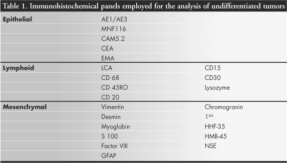
CONTEXT: Undifferentiated head and neck and skull base tumors are not unusual. They can arise in mucosa as well as in salivary glands, soft tissues or lymph nodes. Suitable therapy and prognosis for each case depends upon precise histopathological diagnosis. OBJECTIVE: To evaluate the role of immunohistochemical techniques in determining the conclusive diagnosis. The occurrence of these tumors in our service and the way in which they were distributed according to cell pattern, patient's age and tumor location was also evaluated. TYPE OF STUDY: Cross-sectional study. SETTING: Hospital das Clínicas, Universidade Estadual de Campinas, Campinas, São Paulo, Brazil. PARTICIPANTS: 43 biopsies performed between January 1990 and December 1997, diagnosed as undifferentiated head and neck tumors. PROCEDURES: We applied an immunohistochemical panel in accordance with the avidin-biotin-peroxidase complex method. The final diagnosis was achieved after new analysis in conjunction with biopsies stained using the hematoxylin-eosin technique. MAIN MEASUREMENTS: This study evaluated undifferentiated tumors in head and neck, and the way in which they were distributed, according to cell pattern, patient's age and tumor location. RESULTS: The most frequent locations for undifferentiated tumors were the lymph nodes, 20.9%; pharynx and neck, 16.3%; paranasal sinus, 14%; and nose, 11.6%. They were most prevalent during the seventh decade of life (34.9%), and twice as prevalent in men as in women. The immunohistochemical technique allowed conclusive diagnosis for 60.5% of the tumors and was suggestive for 20.9% of the biopsies. The most prevalent cell pattern was round cells (51.2%), followed by epithelioid cells (20.9%), spindle cells (16.3%), myxoid pattern (9.3%) and pleomorphic cells (2.3%). CONCLUSION: Our results demonstrate the fundamental role of the immunohistochemical technique for conclusive diagnosis of undifferentiated tumors.
Immunohistochemical; Undifferentiated tumors; Diagnosis; Head neck; Avidin-biotin-peroxidase