An exploratory study of the functional significance of the floral secretory structures in two Miconia species (Melastomataceae)
Abstract
Aiming to verify whether the diversity of secretory structures with their respective exudates are or not responsible for the attendance of floral visitors in Miconia species, the floral secretory structures of two Amazonian species of Miconia were described and interpreted from the functional perspectives and observations of floral visitors. Flowers and floral visitors were collected in the field for 16 months. The flowers were subjected to standard anatomical analyzes using light and scanning electron microscopy, and the secretory structures were evaluated using histochemical tests. The insects were photographed, collected and identified by a specialist. Floral secretory structures (stigmatic papillae and trichomes at the apex of the ovary) were found in M. alata and M. ciliata. Trichomes were found at the hypanthium of M. alata, and of the sepals and receptacles in M. ciliata. In both species, different hydrophilic compounds were detected in the exudates. Only phenolic compounds were found in the trichomes of the M. ciliata receptacle. This study provides information that confirms the hypothesis that other secretory structures and exudates are related to floral visitors, and adds new informations about the features of the secretory structures in Miconia.
Key words
Anatomy; chemical compounds; glandular trichomes; papillae
INTRODUCTION
The effort to study biodiversity as integrated sets of organisms is called “interaction biodiversity” sensu Thompson (1997). Currently, this topic is of great interest, especially in describing interactions in tropical forests that present high levels of mutualistic relationships, including the production of food resources mediating pollination services (Quesada et al. 2011). These researches still do not fully reflect the taxonomic diversity of Melastomataceae.
There are several lineages in Melastomataceae with trait combinations indicating pollination strategies but specific studies have not yet been reported (Dellinger et al. 2022). Additional fieldwork is needed to clarify whether a common reproductive strategy is associated with the growth in the moist, shaded herbaceous understory (Dellinger et al. 2022).
In Brazil, 69 % of the genera and 37 % of the species of Melastomataceae are recorded in the Amazon (Clausing & Renner 2001, Goldenberg et al. 2012, Oliveira et al. 2015). Miconia Ruiz & Pav. is one of the most representative genus, with high levels of endemism (Clausing & Renner 2001, Goldenberg et al. 2012, Oliveira et al. 2015). In the Amazon, this genus occurs in both primary and secondary forests and is known for being pioneer plants that rapidly establish themselves and attract foraging animals (Schupp et al. 1989, Denslow et al. 1990, Antonini & Nunes-Freitas 2004, Lima et al. 2014). The genus also shows high seed production, efficient dispersal, high germination rates, and rapid growth (Albuquerque et al. 2013).
Miconia are known for being shrubby to woody species inhabiting the understory of tropical forests, interacting predominantly with generalist insects that occur in this environment (Kriebel & Zumbado 2014, Silva et al. 2021, Dellinger et al. 2022). Some species are known for interacting with many animals, among flies, wasps, and hummingbirds, establishing mutualistic relationships where food resources are offered in exchange for the protection of floral structures, with insects likely being the primary pollinators (Dellinger et al. 2022). About 62 % of the Miconia share floral traits involved with bee pollination (Silva et al. 2021). Among the set of traits promoting bee pollination, one may not forget the anthers with poricidal openings which are especially related to buzz pollination (Harder & Barclay 1994, Brito et al. 2016, Goldenberg et al. 2022). Chemical compounds have the potential to produce a range of effects (attractive or repellent) on insects (Faly et al. 2024). However, the gap in knowledge regarding the secretory structures occurring in Miconia is significant.
At the anatomical level, genera such as Miconia, usually found at lower elevations, make use of nectar rewards, a fact interpreted as a change promoted by the unpredictability of pollinators (Varassin et al. 2008). Notwithstanding, the nectariferous species of Miconia also bear changes in the anthers which lead to both bat and bird pollination, characterizing a more generalist system (Goldenberg et al. 2008). In those cases, other mechanisms for nectar production have already been identified such as the stigmatic secretion of nectar (Stein & Tobe 1989, Goldenberg et al. 2008).
Studies from the last three decades focusing on floral nectaries for this group reported the absence of nectariferous tissues and failed to find a structure related to nectar release (Stein & Tobe 1989, Tobe et al. 1989, Vogel 1997). Thus, the secretory structures are of utmost importance in understanding evolutionary processes beyond ecological relationships. Besides, studies that address and determine the diversity of the floral biology attributes as well as the behavior of insects visiting ruderal plants allow us to better understand insect-plant interactions, especially pollination and genetic dispersal (Jardim & Kageyama 1994).
Therefore, this study aimed to verify whether, in addition to nectariferous structures previously reported, other secretory structures with their respective exudates are or not responsible for the attendance of floral visitors in Miconia species. To this end, an interdisciplinary was set out in order to characterize the floral secretory structures and their respective exudates in Amazonian species of Miconia. Additionally, the structural and histochemical features were interpreted from the perspective of functional information about the species and observations of floral visitors.
MATERIALS AND METHODS
Experimental design and plant material sampling
Based on the distribution and flowering period of Miconia species, as indicated on the online databases of the regional herbaria, IAN and MG (Thiers 2024, updated continuously) two species were selected Miconia alata (Aubl.) DC. and Miconia ciliata (Rich.) DC. Fertile individuals of both species were identified and marked from September to October 2020 along the main access path at the Parque Estadual do Utinga (PEUt), Municipality of Belém, State of Pará, Brazil (01º23’13” to 01º26’32,70”S and 48º26’41,41” to 48º26’47”W). From those, 10 individuals located up to 40 m from the forest edge were selected. Both species occur at the PEUt, and their flowering was compatible with the study period. The collection and focal observations of species were concentrated in places of greatest abundance, forming three collection sites with greater abundance along the forest edge that were 80 × 1000 m, with approximately 200 m between them (Fig. S1 – Supplementary Material).
According to the flowering period of each species, samples of fertile individuals of M. alata were collected from September 2020 to December 2021, while those of M. ciliata were sampled from May to December 2021. Flowers from at least two inflorescences in each specimen were collected, during the pre-anthesis and anthesis.
Anatomy of the floral secretory structures under Light and Scanning Electron Microscopy
Two fixative treatments were used for different sets of samples. The first set was fixed in FAA (formalin: acetic acid: 50% ethanol, 1:1:18 by volume; Johansen 1940) for 24 h and used for the structural characterization as well as to scrutinize for the presence of hydrophilic compounds; the second set was fixed in NBF (neutral buffered formalin) for 48 h (Lillie 1965) and subjected to histochemical tests to scrutinize for the presence of lipophilic substances. Samples from both fixation treatments were used for the scanning electron microscopy (SEM).
Part of the samples fixed in FAA or NBF were also dehydrated in a tert-butanol series (Johansen 1940) and embedded in histological paraffin with Dimethyl sulfoxide (DMSO) (Paraplast®, ©Leica Biosystems, USA). The samples were cross and longitudinally sectioned at 10-12 µm thick using a semi-automatic rotary microtome (Leica®, RM 2245). Part of the sections were stained with Safranin and Astra blue (Gerlach 1969) and mounted in synthetic resin (Permount®, Fisher Scientific, New Jersey, USA), for structural characterization.
For the general characterization of the exudate, the following histochemical tests were carried out: periodic acid–Schiff reagent for detecting the presence of total polysaccharides (McManus 1948), ruthenium red for acidic mucilage (Gregory & Baas 1989), tannic acid/ferric chloride for neutral mucilage (Pizzolato & Lillie 1973), Lugol’s solution for starch (Johansen 1940), Sudan black B for total lipids (Pearse 1985), ferric chloride for total phenolic compounds (Johansen 1940), vanillin-hydrochloric acid for tannins (Mace & Howell 1974), and xylidine Ponceau for total proteins (O’Brien & McCully 1981). Standard control procedures were carried out simultaneously as required for each test. Slides were also mounted according to the protocol described for each test.
Observations and photographs were made with a light microscope (BX53, Olympus Optical, Tokyo, Japan) coupled to a digital camera (DP72, Olympus Optical, Tokyo, Japan).
For scanning electron microscopy (SEM) (Bozzola & Russell 1999), the samples previously fixed were dehydrated in a crescent ethanol series up to 100% ethanol and critical-point dried with CO2 in a 020 CPD dryer (Bal-Tec; Balzers, Liechtenstein). The samples were mounted on stubs and coated with gold (20 nm), for 150 seconds at 25 mA, using an FDU 010 sputter coater (Bal-Tec). Examinations and photographs were made with a Leo 1450VP® (Zeiss, Heidelberg, Germany) at the Scanning Electron Microscopy Laboratory at the Museu Paraense Emílio Goeldi.
Observation of floral visitors
Focal observations of floral visitors were performed in the field on the selected individuals of each Miconia species. The literature available for Miconia indicates that during the daylight flowers are ready to be pollinated, as the stigma may outgrow the corolla even before the flowers are completely open which happens before daybreak (Baumgratz & Silva 1986, Meyer 1998, Santos 2008, Santos et al. 2010). Therefore, observations were made during daylight, from 6:00 a.m. to 2:00 p.m., which also agrees with previous works for Miconia species (Brito et al. 2017). Subsequent observations were done only during the activity period of pollinators, for 30-minute intervals, following Nogueira-Ferreira & Augusto (2007). Sampling and observation of floral visitors were carried out monthly during 6 non-consecutive days of the month, including at least two days per week of observation, from September 2020 to December 2021.
Insects were collected (Process PAE nº 2022/1233245; Permission nº 017/2022) when they were on or after visiting flowers using plastic bags. To record their behavior, these individuals were photographed and filmed. Sampled individuals were fixed in glass vials containing 70% ethanol and were identified later by a specialist at the Entomology Laboratory at the Museu Paraense Emílio Goeldi.
RESULTS
Structural characterization
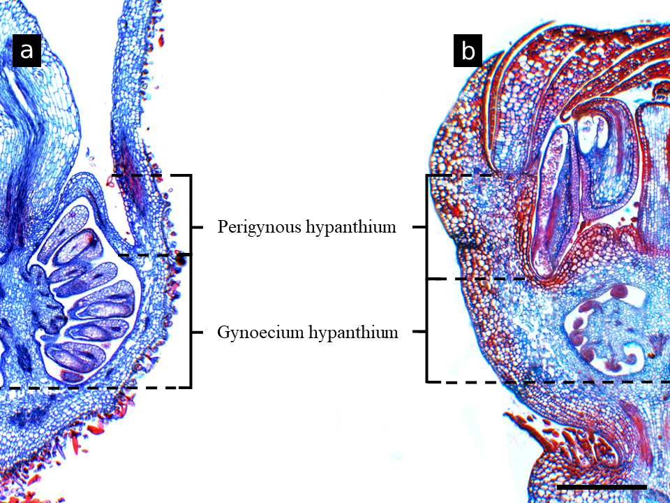
In both species, M. alata and M. ciliata, the surface of the hypanthium, the apex of the ovary and the stigma were the areas where the floral secretory structures were concentrated. For both Miconia, hypanthium could be distinguished into two parts: gynoecial hypanthium, when associated with the gynoecium, and perigynous hypanthium, when it is not associated with the gynoecium (Fig. 1a, b).

Hypanthium regions in Miconia. a: Miconia alata. b: Miconia ciliata. Scale bar: 400 µm (a, b).
Miconia alata had glandular trichomes type capitate on the surface of the hypanthium (both gynoecial and perigynous hypanthium) as well as at the ovary apex (Figs. 2a, b). Papillae was restricted to the stigmatic region (Fig. 2c). In M. ciliata, papillae were observed on both sepals and stigma (Figs. 2d, e, f) while glandular trichomes type capitate were present only on the outer surface of the ovary apex (Figs. 2g, h). In both species, the glandular trichomes type capitate presents a multicellular secretory head, usually with four cells, supported by a single uniseriate stalk (Figs. 2b, g, h). The stigma in both species showed a standard shape (Figs. 2c, e, f).

Secretory structures in Miconia alata and M. ciliata. a-d: Miconia alata. e-h: Miconia ciliata. a, b, g and h: Trichomes on the hypanthium and ovary apex. c, e and f: Stigmatic papillae. d: Papillae on the surface of sepals, note the exudate (asterisk). Arrows: Location of structures. Hyp: hypanthium. Gt: glandular trichomes type capitate. Ov: ovary. Pp: papillae. St: stigma. Sta: stamen. Sty: style. Scale bar: 200 µm (a, e), 100 µm (b, c, f), 50 µm (g, h), 40 µm (d).
Histochemical characterization
In both developmental stages of the flowers of M. alata and M. ciliata (i.e., pre-anthesis and anthesis), the same kinds of secretory structures and exudates were verified (Figs. 3, 4). Positive reactions were observed for total polysaccharides, acidic mucilage, phenolic compounds, and total proteins in almost all secretory structures. The results of all histochemical tests are summarized in Table I.
Results for the histochemical tests to identify compounds in secretions from secretory structures in Miconia alata and M. ciliata. SP: Sepal papillae. PS: Stigmatic papillae. TrH: Hypanthium trichomes. TrO: Ovarium trichomes. (+) positive reaction. (-) negative reaction.
For M. alata, there were positive reactions for total polysaccharides, acidic mucilage, phenolic compounds, and proteins in the trichomes placed on the hypanthium (Fig. 3a, b, c, d) and ovary apex (Fig. 3e, f, g, h, i) (Table I). The stigmatic papillae tested positive for total polysaccharides, phenolic compounds, and proteins (Figs. 3j, k, l, m) (Table I).

Positive histochemical results for the secretions of the floral secretory structures of Miconia alata. a-d: Trichomes on the hypanthium. f-i: Trichomes on the ovary apex. J-M: Stigma. a, e, j: Sections without treatment. Reactions were positive for total polysaccharides as shown by the magenta staining (b, f and k), acidic mucilage, pink to red staining (h), total phenolic compounds, brown to black staining (c, h and l), and total proteins red staining (d, i and m). Gt: glandular trichomes type capitate. Pp: stigmatic papillae. Arrows: reaction sites. Scale bar: 25 µm (a-i), 50 µm (j-m).
For M. ciliata, the stigmatic papillae (Fig. 4a, b, c, d, e), sepal papillae (Figs. 3f, g, h, i, j), and trichomes on the ovary apex (Figs. 4k, l, m, n, o) tested positive for polysaccharides, acidic mucilage, phenolic compounds, and proteins (Table I).

Positive histochemical results for the secretions of the floral secretory structures of Miconia ciliata. a-e: Stigma. f-j: Sepals. k-o: Trichomes on the ovary apex. a, f and k: Sections without treatment. Reactions were positive for total polysaccharides as shown by the magenta staining (b, g and l), acidic mucilage, pink to red staining (c, h and m), total phenolic compounds, brown to black staining (d, i and n), and total proteins, red staining (e, j and o). Gt: glandular trichomes type capitate. Pp: stigmatic papillae. Sp: sepals. Arrows: reaction site. Scale bar: 25 µm (k-o), 75 µm (j), 100 µm (a, c-i), 150 µm (b).
Flower-visiting
For M. alata, only one flower visiting species was observed which belonged to the Curculionidae family (Coleoptera) (Fig. 5a, 6a), that is, true weevils (or “snout beetles”). For the M. ciliata flowers, three Hymenoptera insect species were identified: Augochloropsis sp., Melipona melanoventer Schwarz 1932, and Synoeca virginea Fabricius 1804 (Figs. 5b, c, d, 6b), and an Ocyptamus sp. (Diptera), species without photographic record. The number of M. alata visitors varied between the months of observation, with the highest occurrence being in September (Fig. 6a, b). The number of visitors to M. ciliata had a critical decline in March, with only M. melanoventer being observed. Augochloropsis sp. was predominant during 15 months of observation (Fig. 6b).

Flower-visiting insects of the Miconia species. a: Curculionidae on Miconia alata flowers. Augochloropsis sp. on Miconia ciliata flowers (b). Melipona melanoventer Schwarz, 1932 on Miconia ciliata flowers (c). Synoeca virginea Fabricius, 1804 on Miconia ciliata flowers (d). Arrow: Indication of the visitor. Photos by César Favacho.

Number of Curculionidae visitors on Miconia alata flowers according to the month and year of observation. a. Curculionidae on Miconia alata flowers. b. Three flower-visiting species on Miconia ciliata flowers.
DISCUSSION
Glandular trichomes generally produce, store and secrete substances, and although some compounds are predominant, various groups of chemical-related compounds may be synthetized (Schuurink & Tissier 2020). This is in accordance with the results observed in the present study, since the secretions from glandular trichomes on the hypanthium and ovary apex of M. alata, as well as those from the ovary apex of M. ciliata, reacted positively to different secondary compounds. As a whole, glandular trichomes are responsible for the synthesizes several classes of natural products (Wagner 1991, Zager & Lange 2018, Muravnik 2020), which are involved in an array of ecological functions for plants, with many of these functions related to plant animals interactions (Werker 2000, Belan et al. 2020)
Cells of the sepal and Hypanthium trichomes
The papillae on the sepals as well as the trichomes on the hypanthium show the presence of the same kinds of compounds found at the ovary apex and stigma. In general, glandular trichomes on the sepals and hypanthium are common among the species of Melastomataceae (Goldenberg et al. 2008, Fidanza & Almeda 2011, Michelangeli & Goldenberg 2016, Bécquer et al. 2022, Santos et al. 2022), and the same is true for the genus Miconia (Wurdack 1986, Gonçales et al. 2023a)
Total polysaccharides, phenolic compounds, and total proteins are present in both species, M. alata and M. ciliata, while acid mucilages are only in M. alata. As the hypanthium and sepals are floral parts that are not manipulated by the visiting insects (field observations), such trichomes may aid Miconia species playing a role in anti-herbivory strategies. As mucilages and polysaccharides are water soluble compounds that may form viscose (sticky) colloidal solutions when in contact with water and such sticky secretion could then entrap the herbivores (Read & Gregory 1997, Werker 2000, Rudall 2007, Rocha et al. 2011, Tosif et al. 2021). That mechanism may eventually kill small herbivore insects as entrapped predators often die from desiccation or starvation (Krimmel & Pearse 2013, Riddick & Simmons 2014). Phenolic compounds may also show bioprotectant activity against insects that feed on plants secreting phenolic compounds. Phenols act as a defensive mechanism (antifeedant mechanisms) not only against herbivores but also against microorganisms (Boeckler et al. 2011, War et al. 2012). Therefore, the trichomes on the sepals and hypanthium would be an important tool for protecting M. alata and M. ciliata against herbivory.
Ovarium trichomes
Floral nectary has been reported at the inner face of the ovary apex, close to the style base in Miconia hyemalis A.St.-Hil. & Naudin (Varassin et al. 2008). In M. hyemalis, the ovary apex is rich in papillate trichomes (Varassin et al. 2008). However, in our study, M. alata and M. ciliata present glandular trichomes type capitate instead. Although there is a difference in the type of secretory structure found at the ovary apex in the species we study, such trichomes could be responsible for the secretion of nectar as many studies have reported the floral nectary to be composed of secretory trichomes pollinators (Fahn 1979, Bentley & Elias 1983, Nicolson et al. 2007).
The trichomes on the ovary apex of M. alata and M. ciliata present total polysaccharides, acid mucilages, total proteins as well as phenolic compounds. Total polysaccharides, acid mucilages, and total proteins are commonly reported as rewards for pollinators (Fahn 1979, Bentley & Elias 1983, Nicolson et al. 2007). The trichomes on the ovary apex could also act as food hairs (i.e., edible hairs), in which the hair contents are consumed by the pollinators (Pansarin & Maciel 2017, Lustofin et al. 2020). Mucilages as well as polysaccharides with varied compositions represent rewards for pollinators (Fahn 1979, Roshchina & Roshchina 1993), and nutritious tissues can be found in the petals, as described to Annona L. (Annonaceae Juss.) (Gottsberger & Webber 2018), and as observed in Miconia, in this study.
Secondary metabolites such phenolic compounds are found in the nectar of plants that make use of such compounds as defense against herbivore attack (Nicolson 2022). Although phenolics may be lost during the processing of nectar into honey (Stevenson et al. 2017), phenolics may still be an important component of honey (Becerril-Sánchez et al. 2021). Therefore, the presence of such compounds within the trichomes serving as edible hairs harvested by bees is expected.
Stigmatic papillae
In the Miconia species studied, we verified the presence of papillae on the stigmatic area. The stigma is responsible for pollen capture and adherence, a role that is played by the mechanical interaction between pollen grains and the papillae as well as the interaction with the stigmatic secretion (Heslop-Harrison 1992, Ito & Gorb 2019). The syncarpous gynoecium of Miconia commonly exhibits the stigma papillae that are in continuity with the pollen tube transmitting tract (Gonçales et al. 2023b).
The adherence of pollen would then be guaranteed by the secretion of carbohydrates such as the total polysaccharides and acid mucilage as found in M. alata or total polysaccharides only as found in M. ciliata. The polysaccharides secreted by the stigma are also necessary for pollen tube growth (Ciampolini et al. 1995, Ciampolini & Cresti 1998). Protein-secreting papillae are also present in the stigma of M. alata and M. ciliata. These proteins are involved in the adhesion of the pollen grains, being important for the initial pollen contact (Mattsson et al. 1974, Stead et al. 1980, Safavian & Goring 2013). Stigmatic-released proteins are also involved in the recognition responses of incompatibility systems, being one the most common mechanisms in plants to prevent self-fertilization (Mattsson et al. 1974, Nasrallah 2019).
Phenolic compounds are also present in the stigmatic papillae of the Miconia species studied. However, tests for tannins had negative results. Phenolics are a major group of natural products and have a series of functions mainly related to protecting plants (Furlan et al. 1999, Karabourniotis et al. 2020). In contrast to our results, Ciampolini et al. (1995) described papillate stigma cells of Tibouchina semidecandra Cogn. (Melastomataceae) to be largely occupied by tannin-filled vacuoles whereas Robil & Tolentino (2015) reported that diverse polyphenolic compounds, especially tannins, in the flower structures of Medinilla magnifica Lindl. (Melastomataceae) could indicate their protective role to vital structures against pathogens and herbivores.
However, contradictory functions for this class of compounds are found in the literature as some studies reported flavonoids (a type of phenolic compound) to promote pollen tube development and growth in the same species while inhibitory in others (Cresti et al. 1986, González et al. 1995). Such phenolic compounds may also aid plants in anti-herbivore strategies herbivores avoid plant organs containing contents of phenolic compounds (Croteau et al. 2000, Castro & Demarco 2008, Spiteller 2008). The presence of phenolic compounds in stigmatic papillae is also associated with avoiding contamination caused by floral visitors to the still-functioning stigma (Elzinga et al. 2007, Valentin-Silva et al. 2015).
Flower visitation and natural products
The synthesis of natural products may be accompanied by the production of defense proteins (Yang et al. 2020), which would justify their presence in almost all floral secretory structures of Miconia alata and M. ciliata. The investment in secretory structures and the production of secondary metabolites is high for the plant (Kliebenstein 2013), and for this reason, the greater the number of benefits associated with these compounds, the higher the chance of the species’ survival in the environment.
Among all the flower-visiting insects observed, only two species were predominant and these were observed only on M. ciliata flowers. These flowers were visited by bees (Augochloropsis sp. and Melipona melanoventer) and by the wasp Synoeca virginea. Species of Miconia, such as M. hyemalis A. St.-Hil. & Naudin and M. theaezans (Bonpl.) Cogn., are visited by bees and wasps (Brito et al. 2016) that obtain nectar as a reward (Varassin et al. 2008, Maia et al. 2016). Despite interacting predominantly with generalist insects (Kriebel & Zumbado 2014, Silva et al. 2021, Dellinger et al. 2022), other floral visitors such as flies, wasps and hummingbirds were also reported for Miconia species (Dellinger et al. 2022)
Although in Miconia alata only one species of the family Curculionidae has been observed, and Renner (1989) attributed different pollinators to this species. The presence of these compounds (polysaccharides, acidic mucilage, phenolic compounds, and total proteins) increases visits by pollinating insects searching for nutritional rewards from flowers, such as sugars and proteins (Schupp et al. 1989, Denslow et al. 1990, Antonini & Nunes-Freitas 2004). Thus, more studies are needed to better understand the pollinators of M. alata, in order to understand whether their presence in disturbed areas may be interfering with their floral biology.
This study provides information that confirms the hypothesis that other secretory structures and exudates are related to floral visitors, contributing to the wide geographic distribution of Miconia, even in disturbed areas. We observed that secretory structures on flowers of Miconia species may be involved in attracting and repelling insects, which is evidence of the complexity of the insect-plant interactions within the genus. Furthermore, understanding the insect-plant interactions in Miconia species may help explain the reproductive success of M. alata and M. ciliata. Finally, this study contributes relevant information about the features of the secretory structures in Miconia, such as new data about the floral biology and taxonomy of the genus.
SUPPLEMENTARY MATERIAL
ACKNOWLEDGMENTS
This work was supported by the Coordenação de Aperfeiçoamento de Pessoal de Nível Superior (CAPES) – Financing Code 001. The authors would like to thank the Instituto IDEFLOR-Bio for the authorizations to access the Parque Estadual do Utinga and the Conselho Nacional de Desenvolvimento Científico e Tecnológico (CNPq) for the Master´s (ACSF), and senior postdoctoral grant [ACF(FAPERJ/E-26/203.422/2023)].
REFERENCES
- ALBUQUERQUE LB, AQUINO FG, COSTA LC & MIRANDA ZJG. 2013. Espécies de Melastomataceae Juss. Con potencial para la restauración ecológica de la vegetación riparia del Cerrado/Savana. Polibotánica 35: 1-19.
- ANTONINI RD & NUNES-FREITAS AF. 2004. Estrutura populacional e distribuição espacial de Miconia prasina D.C. (Melastomataceae) em duas áreas de Floresta Atlântica na Ilha Grande. RJ, Sudeste do Brasil. Acta Bot Bras 18: 671-676.
- BAUMGRATZ JFA & SILVA NMFD. 1986. Ecologia da polinização e biologia da reprodução de Miconia stenostachya DC (Melastomataceae). Rodriguésia 38-40: 11-23.
- BECERRIL-SÁNCHEZ AL, QUINTERO-SALAZAR B, DUBLÁN-GARCÍA O & ESCALONA-BUENDÍA HB. 2021. Phenolic compounds in honey and their relationship with antioxidant activity, botanical origin, and color. Antioxidants 10: 1700.
- BÉCQUER ER, BOCHORNY T, GAVRUTENKO M & MICHELANGELI FA. 2022. A revision of the “basal-axile placentation clade” of Miconieae, the newly erected Miconia sect. Liogieria (Melastomataceae: Miconieae) from the Greater Antilles. Willdenowia 52 (3): 387-432.
- BELAN HC, BARÔNIO GJ, KUSTER VC, OLIVEIRA DC & VASCONCELOS HL. 2020. Extranuptial nectaries in flowers: ants increase the reproductive success of the ant‐plant Miconia tococa (Melastomataceae). Scopece G (Ed.). Plant Biol J 22: 917-923.
- BENTLEY B & ELIAS TS. 1983. The biology of nectaries, New York: Columbia University Press, 259 p.
- BOECKLER GA, GERSHENZON J & UNSICKER SB. 2011. Phenolic glycosides of the Salicaceae and their role as anti-herbivore defenses. Phytochemistry 72: 1497-1509.
- BOZZOLA JJ & RUSSELL LD. 1999. Electron microscopy: principles and techniques for biologists, 2nd ed., Sudbury, Mass: Jones and Bartlett, 670 p.
- BRITO VLG, FENDRICH TG, SMID EC, VARASSIN IG & GOLBENBERG R. 2016. Shifts from specialised to generalised pollination systems in Miconieae (Melastomataceae) and their relation with anther morphology and seed number. Plant Biology 4: 585-593.
- BRITO VLG, RECH AR, OLLERTON J & SAZIMA M. 2017. Nectar production, reproductive success and the evolution of generalised pollination within a specialised pollen-rewarding plant family: a case study using Miconia theizans. Plant Syst Evol 303: 709-718.
- CASTRO MM & DEMARCO D. 2008. Phenolic compounds produced by secretory structures in plants: a brief review. Nat Prod Commun 3: 1273-1284.
- CIAMPOLINI F & CRESTI M. 1998. The structure and cytochemistry of the stigma-style complex of Corylus avellana L. “Tonda Gentile delle Langhe” (Corylaceae). Ann Bot 81: 513-518.
- CIAMPOLINI F, FALERI C & CRESTI M. 1995. Structural and cytochemical analysis of the stigma and style in Tibouchina semidecandra Cogn. (Melastomataceae). Ann Bot 76: 421-427.
- CLAUSING G & RENNER SS. 2001. Molecular phylogenetics of Melastomataceae and Memecylaceae: implications for character evolution. Am J Bot 88: 486-498.
- CRESTI M, KEIJZER CJ, TIEZZI A, CIAMPOLINI F & FOCARDI S. 1986. Stigma of Nicotiana: ultrastructural and biochemical studies. Am J Bot 73: 1713-1722.
- CROTEAU R, KUTCHAN TM & LEWIS NG. 2000. Natural products (secondary metabolites). Biochem Mol Biol Plants 24: 1250-1319.
- DELLINGER AS, KOPPER C, KAGERL K & SCHÖNENBERGER J. 2022. Pollination in Melastomataceae: a family-wide update on the little we know and the much that remains to be discovered. In: Goldenberg R et al. (Eds.), Systematics, evolution, and ecology of Melastomataceae, Cham: Springer International Publishing, p. 585-607.
- DENSLOW JS, SCHULTZ JC, VITOUSEK PM & STRAIN BR. 1990. Growth responses of tropical shrubs to treefall gap environments. Ecology 71: 165-179.
- ELZINGA JA, ATLAN A, BIERE A, GIGORD L, WEIS AE & BERNASCONI G. 2007. Time after time: flowering phenology and biotic interactions. Trends Ecol Evol 22: 432-439.
- FAHN A. 1979. Secretory tissues in plants, London: Academic Press, 302 p.
- FALY L, BRYGADYRENKO V & PAULAUSKAS A. 2024. Repellent and Attractant Activities of Organic Compounds on Female and Male Philonthus decorus (Coleoptera, Staphylinidae). Biology 13: 294.
- FIDANZA K & ALMEDA F. 2011. New species of the Brazilian endemic genus Cambessedesia (Melastomataceae). Harv Pap Bot 16: 57-63.
- FURLAN CM, SALATINO A & DOMINGOS M. 1999. Leaf contents of nitrogen and phenolic compounds and their bearing with the herbivore damage to Tibouchina pulchra Cogn. (Melastomataceae), under the influence of air pollutants from industries of Cubatão, São Paulo. Rev Bras Bot 22: 317-323.
- GERLACH D. 1969. Botanische mikrotechnik: eine einführung, Stuttgart: Georg Thieme Verlag, 311 p.
- GOLDENBERG R, BAUMGRATZ JFA & SOUZA MLDR. 2012. Taxonomia de Melastomataceae no Brasil: retrospectiva, perspectivas e chave de identificação para os gêneros. Rodriguésia 63: 145-161.
- GOLDENBERG R, MICHELANGELI FA & ALMEDA F. 2022. Systematics, evolution, and ecology of Melastomataceae, Cham: Springer International Publishing, 793 p.
- GOLDENBERG R, PENNEYS DS, ALMEDA F, JUDD WS & MICHELANGELI FA. 2008. Phylogeny of Miconia (Melastomataceae): patterns of stamen diversification in a megadiverse neotropical genus. Int J Plant Sci 169(7): 963-979.
- GONÇALES R, CARMO AAOD, SANTOS AFD & DE SOUZA LA. 2023a. Floral indumentum of Miconia: in search of diagnostic characters for the tribe Miconieae. Rev Mex Biodiv 94: e945272.
- GONÇALES R, MARTARELLI TMM, DOS SANTOS AF & DE SOUZA LA. 2023b. Some remarks on the taxonomic significance of the floral structure in several Miconia (Melastomataceae) species. Cuad Ed Desar 15: 12708-12730.
- GONZÁLEZ MV, COQUE M & HERRERO M. 1995. Papillar integrity as an indicator of stigmatic receptivity in kiwifruit (Actinidia deliciosa). J Exp Bot 46: 263-269.
- GOTTSBERGER G & WEBBER AC. 2018. Nutritious tissue in petals of Annonaceae and its function in pollination by scarab beetles. Acta Bot Bras 32: 279-286.
- GREGORY M & BAAS P. 1989. A survey of mucilage cells in vegetative organs of the dicotyledons. Isr J Plant Sci 38: 125-174.
- HARDER LD & BARCLAY RMR. 1994. The functional significance of poricidal anthers and buzz pollination: controlled pollen removal from Dodecatheon. Funct Ecol 8: 509.
- HESLOP-HARRISON JS. 1992. Pollen capture, adhesion and hydration. In: Cresti M & Tiezzi A (Eds), Sexual plant reproduction, Berlin, Heidelberg: Springer Berlin Heidelberg, p. 81-88.
- ITO S & GORB SN. 2019. Attachment-based mechanisms underlying capture and release of pollen grains. J R Soc Interface 16: 20190269.
- JARDIM MAG & KAGEYAMA PY. 1994. Fenologia de floração e frutificação em população natural de açaizeiro (Euterpe oleracea Mart.) no estuário amazônico. Bol Mus Para Emílio Goeldi Sér Ciênc Nat 10(1): 77-82.
- JOHANSEN DA. 1940. Plant microtechnique, London: McGraw-Hill Book Company, Incorporated, 523 p.
- KARABOURNIOTIS G, LIAKOPOULOS G, NIKOLOPOULOS D & BRESTA P. 2020. Protective and defensive roles of non-glandular trichomes against multiple stresses: structure–function coordination. J For Res 31: 1-12.
- KLIEBENSTEIN DJ. 2013. Making new molecules—evolution of structures for novel metabolites in plants. Curr Opin Plant Biol 16: 112-117.
- KRIEBEL R & ZUMBADO MA. 2014. New reports of generalist insect visitation to flowers of species of Miconia (Miconieae: Melastomataceae) and their evolutionary implications. Brittonia 66: 396-404.
- KRIMMEL BA & PEARSE IS. 2013. Sticky plant traps insects to enhance indirect defence. Ecol Lett 16: 219-224.
- LILLIE RD. 1965. Histopathologic technic and practical histochemistry, 3rd ed., New York: McGraw-Hill, 715 p.
- LIMA LFGD, SANTOS JUMD, ROSÁRIO ASD & BAUMGRATZ JFA. 2014. Melastomataceae em formações costeiras de restingas no Pará, Brasil. Acta Amaz 44: 45-57.
- LUSTOFIN K, ŚWIĄTEK P, STOLARCZYK P, MIRANDA VFO & PŁACHNO BJ. 2020. Do food trichomes occur in Pinguicula (Lentibulariaceae) flowers? Ann Bot 126: 1039-1048.
- MAIA FR, VARASSIN IG & GOLDENBERG R. 2016. Apomixis does not affect visitation to flowers of Melastomataceae, but pollen sterility does. Plant Biol 18: 132-138.
- MACE ME & HOWELL CR. 1974. Histochemistry and identification of condensed tannin precursors in roots of cotton seedlings. Can J Bot 52: 2423-2426.
- MATTSSON O, KNOX RB, HESLOP-HARRISON J & HESLOP-HARRISON Y. 1974. Protein pellicle of stigmatic papillae as a probable recognition site in incompatibility reactions. Nature 247: 298-300.
- MCMANUS JFA. 1948. Histological and histochemical uses of periodic acid. Stain Technol 23: 99-108.
- MEYER J. 1998. Observations on the reproductive biology of Miconia calvescens DC (Melastomataceae), an alien invasive tree on the island of Tahiti (South Pacific Ocean). Biotropica 30: 609-624.
- MICHELANGELI F & GOLDENBERG R. 2016. Miconia papillosperma (Melastomataceae, Miconieae): a new species from Amazonas, Brazil. PhytoKeys 63: 31-40.
- MURAVNIK LE. 2020. The structural peculiarities of the leaf glandular trichomes: a review. In: Ramawat KG et al. (Eds.), Plant cell and tissue differentiation and secondary metabolites, Cham: Springer International Publishing, p. 1-35.
- NASRALLAH JB. 2019. Self-incompatibility in the Brassicaceae: regulation and mechanism of self-recognition. In: Grossniklaus U (Ed), Plant Development and Evolution, Academic Press, p. 435-452.
- NICOLSON SW. 2022. Sweet solutions: nectar chemistry and quality. Phil Trans R Soc B Biol ScI 377: 20210163.
- NICOLSON SW, NEPI M & PACINI E. 2007. Nectaries and Nectar, Dordrecht: Springer Netherlands, 396 p.
- NOGUEIRA-FERREIRA FH & AUGUSTO SC. 2007. Amplitude de nicho e similaridade no uso de recursos florais por abelhas eussociais em uma área de Cerrado. Biosci J 23: 45-51.
- O’BRIEN TP & MCCULLY ME. 1981. The study of plant structure: principles and selected methods, Melbourne, Australia: Termarcarphi Pty, 357 p.
- OLIVEIRA EK, NAGY AC, BARROS Q, MARTINS B & MURTA JÚNIOR L. 2015. Composição florística e fitossociológica de fragmento florestal no sudoeste da Amazônia. Encicl Biosf 11: 2126-2146.
- PANSARIN ER & MACIEL AA. 2017. Evolution of pollination systems involving edible trichomes in orchids. AoB Plants 9: 1-13.
- PEARSE AGE. 1985. Histochemistry: theoretical and applied, 4th ed., Edinburgh: Livingstone, 1055 p.
- PIZZOLATO P & LILLIE RD. 1973. Mayer’s tannic acid-ferric chloride stain for mucins. J Histochem Cytochem 21: 56-64.
- QUESADA M, ROSAS F, AGUILAR R, ASHWORTH L, ROSAS-GUERRERO VM, SAYAGO R, LOBO JA, HERRERÍAS-DIEGO Y & SÁNCHEZ-MONTOYA G. 2011. Human impacts on pollination, reproduction, and breeding systems in tropical forest plants. In: Dirzo R et al. (Eds.), Seasonally dry tropical forests, Washington, DC: Island Press/Center for Resource Economics, p. 173-194.
- READ DB & GREGORY PJ. 1997. Surface tension and viscosity of axenic maize and lupin root mucilages. New Phytol 137: 623-628.
- RENNER SS. 1989. A survey of reproductive biology in Neotropical Melastomataceae and Memecylaceae. Ann Missouri Bot Gard 76(2): 496-518.
- RIDDICK EW & SIMMONS AM. 2014. Do plant trichomes cause more harm than good to predatory insects? Pest Manag Sci 70: 1655-1665.
- ROBIL JLM & TOLENTINO VS. 2015. Histological localization of tannins at different developmental stages of vegetative and reproductive organs in Medinilla magnifica (Melastomataceae). Flora: Morphol Distrib Funct Ecol Plants 217: 82-89.
- ROCHA JF, PIMENTEL RR & MACHADO SR. 2011. Estruturas secretoras de mucilagem em Hibiscus pernambucensis Arruda (Malvaceae): distribuição, caracterização morfoanatômica e histoquímica. Acta Bot Bras 25: 751-763.
- ROSHCHINA VV & ROSHCHINA VD. 1993. The excretory function of higher plants, Berlin, Heidelberg: Springer Berlin Heidelberg, 314 p.
- RUDALL PJ. 2007. Anatomy of flowering plants: an introduction to structure and development, 3rd ed., Cambridge: Cambridge University Press, 145 p.
- SAFAVIAN D & GORING DR. 2013. Secretory activity is rapidly induced in stigmatic papillae by compatible pollen, but inhibited for self-incompatible pollen in the Brassicaceae. PLoS ONE 8: e84286.
- SANTOS APM. 2008. Biologia reprodutiva de Melastomataceae do Parque Nacional da Serra da Canastra, Minas Gerais (Dissertação, Mestrado em Ecologia e Conservação de Recursos Naturais, Universidade Federal de Uberlândia). Unpublished.
- SANTOS APMD, ROMERO R & OLIVEIRA PEAMD. 2010. Biologia reprodutiva de Miconia angelana (Melastomataceae), endêmica da Serra da Canastra, Minas Gerais. Rev Bras Bot 33: 333-341.
- SANTOS PMLAD, GOLDENBERG R & FRAGA CND. 2022. Melastomataceae of the Parque Natural Municipal de São Lourenço, Espírito Santo, Brazil. Rodriguésia 73: e00822021.
- SCHUPP EW, HOWE HF, AUGSPURGER CK & LEVEY DJ. 1989. Arrival and survival in tropical treefall gaps. Ecology 70: 562-564.
- SCHUURINK R & TISSIER A. 2020. Glandular trichomes: micro‐organs with model status? New Phytol 225: 2251-2266.
- SILVA LB, DA SILVA JB, SOUZA CS, MENCK GUIMARÃES M, SALES MF & DE CASTRO CC. 2021. Plant–animal interactions of understory species in an area of tropical rainforest, north‐eastern Brazil. Austral Ecol 46: 561-573.
- SPITELLER D. 2008. Plant defense strategies. In: Jørgensen SE & Fath BD (Eds.), Encyclopedia of ecology, Volume IV, Oxford: Elsevier, p. 2798-2811.
- STEAD AD, ROBERTS IN & DICKINSON HG. 1980. Pollen-stigma interaction in Brassica oleracea: the role of stigmatic proteins in pollen grain adhesion. J Cell Sci 42: 417-423.
- STEIN BA & TOBE H. 1989. Floral nectaries in Melastomataceae and their systematic and evolutionary implications. Ann Missouri Bot Gard 76: 519-531.
- STEVENSON PC, NICOLSON SW & WRIGHT GA. 2017. Plant secondary metabolites in nectar: impacts on pollinators and ecological functions. Funct Ecol 31: 65-75.
-
THIERS B. 2024. Index Herbariorum - (continuously updated). A global directory of public herbaria and associated staff. Available in: http://sweetgum.nybg.org/ih/
» http://sweetgum.nybg.org/ih/ - THOMPSON JN. 1997. Conserving interaction biodiversity. In: Pickett STA et al. (Eds.), The ecological basis of conservation, Boston, MA: Springer US, p. 285-293.
- TOBE H, HAKKI MI & LANGHAMMER L. 1989. Floral nectary in Medinilla magnifica, an Old World Melastomataceae. Bot Jahrb Syst 111: 57-62.
- TOSIF MM, NAJDA A, BAINS A, KAUSHIK R, DHULL SB, CHAWLA P & WALASEK-JANUSZ M. 2021. A comprehensive review on plant-derived mucilage: characterization, functional properties, applications, and its utilization for nanocarrier fabrication. Polymers 13: 1066.
- VALENTIN‐SILVA A, COELHO VPDM, VENTRELLA MC & VIEIRA MF. 2015. Timing of pollen release and stigma receptivity period of Piper vicosanum: New insights into sexual reproduction of the genus. Am J Bot 102: 626-633.
- VARASSIN IG, PENNEYS DS & MICHELANGELI FA. 2008. Comparative anatomy and morphology of nectar-producing Melastomataceae. Ann Bot 102: 899-909.
- VOGEL S. 1997. Remarkable nectaries: structure, ecology, organophyletic perspectives. I. Substitutive nectaries. Flora 192: 305-333.
- WAGNER GJ. 1991. Secreting glandular trichomes: more than just hairs. Plant Physiol 96: 675-679.
- WAR AR, PAULRAJ MG, AHMAD T, BUHROO AA, HUSSAIN B, IGNACIMUTHU S & SHARMA HC. 2012. Mechanisms of plant defense against insect herbivores. Plant Signal Behav 7: 1306-1320.
- WERKER E. 2000. Trichome diversity and development. Adv Bot Res 31: 1-35.
- WURDACK JJ. 1986. Atlas of hairs for neotropical Melastomataceae. Smithsonian Contr Bot 63: 1-80.
- YANG Y, LUO X, WEI W, FAN Z, HUANG T & PAN X. 2020. Analysis of leaf morphology, secondary metabolites and proteins related to the resistance to Tetranychus cinnabarinus in cassava (Manihot esculenta Crantz). Sci Rep 10: 14197.
- ZAGER JJ & LANGE BM. 2018. Assessing flux distribution associated with metabolic specialization of glandular trichomes. Trends Plant Sci 23: 638-647.