Possible erythropoietin pharmacotherapeutic targets on painful diabetic neuropathy in rats
Abstract
Erythropoietin is a renal cytokine involved in regulating hematopoiesis. Current evidence indicates that erythropoietin exerts pleiotropic effects in animal models. However, its role in painful diabetic neuropathy, as well as the possible action mechanisms are not yet established. Therefore, this was the purpose of our study. Rats were injected with streptozotocin to produce hyperglycemia. The mechanical allodynia was measured by the up-down method using the von Frey filaments in diabetic rats. To determine the action mechanisms of erythropoietin, levels of NF-κB in serum were measured with ELISA and was used L-NAME (Nω-nitro-L-arginine methyl ester hydrochloride, non-selective nitric oxide synthase inhibitor; 0.1-1 mg/kg, i.p.), glibenclamide (ATP-sensitive K+ channels blocker; 1-10 mg/kg, i.p.), methiothepin (non-selective 5-HT receptor antagonist; 0.01-0.1 mg/kg, i.p.) and naloxone (non-selective opioid receptor antagonist; 1 mg/kg). Intraperitoneal administration of erythropoietin (500-4000 UI/kg) prevented allodynia in diabetic rats. Additionally, erythropoietin significantly decreased serum levels of NF-κB during the evaluation of tactile allodynia and L-NAME, glibenclamide and methiothepin, but not naloxone, reverted erythropoietin-induced antiallodynia. These data suggest erythropoietin effect on painful diabetic neuropathy are mediated at least in part, via deactivation of NF-κB, activation of nitric oxide-ATP-sensitive K+ channel pathway as well as the activation of 5-HT receptors.
Key words
Erythropoietin; NF-κB; Nitric oxide; Painful diabetic neuropathy; Potassium channels; Serotoninergic receptor
INTRODUCTION
Diabetic neuropathy is a complication of diabetes affecting 50% of individuals with diabetes. Individuals with diabetic neuropathy describe it as a painful condition, experiencing tingling, numbness, burning sensation, paresthesia, hyperalgesia and allodynia (Boulton 2012). Currently, the most recommended pharmacotherapy for treating of the painful diabetic neuropathy includes anticonvulsants, antidepressants, opioids and capsaicin (Javed et al. 2015). However, the lack of efficacy and adverse effects of these drugs are leading to search for new treatments. The complex nature of the diabetic neuropathy demands for new alternatives, such as neuroprotective strategies, since preventing or retarding nerve fiber loss also alleviate painful symptoms.
Erythropoietin (EPO) is a hormone with erythropoietic activity, produced and secreted by the kidney in the adult and by the liver in the fetus. EPO is exogenously applied for the treatment of the anemia in patients with renal failure or cancer. This hormone is involved in neuroprotective effects through anti-inflammatory, antioxidant and antiapoptotic activities in experimental diabetic neuropathy, nephropathy and retinopathy (Wang et al. 2015, Eren et al. 2016, Bianchi et al. 2004) as well as in ischemia-reperfusion of cerebral, renal or liver and myocardial injuries in rodents (Wang et al. 2014). Likewise, gender differences have been reported regarding the EPO effect on metabolic and ventilatory responses to hypoxia (Ballot et al. 2015, Zhang et al. 2017).
Moreover, reports indicate that systemic or local administration of EPO produces analgesic effects in neuropathic pain models, by inhibiting the activation of nuclear factor kappa B (NF-κB) (Campana et al. 2006, Huang et al. 2018, Jia et al. 2009). NF-κB is involved in the transcription of IL-1β, IL-6, TNF-α, cell adhesion molecules, endothelial nitric oxide synthase (eNOS) and cyclooxygenase 2 (COX-2), all of them mediators related to inflammation and painful sensation (Suryavanshi & Kulkarni 2017). In addition, EPO enhances nitric oxide production in renal ischemia/reperfusion and opening ATP-sensitive K+ channels in cultured tubular renal cells in conditions of hypoxia. In this sense, it is known that nitric oxide directly or indirectly increases cyclic guanosine monophosphate (cGMP) which phosphorylates protein kinase G (PKG) (Fukao et al. 1999) causing the activation of ATP-sensitive K+ channels and thus hyperpolarization and antinociception. On the other hand, the participation of 5-hydroxytryptamine (5-HT, serotonin) receptors in antinociceptive and antiallodynic EPO effects in rats has not yet been studied. It is likely that this drug could modulate some of these molecular targets since they are implicated in the pathophysiology of painful diabetic neuropathy. Therefore, the purpose of this study was to investigate the antiallodynic effect of EPO in diabetic rats of both genders. Additionally, we assessed the role of NF-κB, the nitric oxide-ATP-sensitive K+ channel pathway and 5-HT receptors, in order to explain EPO-induced antiallodynic effect.
MATERIALS AND METHODS
Animals
The experiments were performed on Wistar rats (n = 171) male (n= 159) and female (n = 12) with a body weight range 220-250 g. The animals were housed in laboratory animal center of the Universidad Juárez Autónoma de Tabasco (UJAT), with a 12 h dark-light cycle, at 24 ± 2°C and free access to food and water. All experiments followed the Guidelines on Ethical Standards for Investigation of Experimental Pain in Animals (Zimmermann 1983) and was approved by the local Internal Committee for the care and use of laboratory animals (013-10/CICUAL/DACS) of the UJAT, División Académica de Ciencias de la Salud (DACS). The number of animals used was the minimum possible, this were used once only and immediately euthanized at the end of the experiment.
Induction of diabetes
Experimental diabetes was induced following an overnight fast, by an intraperitoneal injection of 60 mg/kg streptozotocin dissolved in 0.9% sterile saline (Torres-López et al. 2007, Juárez-Rojop et al. 2015), immediately after of administration were given food and water ad libitum. The glucose levels were measured 4 days after streptozotocin administration using a glucometer (Accu-Check Sensor Comfort, Roche, Mexico City). The rats with blood glucose levels ≥250 mg/dL were considered to study. The mortality rate at four weeks of diabetes corresponded to 10% at different days due pathophysiology variability of disease.
Assessment of allodynia
Tactile allodynia was measured in diabetic rats (4 weeks) using the method of Dixon (1980). Von Frey filaments (Stoelting, Wood Dale, IL, USA) were used to assess the 50% paw withdrawal threshold according to the up-down method of Dixon (Chaplan et al. 1994). von Frey filaments were used in consecutive sequence to stimulate the plantar intermediate region of the rat hind paw for 5 s. Lifting of the paw indicated a positive response and prompted the use of the next weaker filament whereas no-paw withdrawal indicated a negative response and prompted the use of the next filament of increasing weight. The up-down process was repeated 4 times after the first change in response, and the 50% paw withdrawal threshold (50% PWT) was determined combining the individual response pattern and the value of the last von Frey filament used; values of 4 or below indicated tactile allodynia.
Drugs
Streptozotocin, EPO, NG-nitro-L-arginine methyl ester (L-NAME), NG-nitro-D-arginine methyl ester (D-NAME), glibenclamide, methiothepin and naloxone were obtained from Sigma® (St. Louis, MO, USA). Streptozotocin, EPO, L-NAME, D-NAME, methiothepin and naloxone were dissolved in 0.9% sterile saline, while glibenclamide was dissolved in 50% dimethyl sulfoxide.
Study design
We evaluated allodynia in non-diabetic (n = 6) o four week-diabetic rats (n = 42) using von Frey filaments. To determine antiallodynic effect of EPO, diabetic male rats (n = 30) received an intraperitoneal injection of vehicle (300μL; saline or dimethyl sulfoxide 50%) or increasing doses of intraperitoneal EPO (500, 1000, 2000, 4000 IU/kg) (Figure 1). The best antiallodynic effect of EPO in diabetic male rats was reached using EPO 4000 UI/kg, this dose was selected to compare EPO effect regarding to the diabetic female rats (n = 12) as well as for the mechanisms action studies (Figure 1).

Graphical representation of experimental design. Abbreviations: EPO, erythropoietin; NF-κB, nuclear factor kappa B; L-NAME (inhibitor of oxide nitric synthase); D-NAME (selective inhibitor of L-NAME); GLB, glibenclamide; MTP, methiothepin; NLX, naloxone.
In order to investigate the possible participation of NF-κB on antiallodynic effect of EPO in diabetic rats (n = 45), blood was drawn from the tail vein of the rats and serum levels of NF-κB were measured. For this we established three experimental groups: (1) the no-diabetic group (n = 15); (2) the diabetic group (n = 15); (3) the diabetic group treaty with EPO (n = 15). The blood samples were collected at 0, 60, 120, 180 and 240 minutes. Three animals were evaluated for each time (Figure 1). Serum obtained from blood collected was stored at -70 °C until used. Serum levels of NF-κB were quantified using a specific enzyme-linked immunosorbent assay (ELISA) kit for rats following the manufacturer’s instructions (BioSource International Inc, United States).
On the other hand, in order to investigate the possible involvement of the nitric oxide-ATP-sensitive K+ channel pathway on antiallodynic effect of EPO in diabetic rats (n = 48), we used the L-NAME (n = 18; Nω-nitro-L-arginine methyl ester hydrochloride, non-selective nitric oxide synthase inhibitor; 0.1-1 mg/kg, i.p.), D-NAME (n = 12; Nω-nitro-D-arginine methyl ester hydrochloride, inactive form of L-NAME; 1 mg/kg, i.p.) and glibenclamide (n = 18; ATP-sensitive K+ channels blocker; 1-10 mg/kg, i.p.) (Figure 1). Finally, to investigate the participation of 5-HT and opioid receptors on antiallodynic effect of EPO in diabetic rats (n = 30), methiothepin (n = 18; non-selective 5-HT receptor antagonist; 0.01-0.1 mg/kg, i.p.) and naloxone (n = 12; non-selective opioid receptor antagonist; 1 mg/kg, s.c.) were used (Figure 1). All antagonists were administered 10 min before EPO injection which was given 30 min before of start the assessment of painful behavior (Figure 1). Doses and route of drugs administration were chosen following reports similar (Campana & Myers 2003, Huang et al. 2018, Quiñonez-Bastidas et al. 2013, Granados-Soto et al. 2010) and previous experiences in our laboratory.
Data analysis and statistics
All results are presented as mean ± S.E.M. for 3 or 6 animals per group (Figure 1). Dose-response curves EPO and it´s coadministrations with the antagonists were built from 50% threshold paw withdrawal vs time; subsequently, area under curve (AUC) of these graphs was computed following the trapezoidal rule. Differences between treatments were determined by one-way analysis of variance (ANOVA), followed by Tukey’s test. Differences were considered to reach statistical significance when p ≤ 0.05.
RESULTS
Antiallodynic effect of EPO in diabetic rats
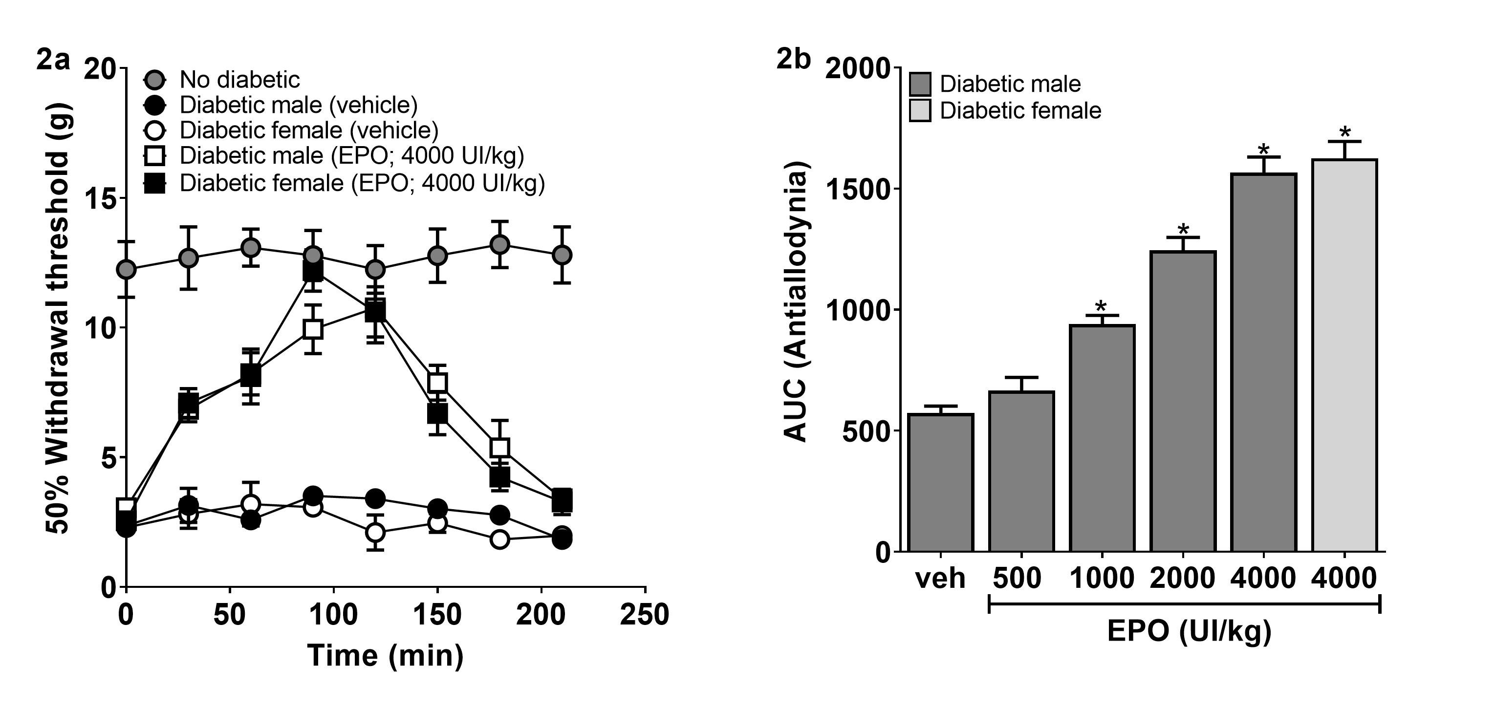
The streptozotocin administration resulted in hyperglycemia within 3-4 days. Blood glucose levels in these rats were 69.3 ± 0.8 mg/dL before the streptozotocin injection and 546.9 ± 9.5 mg/dL 4 weeks after receiving streptozotocin. In addition, diabetic rats produced tactile allodynia 4 weeks after receiving streptozotocin compared with rats who only received distilled water (Figure 2a). On this condition, intraperitoneal administration of EPO (4000 UI/kg), but not the vehicle, increased the withdrawal threshold similarly in male and female diabetic rats (Figure 2a). Furthermore, EPO significantly (p ≤ 0.05) and dose-dependently (1000, 2000 and 4000 UI/kg) reduced streptozotocin-induced tactile allodynia (Figure 2b).

Time course antiallodynic effect produced by a systemic administration of EPO (4000 UI/kg, i.p.) in diabetic rats, male and female (a). Bars show the dose-response curve antiallodynic of EPO (b). In these plots, data are expressed as the area under the curve for the 50% withdrawal threshold against time (AUC). Data are expressed as mean ± S.E.M. for 6 animals per group. *Significantly different from the vehicle group (p ≤ 0.05), as determined by one-way ANOVA followed by Tukey’s test.
Serum concentrations of NF-κB in antiallodynic effect of EPO
The intraperitoneal injection of the most effective dose of EPO (4000 UI/kg) significantly decreased serum levels of NF-κB in diabetic rats compared to the control group from minute 30 to 180 (Figure 3a). Consistently, the antiallodynia caused by EPO was observed at the same times when serum levels of NF-κB decreased (Figure 3b).

Time course of serum levels of NF-κB during antiallodynic effect of EPO (a). Serum levels of NF-κB were congruent with antiallodynic activity of EPO obtained prior from each blood samples collected (b). *p ≤ 0.05 and #p ≤ 0.05 versus Diabetic (vehicle) group, as determined by one-way ANOVA followed by Tukey’s test.
Effect of L-NAME and glibenclamide on EPO-induced antiallodynia
The systemic pre-treatment with L-NAME significantly prevented the antiallodynic effect of EPO (1 mg/kg, Figure 4a), while the systemic pre-treatment with D-NAME did not prevent the antiallodynic effect of EPO in diabetic rats (1 mg/kg, Figure 4b). As well as intraperitoneal injection glibenclamide reverted the antiallodynia of EPO in diabetic rats (1-10 mg/kg, Figure 4c). The administration of L-NAME, D-NAME and glibenclamide per se did not affect the streptozotocin-induced allodynia (Figure 4a-c).

Effect of systemic pretreatment with inhibitor of oxide nitric synthase, L-NAME (a); inactive isoform of the selective inhibitor of L-NAME, D-NAME (b); and the blocker of ATP sensitive K+ channels, glibenclamide (GLB) (c) on antiallodynic effect induced by EPO in diabetic rats. Data are expressed as the area under the curve for the 50% withdrawal threshold against time (AUC). Bars are the mean ± S.E.M. of six animals. $ p ≤ 0.05 versus ND (no diabetic) group, * p ≤ 0.05 versus D (Diabetic) group and # p ≤ 0.05 versus EPO, as determined by one-way ANOVA followed by Tukey’s test.
Effect of methiothepin and naloxone on EPO-induced anti-allodynic effects
The systemic pre-treatment with methiothepin significantly prevented EPO-induced antiallodynia in diabetic rats (0.1 mg/kg, Figure 5a), while naloxone did not prevent this effect of EPO (1 mg/kg, Figure 5b). Moreover, intraperitoneal administration of methiothepin and naloxone per se did not modify streptozotocin-induced allodynia (Figure 5a-b).

Effect of systemic pretreatment with non-selective 5-HT antagonist receptor, methiothepin (MTP) (a); and the non-selective antagonist of opioid receptors, naloxone (NLX) (b) on antiallodynic effect of EPO in diabetic rats. Data are expressed as the area under the curve of the 50% withdrawal threshold against time (AUC). Bars are the mean ± S.E.M. of six animals. $ p ≤ 0.05 versus ND (no diabetic) group, * p ≤ 0.05 versus D (Diabetic) group and # p ≤ 0.05 versus EPO, as determined by one-way ANOVA followed by Tukey’s test.
DISCUSSION
In this study, the systemic administration of EPO reduced tactile allodynia in diabetic rats dose-dependent manner. Our data agree with Bianchi et al. (2004) showing that chronic intraperitoneal administration of EPO (40 μg/kg three times a week) reverses mechanical and thermal nociception in diabetic rats. Moreover, it has been reported that therapy using EPO reduces mechanical allodynia and thermal hyperalgesia in models of neuropathic pain caused by chronic constriction injury (Campana et al. 2006, Huang et al. 2018) and L5 spinal nerve transection (Jia et al. 2009). Likewise, our results suggest that gender is not an important factor in the antiallodynic effect of EPO. In contrast, gender differences of EPO effects have been previously observed in cisplatin-induced nephrotoxicity where the effect was stronger in male rats than in female rats (Eshraghi-Jazi et al. 2013), while after neonatal stroke the neuroprotective effect of EPO was greater in female rats than in male rats (Wen et al. 2006). These studies suggest estrogen as possible factor responsible in the difference responses to EPO according to gender. Our results could differ from other studies due to the variables measured, sites of injections, doses and/or evaluating other animal species. All in all, data suggest that systemic EPO exerts antiallodynic effect in diabetic rats, without showing different effect due to gender.
The mechanism that causes antiallodynia in diabetic rats after receiving EPO is still unknown. However, considering the pleiotropic effect of EPO in different pathological conditions, we decided to assess the role of NF-κB, the nitric oxide-ATP-sensitive K+ channel pathway, as well as the serotoninergic and opioidergic systems in this EPO effect. In this sense, we found significantly decreased serum levels of NF-κB during the antiallodynic effect of the most effective dose of EPO. It has been reported that the systemic administration of EPO decreased pro-inflammatory cytokines (TNF‐α, IL‐1β, and IL‐6) via inhibiting glial activation and the NF-κB signaling pathway in models of neuropathic pain (Jia et al. 2009, Huang et al. 2018, Campana et al. 2006). Our data suggest that the effect of EPO reducing tactile allodynia in diabetic rats, may be due to inhibiting the activation of NF-κB (Figure 6 a, b). In this sense, the activation of the p50 subunit of NF-κB by EPO is implicated in erythropoiesis, as well as in neuronal development and survival (Gutierrez & Davies 2011). On the contrary, the anti-inflammatory effect of EPO is due to the inhibition of the activation of the p65 subunit of NF-κB (Nairz et al. 2011). This is consistent with the fact that in diabetic neuropathy undergoes an inflammatory process through NF-κB activation from glucose-induced pathways such as protein kinase C (PKC), advanced glycation end products (AGEs) and increased of oxidative stress (Hosseini & Abdollahi 2013) (Figure 6a).

Diagram representing the action mechanism of EPO on painful diabetic neuropathy. Peripheral sensitization (a), central sensitization (b) and descending serotoninergic pain modulation (c). The inhibition of NF-κB activation, activation of the nitric oxide-ATP-sensitive K+ channel pathway, and 5-HT1/5 receptors activation, reverted tactile allodynia in streptozotocin-induced diabetic rats. Red: inhibition; Green: activation; Blue: antiallodynic activity of EPO. Abbreviations: EPO, erythropoietin; F-6-P, fructose 6-phosphate; G-3-P, glyceraldehyde 3-phosphate; PKC, protein kinase C; AGEs, advanced glycation end-products; NF-κB, nuclear factor-κB; IL, interleukin; TNF-α, tumor necrosis factor alpha; COX-2, cyclooxygenase-2; NO, nitric oxide; nNOS, neuronal nitric oxide synthase; cGMP, cyclic guanosine monophosphate; ROS, reactive oxygen species; SP, substance P; PKA, protein kinase C; IP3, inositol 1, 4, 5-trisphosphate; NMDA, N-methyl-D-aspartic acid; AMPA, α-amino-3-hydroxy-5-methyl-4-isoxazole propionate; NK-1, neurokinin 1; PGE2, prostaglandin E2; cAMP, cyclic adenine monophosphate; 5-HT1/2/3/5/7, subtype of serotonin receptors.
On the other hand, a decrease of neuronal nitric oxide synthase (nNOS) expression and consequently a decrease in the activity of nitric oxide-GMPc-ATP-sensitive K+ channel pathway has been reported in DRG and nerve of rats with painful diabetic neuropathy (Araiza-Saldaña et al. 2005, Zochodne et al. 2000) (Figure 6a). In this sense, EPO enhanced nitric oxide production in wound healing during flap surgery (Sorg et al. 2013) as well as in cytoprotective effect against renal injury by ischemia/reperfusion (Elshiekh et al. 2017) and hemorrhagic shock (Ranjbaran et al. 2017). Moreover, EPO was associated with the opening of ATP-sensitive K+ channels in cultured tubular renal cells induced by hypoxia (Yilmaz et al. 2015) and renal damage induced by ischemia/reperfusion (Yazihan et al. 2008). We suggest that the nitric oxide-ATP-sensitive K+ channel pathway could be associated to the antiallodynic effect of this drug. Since we found that the non-selective nitric oxide synthesis inhibitor L-NAME prevented EPO-induced antiallodynia. Likewise, the ATP-sensitive K+ channels blocker glibenclamide, blocked EPO effect. Therefore, we hypothesized that EPO could modulate nitric oxide and opening ATP-sensitive K+ channels to hyperpolarize neurons, and so produce antiallodynia (Figure 6a, b). Nitric oxide may have a dual effect (pronociceptive or antinociceptive) dependent not only nitric oxide levels, but on other factors such as of the experimental model, dose, and administration route.
In the present study we also found that the administration of methiothepin, serotonin receptors antagonist with moderate affinity to 5HT1/2/5/7 receptor subtypes, significantly blocked EPO-induced antiallodynia; these results suggest that the serotoninergic system is implicated in EPO-induced antiallodynia in diabetic rats (Figure 6c). Evidence exists that activation of 5-HT1/5 receptors produce antinociceptive effects (Granados-Soto et al. 2010), whereas the activation of 5-HT2/7 receptors tends to promote nociception (Pineda-Farias et al. 2015). Thus, it could be assumed that EPO reduces the allodynia in diabetic rats by activation of spinal 5-HT1/5 receptors. In contrast to previous antagonist, naloxone did not modify antiallodynic effect of EPO in diabetic rats, this could be to an down-regulation or desensitization of opioid receptors as previously suggested in other neuropathic pain models (Porreca et al. 1998); our data suggest that antiallodynic effect of EPO is independent of opioid receptors.
Recent studies indicate that EPO could be an effective therapy to treat pain, due to its neuroprotective mechanisms. In this sense, 1) EPO increases glutathione levels and decreases levels of reactive oxygen species in Schwann cells isolated from the sciatic nerve of diabetic rats (Yu et al. 2014); 2) EPO increases the expression of superoxide dismutase, glutathione peroxidase and catalase in retinal ganglion cells exposed to high glucose (Wang et al. 2015); 3) EPO increases the expression of glutamate transporter-1 (GLT-1) and glutamate aspartate transporter (GLAST) in astrocytes during ischemia/reperfusion cerebral injury (Yu et al. 2016). Therefore, these mechanisms into an antiallodynic effect of EPO in diabetic rats should be investigated in the future.
CONCLUSIONS
Our results indicate that the systemic administration of EPO is able to reverse allodynia in diabetic rats regardless of the genders. Since EPO decreased the serum levels of NF-κB, and L-NAME, glibenclamide, and methiothepin blocked the antiallodynic activity of EPO; these data suggest the combination of NF-κB inhibition, the activation of the nitric oxide-ATP-sensitive K+ channel pathway and the activation of 5-HT1/5 receptors, could be in together the mechanism of action antiallodynic of EPO.
ACKNOWLEDGMENTS
Samuel Suarez-Mendez is a CONACyT fellow (fellowship 744091) and this study is part of his Ph.D. dissertation. This research did not receive any specific grant from funding agencies in the public, commercial, or not-for-profit sectors. The authors declare no potential conflicts of interest.
REFERENCES
- ARAIZA-SALDAÑA CI, REYES-GARCIA G, BERMUDEZ-OCANA DY, PEREZ-SEVERIANO F & GRANADOS-SOTO V. 2005. Effect of diabetes on the mechanisms of intrathecal antinociception of sildenafil in rats. Eur J Pharmacol 527: 60-70.
- BALLOT O, JOSEPH V & SOLIZ J. 2015. Endogenous brain erythropoietin is a potent sex-specific respiratory stimulant in adult and newborn mice. J Appl Physiol 118: 1386-1395.
- BIANCHI R ET AL. 2004. Erythropoietin both protects from and reverses experimental diabetic neuropathy. Proc Natl Acad Sci U S A 101: 823-828.
- BOULTON AJ 2012. Diabetic neuropathy: is pain God’s greatest gift to mankind? Semin Vasc Surg 25: 61-65.
- CAMPANA WM, LI X, SHUBAYEV VI, ANGERT M, CAI K & MYERS RR. 2006. Erythropoietin reduces Schwann cell TNF-alpha, Wallerian degeneration and pain-related behaviors after peripheral nerve injury. Eur J Neurosci 23: 617-626.
- CAMPANA WM & MYERS RR. 2003. Exogenous erythropoietin protects against dorsal root ganglion apoptosis and pain following peripheral nerve injury. Eur J Neurosci 18: 1497-1506.
- CHAPLAN SR, BACH FW, POGREL JW, CHUNG JM & YAKSH TL. 1994. Quantitative assessment of tactile allodynia in the rat paw. J Neurosci Methods 53: 55-63.
- DIXON WJ. 1980. Efficient analysis of experimental observations. Annu Rev Pharmacol Toxicol 20: 441-462.
- ELSHIEKH M, KADKHODAEE M, SEIFI B, RANJBARAN M & ASKARI H. 2017. Up-regulation of nitric oxide synthases by erythropoietin alone or in conjunction with ischemic preconditioning in ischemia reperfusion injury of rat kidneys. Gen Physiol Biophys 36: 281-288.
- EREN Z ET AL. 2016. Pleiotropic and Renoprotective Effects of Erythropoietin Beta on Experimental Diabetic Nephropathy Model. Nephron 132: 292-300.
- ESHRAGHI-JAZI F, NEMATBAKHSH M, PEZESHKI Z, NASRI H, TALEBI A, SAFARI T, MANSOURI A, MAZAHERI S & ASHRAFI F. 2013. Sex differences in protective effect of recombinant human erythropoietin against cisplatin-induced nephrotoxicity in rats. Iran J Kidney Dis 7: 383-389.
- FUKAO M, MASON HS, BRITTON FC, KENYON JL, HOROWITZ B & KEEF KD. 1999. Cyclic GMP-dependent protein kinase activates cloned BKCa channels expressed in mammalian cells by direct phosphorylation at serine 1072. J Biol Chem 274: 10927-10935.
- GRANADOS-SOTO V, ARGUELLES CF, ROCHA-GONZALEZ HI, GODINEZ-CHAPARRO B, FLORES-MURRIETA FJ & VILLALON CM. 2010. The role of peripheral 5-HT1A, 5-HT1B, 5-HT1D, 5-HT1E and 5-HT1F serotonergic receptors in the reduction of nociception in rats. Neuroscience 165: 561-568.
- GUTIERREZ H & DAVIES AM. 2011. Regulation of neural process growth, elaboration and structural plasticity by NF-kappaB. Trends Neurosci 34: 316-325.
- HOSSEINI A & ABDOLLAHI M. 2013. Diabetic neuropathy and oxidative stress: therapeutic perspectives. Oxid Med Cell Longev 2013: 168039.
- HUANG CT, CHEN SH, LIN SC, CHEN WT, LUE JH & TSAI YJ. 2018. Erythropoietin reduces nerve demyelination, neuropathic pain behavior and microglial MAPKs activation through erythropoietin receptors on Schwann cells in a rat model of peripheral neuropathy. Glia 66: 2299-2315.
- JAVED S, ALAM U & MALIK RA. 2015. Treating Diabetic Neuropathy: Present Strategies and Emerging Solutions. Rev Diabet Stud 12: 63-83.
- JIA H, FENG X, LI W, HU Y, ZENG Q, LIU J & XU J. 2009. Recombinant human erythropoietin attenuates spinal neuroimmune activation of neuropathic pain in rats. Ann Clin Lab Sci 39: 84-91.
- JUÁREZ-ROJOP IE, MORALES-HERNÁNDEZ PE, TOVILLA-ZÁRATE CA, BERMÚDEZ-OCAÑA DY, TORRES-LOPEZ JE, BLE-CASTILLO JL, DÍAZ-ZAGOYA JC & GRANADOS-SOTO V. 2015. Celecoxib reduces hyperalgesia and tactile allodynia in diabetic rats. Pharmacol Rep 67: 545-552.
- NAIRZ M ET AL. 2011. Erythropoietin contrastingly affects bacterial infection and experimental colitis by inhibiting nuclear factor-kappaB-inducible immune pathways. Immunity 34: 61-74.
- PINEDA-FARIAS JB, VELAZQUEZ-LAGUNAS I, BARRAGAN-IGLESIAS P, CERVANTES-DURAN C & GRANADOS-SOTO V. 2015. 5-HT2B Receptor Antagonists Reduce Nerve Injury-Induced Tactile Allodynia and Expression of 5-HT2B Receptors. Drug Dev Res 76: 31-39.
- PORRECA F, TANG QB, BIAN D, RIEDL M, ELDE R & LAI J. 1998. Spinal opioid mu receptor expression in lumbar spinal cord of rats following nerve injury. Brain Res 795: 197-203.
- QUIÑONEZ-BASTIDAS GN, CERVANTES-DURÁN C, ROCHA-GONZÁLEZ HI, MURBARTIÁN J & GRANADOS-SOTO V. 2013. Analysis of the mechanisms underlying the antinociceptive effect of epicatechin in diabetic rats. Life Sci 93: 637-645.
- RANJBARAN M, KADKHODAEE M, SEIFI B, ADELIPOUR M & AZARIAN B. 2017. Erythropoietin attenuates experimental haemorrhagic shock-induced renal damage through an iNOS- dependent mechanism in male Wistar rats. Injury 48: 262-269.
- SORG H, HARDER Y, KRUEGER C, REIMERS K & VOGT PM. 2013. The nonhematopoietic effects of erythropoietin in skin regeneration and repair: from basic research to clinical use. Med Res Rev 33: 637-664.
- SURYAVANSHI SV & KULKARNI YA. 2017. NF-κβ: A Potential Target in the Management of Vascular Complications of Diabetes. Front Pharmacol 8: 798.
- TORRES-LÓPEZ JE, JUÁREZ-ROJOP IE, GRANADOS-SOTO V, DIAZ-ZAGOYA JC, FLORES-MURRIETA FJ, ORTÍZ-LÓPEZ JU & CRUZ-VERA J. 2007. Peripheral participation of cholecystokinin in the morphine-induced peripheral antinociceptive effect in non-diabetic and diabetic rats. Neuropharmacology 52: 788-795.
- WANG L, DI L & NOGUCHI CT. 2014. Erythropoietin, a novel versatile player regulating energy metabolism beyond the erythroid system. Int J Biol Sci 10: 921-939.
- WANG Y, ZHANG H, LIU Y, LI P, CAO Z & CAO Y. 2015. Erythropoietin (EPO) protects against high glucose-induced apoptosis in retinal ganglional cells. Cell Biochem Biophys 71: 749-755.
- WEN TC, ROGIDO M, PENG H, GENETTA T, MOORE J & SOLA A. 2006. Gender differences in long-term beneficial effects of erythropoietin given after neonatal stroke in postnatal day-7 rats. Neuroscience 139: 803-811.
- YAZIHAN N, ATAOGLU H, KAVAS GO, AKYUREK N, YENER B & AYDM C. 2008. The effect of K-ATP channel blockage during erythropoietin treatment in renal ischemia-reperfusion injury. J Invest Surg 21: 340-347.
- YILMAZ TU, YAZIHAN N, DALGIC A, KAYA EE, SALMAN B, KOCAK M & AKCIL E. 2015. Role of ATP-dependent K channels in the effects of erythropoietin in renal ischaemia injury. Indian J Med Res 141: 807-815.
- YU D, FAN Y, SUN X, YAO L & CHAI W. 2016. Effects of erythropoietin preconditioning on rat cerebral ischemia-reperfusion injury and the GLT-1/GLAST pathway. Exp Ther Med 11: 513-518.
- YU T, LI L, BI Y, LIU Z, LIU H & LI Z. 2014. Erythropoietin attenuates oxidative stress and apoptosis in Schwann cells isolated from streptozotocin-induced diabetic rats. J Pharm Pharmacol 66: 1150-1160.
- ZHANG Y, ROGERS HM, ZHANG X & NOGUCHI CT. 2017. Sex difference in mouse metabolic response to erythropoietin. FASEB J 31: 2661-2673.
- ZIMMERMANN M. 1983. Ethical guidelines for investigations of experimental pain in conscious animals. Pain 16: 109-110.
- ZOCHODNE DW, VERGE VM, CHENG C, HOKE A, JOLLEY C, THOMSEN K, RUBIN I & LAURITZEN M. 2000. Nitric oxide synthase activity and expression in experimental diabetic neuropathy. J Neuropathol Exp Neurol 59: 798-807.