Validation of cervical cancer genetic signature in minimally invasive samples
Abstract
Cervical cancer remains the leading cause of cancer death in 36 countries, and high-risk human papillomavirus types are responsible for most cases. Identifying strategies to make treatment more targeted and effective has become a priority. This study aims to validate a set of differentially expressed genes previously identified in cervical cancer stem cells as predictive biomarkers for response to chemoradiotherapy using minimally invasive samples. Additionally, it aims to elucidate the relationship between high-risk human papillomavirus infection and cervical cancer patients’ response to treatment. Gene expression for three differentially expressed genes (COPZ1, ILF2, and SNX2) was evaluated from 20 cervical cancer patients’ cervical cytology brushes. Unmapped reads from the same transcriptome were used to evaluate the presence of human papillomavirus in tumor tissue through qualitative screening of 13 high-risk human papillomavirus types. Our study did not clarify the relationship between high-risk human papillomavirus infection and the treatment response. However, we found downregulation of COPZ1 in patients who responded to treatment compared to non-responders, and ILF2 in patients with more advanced tumor stages. This suggests that COPZ1 and ILF2 expressions are potential cervical cancer prognostic biomarkers that can be assessed using samples commonly used in clinical practice.
Key words
cervical cancer; gene expression profiling; human papillomavirus; tumor biomarker
INTRODUCTION
Cervical Cancer (CC) remains the leading cause of cancer death in 36 countries around the world (Sung et al. 2021). With the significant influence of the Human Development Index (HDI) and poverty rates, women living in areas of lower socioeconomic status are the most affected (Sung et al. 2021, Singh et al. 2012). The estimated risk of CC for the triennium 2023-2025 in Brazil is 13.25/cases for every 100,000 women (Santos et al. 2023). In addition, in 2020, the CC was responsible for 4.60 deaths per 100,000 women affected by the disease in the country (Santos 2020).
Worldwide, Human Papillomavirus (HPV) is responsible for most cases (Schiffman et al. 2016). HPV is a double-stranded circular DNA virus with high specificity that infects cutaneous and mucosal epithelial tissues, causing infections that can last long periods (Szymonowicz & Chen 2020, Chen et al. 2020). Due to its great clinical importance, it has been extensively studied, and the strains described so far have already had their genetic characteristics identified through next generation sequencing (NGS) (van Doorslaer 2013). They are classified into low-risk HPV (LR-HPV) - which causes the appearance of genital warts but does not cause cancer (HPV 6 and 11) - and high-risk HPV (HR-HPV) - which can generate cellular changes favoring the development of cancer (HPV 16, 18, 33) (Farmer et al. 2021).
Cervical carcinogenesis begins with the infection of HR-HPV. Later, with the persistence of the virus, there may be progression to precancerous lesions and invasive cancer (Petca et al. 2020). However, despite the pathogenic effect of HR-HPV being well established in CC, the prognosis related to the infection positivity in patients with this neoplasm is still unclear, since there is a heterogeneity in the results of the relationship between HPV status and their different genotypes with disease prognosis (Chen et al. 2020). Previous studies have demonstrated that HPV-negative CC patients have poor progression-free and global survival results compared with HPV-positive ones. In contrast, other studies showed that patients with some specific HPV genotypes had worse progression-free and global survival after chemoradiotherapy treatment than HPV 16-positive CC patients (Ruiz et al. 2021).
In Brazil, the public health system has a triple-intervention strategy involving prevention (HPV vaccination program), screening (Pap smears), and treatment (surgery, and chemoradiotherapy) of CC. However, it has not efficiently reduced CC mortality rates in the country. Identifying strategies to make cancer treatment more targeted and effective for patients reducing mortality rates has become a worldwide priority. In this context, the use of differentially expressed genes (DEGs) as predictive and prognosis tumor biomarkers have been suggested (Rodrigues et al. 2022). In this context, this study aims to validate a set of DEGs, previously identified in CC tumor biopsies stem cells (CCSC), as predictive biomarkers of response to chemoradiotherapy treatment using minimally-invasive samples. Furthermore, we aim to elucidate the relationship between HR-HPV infection and CC patients’ response to treatment.
MATERIALS AND METHODS
Study population
Samples from 20 women who underwent treatment for cervical cancer at the Instituto Mário Penna (Belo Horizonte, Brazil) between August 2017 and May 2019 were used in this study. A non-probabilistic convenience sampling method was applied, with the following inclusion criteria: diagnosis of cervical cancer (either squamous cell carcinoma or adenocarcinoma) and no prior history of other neoplasms. The samples were collected using a cytological brush at the first medical appointment. In addition, four volunteers without the disease (confirmed by Pap smear) were included as controls and their cervical cytological smears were collected. Patients were treated with chemoradiotherapy and classified into two groups, Responders (patients with undetected cervical lesions) and Non-responders (patients with tumor progression, partial response or stable disease) after 8 months of treatment, according to the protocols of the Onco-Gynecological department; patients’ outcomes at 12 months post-treatment were also evaluated. The study was approved by the local Research Ethics Committee (CAEE: 41114915.5.0000.5121), and signed informed consent was obtained from all participants.
Nucleic Acids extraction
Due to the high prevalence of vaginal bleeding in women with this tumor type, the Gentra Puregene® blood kit (Qiagen, Hilden, Germany) was used to lyse blood cells before acid nucleic extraction. Subsequently, samples were divided into two tubes. In the first tube, Gentra Puregene® protocol was followed to extract DNA, and in the second tube, TRIzol™ reagent (Invitrogen, Waltham, Massachusetts, USA) was added to isolate RNA, following the manufacturer’s instructions. Samples quantification was performed using the Nanovue™ spectrophotometer (GE Healthcare, Chicago, Illinois, USA).
cDNA synthesis
An input of 100 ng of RNA was used for the cDNA synthesis reaction. Reverse transcription (RT) was carried out using the SuperScript™ IV VILO MasterMix with ezDNase™ (Invitrogen, Waltham, Massachusetts, USA) following the manufacturers’ guidelines. No-RT controls were performed without RT enzyme. Finally, cDNAs and no-RT controls were stored at -20°C for later use.
qPCR for Gene Expression Analysis
TaqPath™ qPCR Master Mix (Applied Biosystems, Waltham, Massachusetts, USA) was used for the relative expression assay of COPZ1, ILF2, and SNX2 genes (Table SI – Supplementary Material) using TaqMan™ Assays according to manufacturer’s procedures on the 7500 Real-Time PCR System (Applied Biosystems) instrument. These genes were DEGs associated with the response to chemoradiotherapy in CCSC, as initially identified in the transcriptomic analysis conducted by Zuccherato et al. (2021). The TATA-Box Binding Protein (TBP) gene was used as endogenous control. The four neoplasm-free cervical cytology brush samples were pooled for testing as an internal control. Quantification data were analyzed using Applied Biosystems™ Analysis Software, using the 2-∆∆Ct relative quantification method.
Transcriptome bioinformatic analysis
Transcriptome reads unmapped to the human genome (GRCh38) were used to evaluate the presence of HPV virus sequences in the CC patient’s tumor stem cells. To prevent low-quality or multiple regions mapping sequences, only reads with a range size from 40 to 100 nucleotides, a phred score ≥ 30, and without highly repetitive regions were retained in the multi-fasta file. Only sequences identified exclusively for one single HPV type were considered valid.
HR-HPV Screening
13 high-risk HPV real-time PCR kit (Hybribio, Hong Kong, China) was used for qualitative screening of the types 16, 18, 31, 33, 35, 39, 45, 51, 52, 56, 58, 59, and 68. The in vitro detection was performed using Taqman® Technology to DNA. The assay utilized a positive control (non-infectious plasmid DNA), negative control, and cellular internal control provided by the manufacturer. Samples with a Ct value between 15 and 40 were considered positive results.
Statistical analyses
Statistical analyses were conducted using GraphPad Prism 8.0.2 software. Clinicopathological data were evaluated using Fisher’s Exact Test to investigate potential confounding factors between the study groups. The Kaplan-Meier method was employed to assess overall patient survival and progression-free survival, with comparisons performed using Log-rank tests. The non-parametric Mann-Whitney or parametric t-test was used to evaluate the relative quantification of gene expression. Furthermore, Spearman’s non-parametric test was applied to analyze the correlation between two variables. P value <0.05 was considered statistically significant.
RESULTS
Clinicopathological features and survival outcomes of patients
The clinicopathological characteristics of the patients are summarized in Table I. Considering the patient’s follow-up eight months after chemoradiotherapy treatment, nine subjects (45%) were classified as Non-responders (NR), and 11 subjects (55%) were classified as Responders (R). Age categories were defined based on the average age of the study cohort, calculated at 48.35 years (range 24 to 81). No statistical differences were observed in the clinicopathological characteristics between patients who responded to chemoradiotherapy treatment and those who did not. Patients’ outcomes at 12 months post-treatment were evaluated in Kaplan-Meier curves (Figure 1). R patients presented higher overall survival rates compared to NR patients (Hazard Ratio [HR]= 0.1014, 95% CI of ratio 0.02201 to 0.4672, p=0.0074**) (Figure 1a). Furthermore, the Progression-free Survival test was able to stratify R and NR patients with statistical significance (p value=p=0.0002***) (HR= 0.07005, 95% CI of ratio 0.01962 to 0.2501) (Figure 1b).

The outcomes at 12 months post-treatment in cohort patients. a) Kaplan-Meier Curve indicating the survival percentage in R and NR patients. Long-rank (Mantel-Cox) test was used to determine statistical significance (HR= 0.1014, 95% CI of ratio 0.02201 to 0.4672, p value=0.0074); b) Progression-free Survival stratified R and NR patients. A Long-rank (Mantel-Cox) test was used to determine statistical significance (HR= 0.07005, 95% CI of ratio 0.01962 to 0.2501, p value=0.0002). PFS, Progression-free survival.
Gene expression analysis
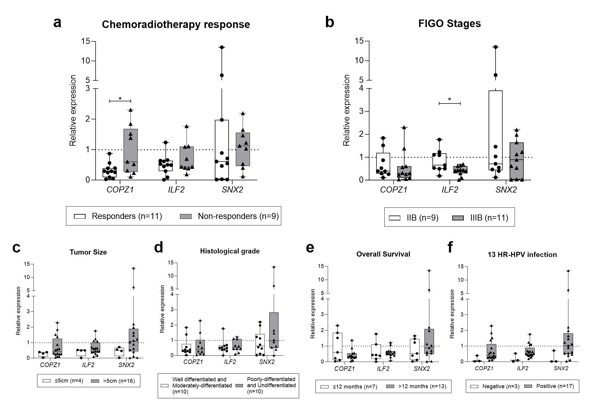
The relative expressions of COPZ1, ILF2, and SNX2 were examined as potential biomarkers for CC in a minimally invasive sample using a cytological brush collected from the patient cohort. The results revealed the relative quantification means (RQ) of COPZ1, ILF2, and SNX2 gene expressions as 0.6129 (range 0.025-2.303), 0.5995 (range 0.01-1.768), and 1.743 (range 0.015-13.53), respectively. A moderate negative Spearman correlation was observed between COPZ1-ILF2 (0.501) and SNX2-ILF2 (0.568) with statistical significance (p-value=0.025* and 0.009**, respectively). These targets exhibited underexpression (RQ≤1,0) in the majority of samples (Figure 2). Statistical analysis revealed a significant difference in COPZ1 expression levels between NR and R patients (p=0.0168*) (Figure 2a), as well as in ILF2 expression levels between patients at FIGO stages IIB and IIIB (p=0.0159*) (Figure 2b). SNX2 expression was not significant for any of the clinical parameters evaluated. Regarding tumor size (≤5 cm or >5 cm) (Figure 2c), the tumor histological grade (well-differentiated, moderately-differentiated, poorly-differentiated or undifferentiated) (Figure 2d) and patient survival (Figure 2e), no statistically significant differences were observed for these targets’ expression. Although the patients with negative HR-HPV exhibited underexpression of all evaluated genes, the differences did not reach statistical significance between groups (Figure 2f).

The relationship between the expression of COPZ1, ILF2, and SNX2 genes and clinicopathological characteristics of CC patients. The TBP gene and the expression level of the calibrator were used as data normalizers, with the RQ value equal to 1. a) COPZ1 gene expression between non-responders (n=9) and responders (n=11); b) Differential expression of ILF2 between patients at FIGO stages IIB (n=9) and IIIB (n=11); c) Assessment of gene expression among patients with tumor size less than or equal to 5 cm or greater than 5 cm at the time of diagnosis; d) Genes expression profile in patients with histological grades well differentiated with moderately-differentiated and poorly-differentiated with undifferentiated; e) Gene expression in groups with survival less than 12 months (n=7) and survival greater than 12 months (n=13); f) Expression of targets related to the negative (n=3) and positive (n=17) groups for detection of 13 HR-HPV. Specifically, the significant downregulation of COPZ1 (p=0.0168) has been highlighted in treatment responders compared to non-responders, while differential expression of ILF2 (p=0.0159) was observed between patients at FIGO stages IIB and IIIB.
HR-HPV status in CC stem cells, and tumor bulk
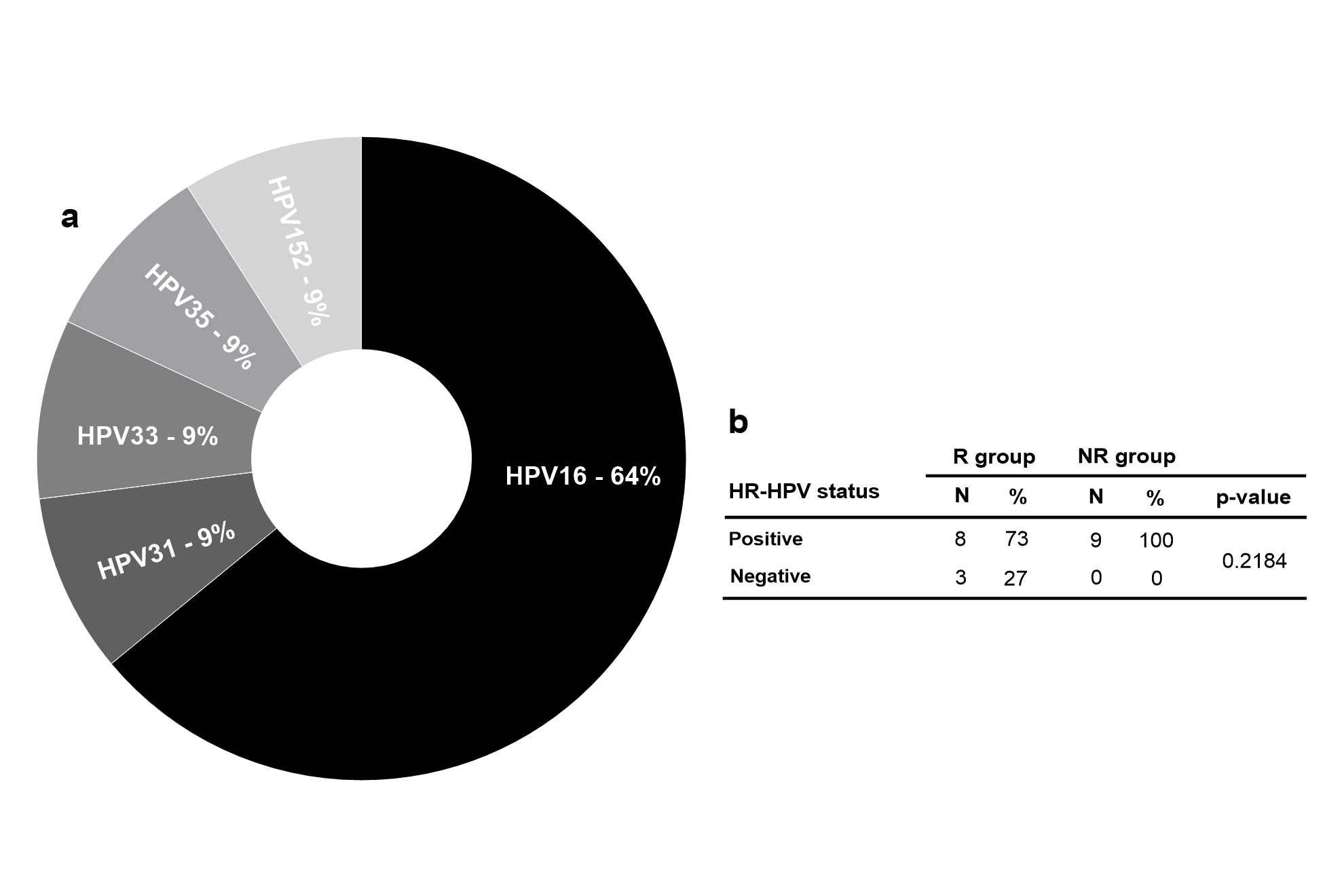
The RNA-seq results from 11 patients were analyzed to identify HPV sequences in CC stem cells. Unmapped sequences in the human genome (GRCh38) were aligned with the HPV genome. The multi-fasta file containing 220 HPV genomes (https://pave.niaid.nih.gov/) was obtained, and only the uniquely mapped reads were tallied. The most prevalent HPV types identified were 16, 31, 33, 35 and 152 (Figure 3a). In parallel, the detection of 13 types of HR-HPVs in 20 patient samples revealed a positivity rate of 73% in the responder (R) group and 100% in the non-responder (NR) group. All negative results for HR-HPVs (27%) were observed in the R group. No significant association was found between HR-HPV presence and treatment response (p=0.2184) (Figure 3b). One of these patients, with a negative result in the qPCR test, presented a single HPV31 sequence in the RNA-seq data.

HPV prevalence in the patient cohort. a) Most prevalent HPV types identified in RNA-seq results performed from CC stem cells (n=11); b) The HR-HPV detection results obtained through qPCR using DNA extracted from cytological brushes of R and NR patients. Fisher test was performed to evaluate statistical significance (p=0.2184). R, Responders; NR, Non-responders.
DISCUSSION
Globally, CC is the fourth most prevalent cancer among women. Despite prevention with HPV vaccination and screening initiatives to detect pre-malignant and early-stage cancers (e.g., Pap smears programs), the majority of CC-diagnosed patients are at intermediate to advanced stages, comprising 35% in stage II, 44% in stage III, and 8% in stage IV. The late diagnosis of CC is associated with a poor prognosis and chemoradiation and surgical treatment refractories cases (Hill 2020, Gopu et al. 2021). Therefore, further improving care for women with this disease is necessary. An example of this is recognizing the associated molecular agents in CC and transforming this into a more effective disease-management tool (Adiga et al. 2021).
This study used cytological brush samples to validate three DEGs (COPZ1, ILF2, and SNX2) previously identified in the CCSC transcriptome analysis from the same tumor tissue samples. Zuccherato et al. (2021) selected 21 genes as differentially expressed transcripts in CCSC, including these targets. We selected them as validators due to the higher partial area under the ROC curve (pAUC, 0.1) values found when grouping patients by treatment status in R and NR. Then, we considered these genes due to their potential as biomarkers for assessing the response to chemoradiotherapy treatment in patients with CC. Our data revealed COPZ1 downregulated in patients who responded to treatment compared to NR, indicating an association between the differential expression of COPZ1 and the response to chemoradiotherapy.
COPZ1 is a constituent of the coatomer protein complex I and plays a role in intracellular trafficking, endosome maturation, lipid homeostasis, and autophagy (Zhang et al. 2021). The knockdown of this gene has demonstrated the ability to prevent proliferation, induce autophagy, and stimulate apoptosis in various cancer cells, including those of the breast, thyroid, and glioblastoma (Zhang et al. 2021, Chen et al. 2022, Anania at al. 2020). Corroborating our results, the upregulation of this gene has been linked to a poor prognosis and the progression of glioblastoma, demonstrating possible potential as a predictive biomarker.
Although ILF2 expression showed no significant difference between the R and NR groups, it distinguished the FIGO stage of CC, with the more advanced stage exhibiting lower gene expression. ILF2 regulates gene expression at different levels, including RNA transcription, processing, and translation. Additionally, it is involved in processes such as the replication of various types of RNA viruses, and like COPZ1, it has been studied as a prognostic biomarker and therapeutic target in different types of tumors (Yin et al. 2017). In contrast to our findings, ILF2 upregulation has been associated with negative outcomes in patients with breast, gastric, liver, lung, and CC, being related to increased tumor cell proliferative rate, inhibition of apoptosis, greater depth of invasion, more advanced pathological stage, histological differentiation and anchorage independence (Yin et al. 2017, Du et al. 2019, Ni et al. 2015, Jin et al. 2018, Cheng et al. 2016, Shi et al. 2017, Li et al. 2019).
Unlike the other two DEGs, SNX2 did not show differential expression among the evaluated groups, which could be attributed to the high variability in expression levels among patients. Data related to this gene expression in cancer are still incipient (Duclos et al. 2017). SNX2 appears to influence sensitivity to anticancer drugs targeting c-Met and EGFR in lung cancer cell lines when it is silenced (Ogi et al. 2013).
In addition to validating DEGs, we utilized transcriptome data to identify HPV sequences in CCSCs, concurrently with qPCR detection of 13 HR-HPV in cytology brush samples. Despite nearly all cases of CC being attributed to HPV, the impact of the virus on CC prognosis is not yet clear (Lei et al. 2018). In this study, all the negative results obtained by the HR-HPV test were in the R group, which has lower mortality, but this was not enough to confirm the relationship between the infection and a favorable treatment response to the disease. In other more expressive cohorts, the absence of HR-HPV was highly associated with a better prognosis and a lower mortality rate in CC patients (Lei et al. 2018, 2020).
Furthermore, in our findings, one of the patients with a negative result in the qPCR test presented a HR-HPV single sequence in the transcriptome data. Typically, PCR methods for HPV detection are based on designed primers and probes to recognize the most conserved gene in the HPV genome, the L1 gene. Samples containing HPV types with nucleotide sequences differing from those of the primer/probe sequences may go undetected, potentially leading to a false negative result (Arroyo Mühr et al. 2020). Therefore, the discrepancies observed in results may be attributed to the increased sensitivity of the sequencing method compared to qPCR.
Although our study was unable to establish an association between HPV infection and response to therapy or indicate if SNX2 expression levels could be considered a prognostic biomarker for CC, it is important to highlight that these results are not conclusive. An extensive and comprehensive assessment and a larger cohort are necessary to validate these data. Despite these limitations, we demonstrated significant relationships between COPZ1 and ILF2 expression levels with staging and response to therapy. These findings suggest that these genes have potential as auxiliary diagnostic and prognostic biomarkers for CC. Finally, it is worth demonstrating that these results were obtained using a sample commonly used in clinical practice, reinforcing the translational features of these biomarkers.
SUPPLEMENTARY MATERIAL
ACKNOWLEDGMENTS
We thank all the patients for participating in the study. We are also grateful to the Agências Brasileiras de Financiamento do Ministério da Saúde (Pronon – Programa Nacional de Apoio à Atenção Oncológica; Grant number: NUP:25000.079266/2015-09) and Fundação de Amparo à Pesquisa do Estado de Minas Gerais (Programa de Apoio a Instalações Multiusuários; APQ-02564-22).
REFERENCES
- ADIGA D, ESWARAN S, PANDEY D, SHARAN K & KABEKKODU SP. 2021. Molecular landscape of recurrent cervical cancer. Crit Rev Oncol Hematol 157: 103178.
- ANANIA MC, DI MARCO T, MAZZONI M & GRECO A. 2020. Targeting Non-Oncogene Addiction: Focus on Thyroid Cancer. Cancers (Basel) 12: 129.
- ARROYO MÜHR LS, LAGHEDEN C, EKLUND C, LEI J, NORDQVIST-KLEPPE S, SPARÉN P, SUNDSTRÖM K & DILLNER J. 2020. Sequencing detects human papillomavirus in some apparently HPV-negative invasive cervical cancers. J Gen Virol 101: 265-270.
- CHEN S, LI H, CHEN S, WANG B & ZHANG K. 2022. BMI1 promotes the proliferation and inhibits autophagy of breast cancer cells by activating COPZ1. Clin Transl Oncol 24: 2166-2174.
- CHEN X ET AL. 2020. Better or Worse? The Independent Prognostic Role of HPV-16 or HPV-18 Positivity in Patients With Cervical Cancer: A Meta-Analysis and Systematic Review. Front Oncol 10: 1733.
- CHENG S ET AL. 2016. Expression and Critical Role of Interleukin Enhancer Binding Factor 2 in Hepatocellular Carcinoma. Int J Mol Sci 17: 1373.
- DU H, LE Y, SUN F, LI K & XU Y. 2019. ILF2 Directly Binds and Stabilizes CREB to Stimulate Malignant Phenotypes of Liver Cancer Cells. Anal Cell Pathol (Amst) 2019: 1575031.
- DUCLOS CM, CHAMPANHE A, CARRIER JC, SAUCIER C, LAVOIE CL & DENAULT J. 2017. Caspase-mediated proteolysis of the sorting nexin 2 disrupts retromer assembly and potentiates Met/hepatocyte growth factor receptor signaling. Cell Death Discov 3: 16100.
- FARMER E, CHENG MA, HUNG CF & WU TC. 2021. Vaccination Strategies for the Control and Treatment of HPV Infection and HPV-Associated Cancer. Recent Results Cancer Res 217: 157-195.
- GOPU P, ANTONY F, CYRIAC S, KARAKASIS K & OZA AM. 2021. Updates on systemic therapy for cervical cancer. Indian J Med Res 154: 293-302.
- HILL EK. 2020. Updates in Cervical Cancer Treatment. Clin Obstet Gynecol 63: 3-11.
- JIN Z ET AL. 2018. Interleukin enhancer binding factor 2 is a prognostic biomarker for breast cancer that also predicts neoadjuvant chemotherapy responses. Am J Transl Res 10: 1677-1689.
- LEI J, PLONER A, ELFSTRÖM KM, WANG J, ROTH A, FANG F, SUNDSTRÖM K, DILLNER J & SPARÉN P. 2020. HPV Vaccination and the Risk of Invasive Cervical Cancer. N Engl J Med 383: 1340-1348.
- LEI J ET AL. 2018. High-risk human papillomavirus status and prognosis in invasive cervical cancer: A nationwide cohort study. PLoS Med 15: 1002666.
- LI N ET AL. 2019. ILF2 promotes anchorage independence through direct regulation of PTEN. Oncol Lett 18: 1689-1696.
- NI T, MAO G, XUE Q, LIU Y, CHEN B, CUI X, LV L, JIA L, WANG Y & JI L. 2015. Upregulated expression of ILF2 in non-small cell lung cancer is associated with tumor cell proliferation and poor prognosis. J Mol Histol 46: 325-335.
- OGI S ET AL. 2013. Sorting nexin 2-mediated membrane trafficking of c-Met contributes to sensitivity of molecular-targeted drugs. Cancer Sci 104: 573-583.
- PETCA A, BORISLAVSCHI A, ZVANCA ME, PETCA RC, SANDRU F & DUMITRASCU MC. 2020. Non-sexual HPV transmission and role of vaccination for a better future (Review). Exp Ther Med 20: 186.
- RODRIGUES AN ET AL. 2022. Characteristics of patients diagnosed with cervical cancer in Brazil: preliminary results of the prospective cohort EVITA study (EVA001/LACOG 0215). Int J Gynecol Cancer 32: 141-146.
- RUIZ FJ ET AL. 2021. HPV transcript expression affects cervical cancer response to chemoradiation. JCI Insight 6: 138734.
- SANTOS MO. 2020. Estimativa/2020 – Incidência de Câncer no Brasil. Rev Bras Cancerol 66: e-00927.
- SANTOS MO, LIMA FCS, MARTINS LFL, OLIVEIRA JFP, ALMEIDA LM & CANCELA MC. 2023. Estimativa de Incidência de Câncer no Brasil, 2023-2025. Rev Bras Cancerol 69: e-213700.
- SCHIFFMAN M, DOORBAR J, WENTZENSEN N, SANJOSÉ S, FAKHRY C, MONK BJ, STANLEY MA & FRANCESCHI S. 2016. Carcinogenic human papillomavirus infection. Nat Rev Dis Primers 2: 16086.
- SHI C, YANG Y, YU J, MENG F, ZHANG T & GAO Y. 2017. The long noncoding RNA LINC00473, a target of microRNA 34a, promotes tumorigenesis by inhibiting ILF2 degradation in cervical cancer. Am J Cancer Res 7: 2157-2168.
- SINGH GK, AZUINE RE & SIAHPUSH M. 2012. Global Inequalities in Cervical Cancer Incidence and Mortality are Linked to Deprivation, Low Socioeconomic Status, and Human Development. Int J MCH AIDS 1: 17-30.
- SUNG H, FERLAY J, SIEGEL RL, LAVERSANNE M, SOERJOMATARAM I, JEMAL A & BRAY F. 2021. Global Cancer Statistics 2020: GLOBOCAN Estimates of Incidence and Mortality Worldwide for 36 Cancers in 185 Countries. CA Cancer J Clin 71: 209-249.
- SZYMONOWICZ KA & CHEN J. 2020. Biological and clinical aspects of HPV-related cancers. Cancer Biol Med 17: 864-878.
- VAN DOORSLAER K. 2013. Evolution of the papillomaviridae. Virology 445: 11-20.
- YIN ZH, JIANG XW, SHI WB, GUI QL & YU DF. 2017. Expression and Clinical Significance of ILF2 in Gastric Cancer. Dis Markers 2017: 4387081.
- ZHANG Y ET AL. 2021. Loss of COPZ1 induces NCOA4 mediated autophagy and ferroptosis in glioblastoma cell lines. Oncogene 40: 1425-1439.
- ZUCCHERATO L ET AL. 2021. Cervical Cancer Stem-Like Cell Transcriptome Profiles Predict Response to Chemoradiotherapy. Front Oncol 11: 639339.