Antileishmanial Activity of Oseltamivir and Acyclovir: Modulation of Macrophage Phagocytosis and Cytotoxicity Assessment
Abstract
Leishmaniasis is a group of diseases caused by protozoa of the genus <italic>Leishmania</italic>. Current treatments exhibit high toxicity, high costs, and parasite resistance. Consequently, the search for new drugs is of utmost importance, and drug repositioning may offer an alternative for combating the disease. This study aimed to evaluate the antileishmanial and cytotoxic activities, as well as macrophage activation parameters, of oseltamivir (OSV) and acyclovir (ACV). The substances demonstrated antileishmanial activity. The median inhibitory concentration (IC50) values of OSV against different species (<italic>L. amazonensis, L. braziliensis, L. infantum, and L. major</italic>) were 252.321, 268.161, 29.207, and 143.859 μg/mL, respectively. For ACV, the IC50 values were 25.726, 45.552, 580.741, and 608.981 μg/mL, respectively. The median cytotoxic concentrations (CC50) of OSV and ACV were 512.17 and 2568.75 μg/mL, respectively. Both OSV and ACV significantly reduced the percentage of infected macrophages, with a selectivity index (SI) >20. Additionally, OSV and ACV increased phagocytic capacity but did not enhance lysosomal activity or significantly induce nitric oxide synthesis. Further investigations are necessary to evaluate these substances in experimental <italic>in vivo </italic>infection models.
Key words
Antivirals; Drug replacement; Neuraminidase; Sialic acid; Leishmania amazonensis
INTRODUCTION
Leishmaniasis comprises a group of chronic infectious diseases caused by an obligate intracellular protozoan belonging to the class Kinetoplastida, family Trypanosomatidae, and genus Leishmania. This disease is transmitted to definitive hosts during the blood meal of infected female sandflies. It can affect the skin, mucosa, and viscera, depending on the species of the parasite responsible for the infection and the immune response of the infected host (Bichiou et al. 2021, World Health Organization 2023).
Leishmaniasis occurs in 92 countries across four of the six continents. According to data from the World Health Organization (WHO), approximately 1 billion people live in endemic areas and are at risk of contracting the disease. The annual incidence includes about 1 million new cases of Cutaneous Leishmaniasis and 90,000 cases of Visceral Leishmaniasis, with an estimated 20,000 to 30,000 deaths among affected individuals (Sasidharan & Saudagar 2021, World Health Organization 2023).
Leishmaniasis is treated with pentavalent antimonials, amphotericin B, paromomycin, miltefosine, sodium stibogluconate, meglumine antimoniate, and pentamidine (Singh et al. 2023). All current therapies for the treatment of leishmaniasis have significant limitations, either due to cost or toxicity (Pradhan et al. 2022, World Health Organization 2023). Due to the challenges associated with treatment, the WHO encourages the search for new therapeutic alternatives, such as drug repositioning, drug combinations, pharmacological rejuvenation, and the use of natural products (World Health Organization 2023).
The development of new drugs requires time and significant financial investment, and, in view of this limitation, drug repositioning has garnered the interest of researchers. It is a strategy aimed at discovering new applications for existing drugs that were not previously identified and are not currently prescribed or investigated (Jourdan et al. 2020).
Oseltamivir (OSV) is an antiviral, a selective oral neuraminidase inhibitor, used for the prophylaxis and treatment of influenza A and B. It is a prodrug that is rapidly hydrolyzed by hepatic esterases to its active metabolite, oseltamivir carboxylate. This metabolite inhibits the neuraminidase enzyme, which is expressed on the surface of the virus (Tao et al. 2022).
Acyclovir (ACV) has selective action in inhibiting viral replication and exhibits low toxicity in uninfected host cells due to a sequential phosphorylation mechanism, converting it into its active form, acyclovir triphosphate, which occurs only in infected cells (Kausar et al. 2021). The high selectivity of ACV in inhibiting viral DNA replication is due to the increased uptake of the active compound by infected cells, phosphorylation mechanisms via viral thymidine kinase, and the specificity of acyclovir triphosphate for viral DNA polymerase. The inhibition of viral DNA polymerase by acyclovir triphosphate is 10 to 30 times more potent than the inhibition of human DNA polymerase (Kłysik et al. 2020).
As previously mentioned, current treatments for leishmaniasis have significant limitations, and some can be toxic to patients. Therefore, drug repositioning of existing therapies could potentially reduce adverse effects and lower treatment costs, while also exhibiting low toxicity in uninfected cells.
Thus, the present study aimed to evaluate the antileishmanial and cytotoxic activities, as well as macrophage activation parameters, of OSV and ACV as a new potential for drug repositioning in antileishmanial therapy.
Abbreviations
IC50: Mean inhibitory concentration.
CC50: Mean cytotoxic concentration.
DMSO: Dimethylsulfoxide.
MTT: 3-(4,5-dimethylthiazol-2-yl)-2,5-diphenyltetrasodium bromide.
PBS: Phosphate-Buffered Saline.
RPMI: Roswell Park Memorial Institute.
FBS: Fetal bovine serum.
MATERIALS AND METHODS
Place of execution and acquisition of the drugs
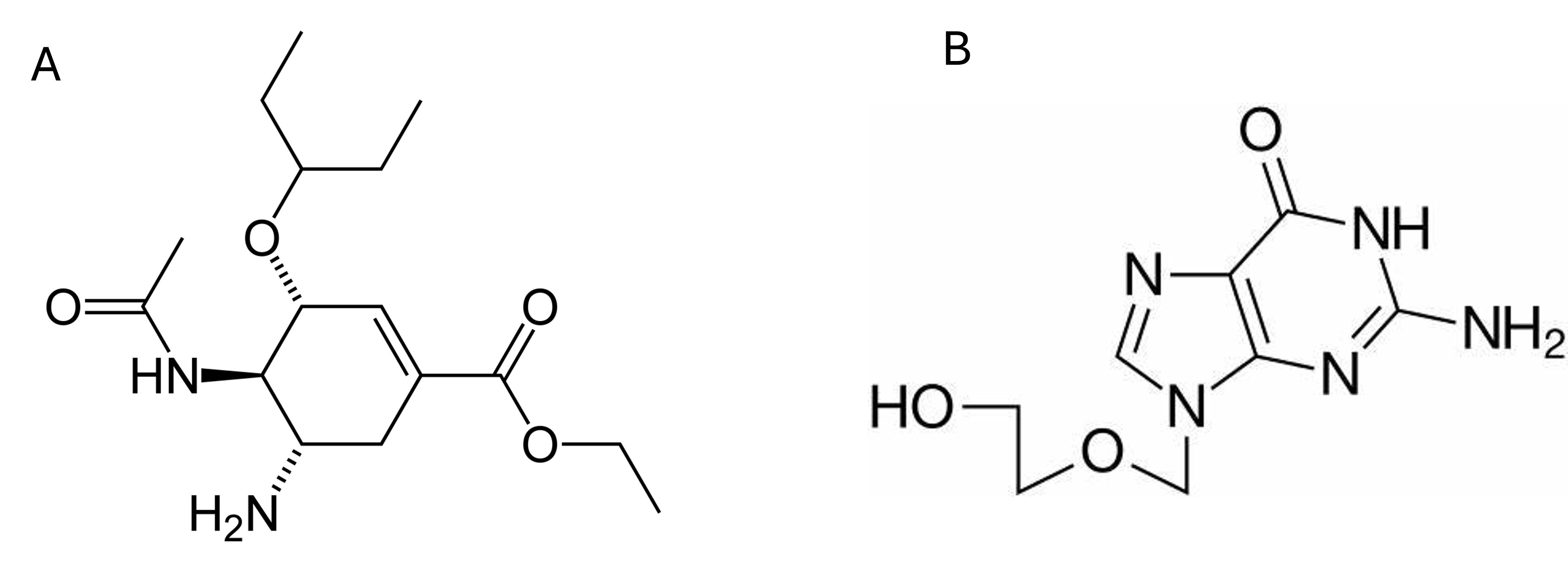
The experiments were performed at the Federal University of Piauí, in the Laboratory of Antileishmania Activity of the Medicinal Plants Research Center, Center of Health Sciences - NPPM/CCS/UFPI and the Physiology laboratory of the Department of Veterinary Morphophysiology of the center of Agricultural Sciences (DMV/CCA/UFPI). The antivirals, Oseltamivir and Aciclovir (Figure 1) were purchased from Merk, Rio de Janeiro, Brasil.

Parasites and cells
Parasites of the species Leishmania amazonensis (IFLA/BR/67/PH8), Leishmania infantum (MHOM/5745), Leishmania major (MHOM/IL/80/Friedlin), and Leishmania brasiliensis (10CL566), kept at the Antileishmanial Activity Laboratory, Medicinal Plant Research Center, Federal University of Piauí, were used. They were cultivated in Schneider’s medium (Sigma, USA), supplemented with 10% fetal bovine serum (FBS) (Merk, Rio de Janeiro, Brasil) and 10,000 IU/10 mg of penicillin-streptomycin (Merk, Rio de Janeiro, Brasil), at 26 °C in a biological oxygen demand (B.O.D.) incubator.
The macrophages were obtained from the peritoneal cavity of BALB/c mice, followed by subsequent euthanasia. These animals were immersed in 70% alcohol, where the macrophages were removed in a laminar flow hood with the animals positioned in dorsal decubitus attached to a cork board, 6 mL of phosphate buffered saline (PBS - NaCl 145 mmol/L, Na2HPO4 9 mmol/L, Na2HPO41 mmol/L, pH 7.4) at 4 °C, sterile, was applied to the abdominal region. Afterwards, the abdominal cavity was gently massaged and the PBS solution that had been injected was aspirated using a needle attached to a sterile syringe.
The entire sample was transferred into a 50 mL polystyrene conical tube and centrifuged at 100 rpm for 10 min. It was then washed three times with sterile PBS at 4 °C. Cell viability was assessed by counting cells in a Neubauer chamber, diluted in Trypan blue dye (Merk, Rio de Janeiro, Brasil).
For the hemolytic activity test, sheep blood was collected at the center of Agricultural Sciences Center of the federal University of Piaui (CCA/UFPI) located in Teresina, PI, Brazil. The project was submitted to the Ethics committee on Animal Experimentation for appraisal and was approved number no 750/2022.
Evaluation of antileishmanial activity against promastigote forms
Promastigote forms of L. amazonensis, L. major, L. brasiliensis, and L. infantum in the logarithmic growth phase were distributed in culture plates (1 x 10⁶ per well) containing oseltamivir (OSV) and acyclovir (ACV) in serial dilutions ranging from 800 to 6.25 μg/mL. The plates were incubated in a B.O.D. incubator at 26 °C ± 1 °C for 48 hours. After this period, 20 µL of resazurin (1 mmol/L) was added to each well, and the plates were incubated for an additional 6 hours. The plates were then read using an ELISA reader (Biotek Elx800) at 550 nm to monitor the growth and viability of the Leishmania parasites (Alves et al. 2017).
Evaluation of cytotoxicity on murine peritoneal macrophages
Approximately 2 x 10⁵ cells per well were added to a 96-well plate with RPMI medium (supplemented with 10% FBS and 10,000 IU of penicillin and 1,000 IU of streptomycin) and incubated at 37 °C with 5% CO₂ for 4 hours to allow cell adhesion to the plate. After washing to remove non-adherent cells, oseltamivir (OSV) and acyclovir (ACV), were added to a final volume of 100 μL per well at concentrations of 800, 400, 200, 100, 50, 25, 12.5, and 6.25 μg/mL, in triplicate, and incubated for 48 hours. After this period, 10 μL of MTT diluted in PBS at a concentration of 5 mg/mL (10% of the well volume, i.e., 10 μL per 100 μL) was added, and the plates were incubated again for 4 hours at 37 °C with 5% CO₂. The supernatant was then discarded, and 100 μL of dimethyl sulfoxide (DMSO) was added to each well. The plate was shaken for approximately 30 minutes on a Kline shaker (model AK 0506) at room temperature to ensure complete dissolution of the formazan. Finally, the absorbance was measured at 550 nm using a plate reader (Alves et al. 2017).
Cytotoxicity on sheep erythrocytes
To assess the hemolytic activity of drugs, sheep erythrocytes collected with an anticoagulant (EDTA) were used. After collection, the erythrocytes were diluted in 80 µL of PBS, adjusting the blood concentration to 5% hematocrit. Serial concentrations of the antivirals, ranging from 800 to 6.25 µg/mL, were prepared in 20 µL of PBS. The samples were incubated for 1 hour at 37 °C, and the reaction was halted by adding 200 µL of PBS. The suspensions were then centrifuged at 1500 RPM for 10 minutes at room temperature. The supernatant was analyzed using spectrophotometry at a wavelength of 550 nm to quantify hemolytic activity. The absence of hemolysis (negative control) and 100% hemolysis (positive control) were determined by replacing the tested sample solution with an equal volume of PBS and sterile Milli-Q water, respectively. The results were expressed as percentages and as the mean hemolytic concentration (CH₅₀), considering the positive control as 100% hemolysis (Sæbø et al. 2023).
Investigation of Drug Effects on Macrophages Infected with Leishmania amazonensis
Macrophages were plated in 24-well culture plates at a concentration of 2 x 10⁵ cells/well in 500 µL of supplemented RPMI medium (containing 10% FBS, 10,000 IU of penicillin, and 1,000 IU of streptomycin). Sterile 13-mm round coverslips were placed in each well. The plates were incubated at 37 °C with 5% CO₂ for 4 hours to allow for cell adhesion. Adhered macrophages were subsequently incubated with fresh medium containing promastigote forms of Leishmania amazonensis at a 10:1 amastigote-to- macrophage ratio at 37 °C with 5% CO₂ overnight. After this incubation period, the medium was aspirated to remove non-internalized parasites. The infected cultures were treated with Oseltamivir and Acyclovir at concentrations of 126, 252, and 504 μg/mL (concentrations determined from the antileishmanial activity test on promastigote forms) and Amphotericin B at 0.2 μg/mL. Following this treatment, the coverslips were removed and stained using Panótico Rápido® (Laborclin, Brazil). For each treatment, the percentage of infected macrophages (% infection) and the parasitic load (number of surviving amastigotes) were assessed by scanning the sample fields using an optical microscope until 100 macrophages were counted (Nunes et al. 2021).
Evaluation of macrophage activation parameters
Preparing solutions
The stock solution of neutral red dye (Sigma-Aldrich, St. Louis, USA) was prepared by dissolving 0.002 g of the dye in 1 mL of DMSO. The extraction solution used for assessing phagocytic capacity and lysosomal activity consisted of 96% glacial acetic acid (1% v/v) and PA ethanol (50% v/v) dissolved in double-distilled water. Zymosan (Sigma-Aldrich, St. Louis, USA), used for the phagocytic capacity assays, was prepared by diluting 0.3 mL of the neutral red stock solution and 0.02 g of zymosan in 3 mL of PBS. The fixative used was Baker’s calcium-formaldehyde, composed of 4% (v/v) formaldehyde, 2% (w/v) sodium chloride, and 1% (w/v) calcium acetate in distilled water (Nunes et al. 2021).
Evaluation of lysosomal activity
Peritoneal macrophages were plated at a density of 2×10⁵ cells per well in triplicate on a 96-well plate. The cells were incubated with five final concentration ranges of the antivirals [100 to 6.25 μg/mL]. After 48 hours of incubation at 37 °C with 5% CO2, 10 μL of a 2% neutral red solution prepared in DMSO was added to each well, followed by a 30-minute incubation. Subsequently, the supernatant was discarded, and the wells were washed with 0.9% saline at 37 °C. A volume of 100 μL of extraction solution was then added to solubilize the neutral red retained within the lysosomal secretion vesicles. After 30 minutes of agitation on a Kline shaker, the plate was read using a Biotek ELISA reader (ELx800) at 550 nm (Nunes et al. 2021).
Determination of phagocytic capacity
Peritoneal macrophages were plated at a density of 2×10⁵ cells per well in triplicate on a 96-well plate. The cells were incubated with five final concentration ranges of the antivirals [100 to 6.25 μg/mL]. After 48 hours of incubation at 37 °C and 5% CO2, 10 μL of stained zymosan solution was added to each well, followed by an additional 30-minute incubation at 37 °C. Subsequently, 100 μL of Baker’s fixative was added to halt the phagocytosis process. After 30 minutes, the wells were washed with 0.9% saline to remove unfagocytosed zymosan and neutral red. The supernatant was discarded, and 100 μL of extraction solution was added to each well. After solubilization using a Kline shaker, the plate was analyzed using a Biotek ELISA reader (ELx800) at 550 nm (Nunes et al. 2021).
Evaluation of the induction of nitric oxide synthesis
Peritoneal macrophages were plated at a density of 2×10⁵ cells per well in triplicate on a 96-well plate. The cells were incubated with five final concentration ranges of the antivirals [100 to 6.25 μg/mL]. Following 24 hours of incubation at 37 °C and 5% CO2, the supernatants were transferred to a new 96-well plate for nitrite quantification. A standard curve was prepared using sodium nitrite dissolved in Milli-Q® water, with concentrations ranging from 1, 5, 10, 25, 50, 75, 100, to 150 µmol/L, diluted in RPMI 1640 medium. For the assay, equal parts of each sample or standard solution were mixed with an equal volume of Griess reagent (1% sulfanilamide in 10% (v/v) H3PO(4), mixed in equal parts with 0.1% naphthyl ethylenediamine in Milli-Q® water). Absorbance readings were taken using a microplate reader at 550 nm (Nunes et al. 2021).
Statistical Analysis
All tests were carried out in triplicate in three independent experiments. The mean inhibitory concentration (IC50), mean cytotoxic concentration (CC50) with a 95% confidence limit, were calculated using probit regression. The selectivity index(SI) was calculated by dividing the CC50 by the CI50. Data were analyzed using analysis of variance (ANOVA), followed by Bonferroni’s post test, with a significance level set at p<0.05.
RESULTS
Evaluation of antileishmanial activity against different promastigote forms
The drugs OSV and ACV showed antileishmanial potential in a concentration- dependent manner. Both compounds exhibited activity against all tested leishmania species. Regarding the promastigote forms of L. amazonensis, L. braziliensis, L. infantum and L. major, OSV showed growth inhibition above 60% at a concentration of 800 μg/mL with inhibition percentages of 78%, 70%, 75% and 83% respectively. At concentrations of 400 μg/mL and 200 μg/mL, growth inhibition for L. amazonensis and L. braziliensis ranged from 50% to 45%. For L. infantum and L. major, growth inhibition at these concentrations ranged from 50% to 70%. ACV at concentrations of 800 μg/mL and 400 μg/mL exhibited 100% inhibition of the growth of the promastigote forms of L. amazonensis and at concentrations of 200 and 100 the inhibition was above 50%, with values of 90% and 88% respectively. For the promastigote form of L. braziliensis, 100% inhibition was observed at 800 μg/mL and inhibition percentages of above 50% was observed at concentrations of 400 μg/mL, 200 μg/mL, 100 μg/mL, and 50 μg/mL. However, for L. infantum and L. major, growth inhibition was slightly above 50% at 800 μg/mL, with inhibition values of 52% and 60%, respectively (Figure 2). The calculated IC50 values for OSV, ACV, and Amphotericin B (Amph B) were 252.321 μg/mL, 25.726 μg/mL, and 1.72 μg/mL, respectively (Table I).
Anti-leishmanial activity OSV, ACV and Anf B against promastigote forms of different species.

Graphical representation of the antileishmanial activity of Oseltamivir and Acyclovir against different species of Leishmania. The strains were incubated for 48 hours with the antivirals and antileishmanial activity was assessed using the resazurin method. Statistical significance *P<0.05;** p < 0.01; *** p < 0.001; **** p < 0.0001.
Evaluation of cytotoxicity on murine peritoneal macrophages and sheep erythrocytes
In their action on macrophages, OSV exibited significant cytotoxicity from the concentration of 100 μg/mL, with a CC50 value 512.172 μg/mL, while ACV showed a significant reduction in macrophage viability starting at a concentration of 50 μg/mL, resulting in a CC50 value of 2568.747 μg/mL (Figure 3a).

Graphical representation of the cytotoxic effect of Oseltamivir and Acyclovir against murine peritoneal macrophages and erythrocyte. The macrophages (a) were incubated for 48 hours with different concentrations of Oseltamivir and Acyclovir. Macrophage viability was measured using tetrazolic salt (MTT). Hemolytic activity (b) was assessed by incubating 5% sheep erythrocyte solution (collected in EDTA), with different concentrations of the antivirals [800 to 6.25 µg/mL] at 37oC for 1 hour. Results represent mean ± SEM of three experiments carried out in triplicate. * p < 0.05; ** p < 0.01; *** p < 0.001; **** p < 0.0001 when compared to the control. The absorbance was measured using a spectrophotometer at 550 nm.
Another way to assess the in vitro toxicity of the compounds is to evaluate their ability to hemolyze erythrocytes. In the evaluation of the cytotoxic effect of Oseltamivir and Acyclovir, cytotoxicity was observed; however, a reduction in cell viability was noted for both Oseltamivir and Acyclovir at doses ranging from 6.25 to 800 μg/mL, with the greatest effect observed at the dose of 800 μg/mL. However, it was not possible to calculate the CH50 for either of the substances tested (Figure 3b).
Effects on infected macrophages and determination of the survival index of internalized amastigotes and the Selectivity Index (SI)
The control group showed 100% parasitized cells, while Amphotericin B reduced this number to approximately 71%. The reduction in parasitized cells treated with OSV and ACV was concentration-dependent. The parasitized cells treated with OSV showed reductions of approximately 58%, 45%, and 35% at concentrations of 126, 252, and 504 μg/mL, respectively. In contrast, cells treated with ACV exhibited reductions of approximately 68%, 60%, and 58% at concentrations of 12.5, 25, and 50 μg/mL, respectively (Figure 4a).

Effects of OSV and Amphotericin B (as a positive control) to evaluate the percentage of infected macrophages (a) and the index of internalized amastigotes (b) in the treatment of murine macrophages infected with L. amazonensis. The cells were treated for 48 hours of incubation with concentrations of 6.25,12.5 and 25 μg/mL of OSV and ACV. Amphotericin B was used at a concentration of 2 μg/mL. The results represent the mean ± SEM of experiments performed in triplicate. * P < 0.05; ** P < 0.01; *** P < 0.001; **** P < 0.0001.

Macrophages infected with L. amazonensis. Anf B was used as a positive control (Anf B). For treatment with OSV, concentrations of 6.25 (A); 12.5 (B) and 25 (C) μg/mL were used. ACV was used at concentrations of 6.25 (D); 12.5 (E) and 25 (F) μg/mL. The arrows indicate the internalized amastigote forms. Magnification 1000x
Analyzing the survival index of internalized amastigotes, it was observed that the control group had an average of 14.0 amastigotes/macrophage. Amphotericin B reduced this number to approximately 3.8 amastigotes/macrophage. Regarding OSV treatment, the number of amastigotes decreased in a concentration-dependent manner to 3, 2, and 1 amastigotes/macrophage at concentrations of 126, 252, and 504 μg/mL, respectively. In the ACV treatment, the number of amastigotes/macrophage was 5.2, 3.8, and 4.0 for concentrations of 12.5, 25, and 50 μg/mL, respectively (Figure 4b).
In the evaluation of the activity of Oseltamivir and Acyclovir on the intracellular amastigote form, they displayed IC50 values of 5.436 and 6.845 μg/mL, respectively (Table II). Oseltamivir and Acyclovir were able to reduce the percentage of macrophages parasitized by L. amazonensis, as shown in Figure 4 with the infected cells.
Cytotoxic effects on mammalian cells and selectivity index values calculated for OSV and ACV.
The cytotoxicity parameters in murine peritoneal macrophages and the activity on the intracellular amastigote form of Leishmania amazonensis were used to determine the selectivity index (SI), which represents the extent to which the toxicity value of the substance is greater for the parasite than for the macrophage. Both OSV and ACV demonstrated greater selectivity for the promastigote forms of L. amazonensis than for murine macrophages. The results obtained for OSV and ACV were 94.21 and 375.27 respectively, indicating that both compounds are more selective for the protozoan than for mammalian cells (Table II).
Evaluation of macrophage activation parametersActivation of Phagocytic Capacity and Lysosomal Activity
Macrophage activation parameters such as phagocytosis capacity and increased lysosomal volume were assessed based on the retention of Zymozan and neutral red particles by the macrophages. OSV significantly induced phagocytosis capacity at concentrations 6.25 and 12.5 µg/mL (Figure 6a), while ACV also induced phagocytosis capacity at a concentration of 6.25 µg/mL. Meanwhile, neither OSV nor ACV significantly induced an increase in lysosomal volume of the macrophages (Figure 6b).

Action of OSV and ACV on Phagocytic Activity (a) and Lysosomal Activity (b). Murine peritoneal macrophages were incubated with serial concentrations of substances for 48 hours. Phagocytic capacity and lysosomal volume were analyzed by spectrophotometry through quantification enhanced by the neutral red dye, followed by solubilization using the extraction solution. Phagocytosis was analyzed by the incorporation of Zymosan particles with neutral red, which was solubilized by the extraction solution. Results represent the mean ± SEM of three experiments performed in triplicate. * P < 0.05; ** P < 0.01 vs. Control.
Evaluation of the induction of nitric oxide synthesis
The production of nitric oxide (NO), a marker of macrophage activation, was quantified by measuring nitrite concentrations through the incubation of macrophages with the substances OSV and ACV. Analysis of the results revealed that neither of the substances demonstrated a significant increase in nitrite synthesis at any of the tested concentrations (Figure 7).

Nitric oxide production by nitrite quantification of murine peritoneal macrophages treated with OSV and ACV for 24 hours. After this period, the culture supernatant was mixed in equal parts with Griess reagent. LPS - bacterial lipopolysaccharide (2 μg/mL). * P < 0.05; ** P < 0.01; *** p < 0.001; ****P < 0.001.
DISCUSSION
All currently available therapies for the treatment of leishmaniasis have significant limitations, whether due to cost or toxicity (World Health Organization 2023, Pradhan et al. 2022). Due to the challenges associated with treatment, the WHO encourages the exploration of new therapeutic alternatives for the treatment of leishmaniasis, including drug repositioning, drug combinations, pharmacological rejuvenation, and the use of natural products (World Health Organization 2023).
Drug repositioning involves exploring new uses for approved drugs (Hamid et al. 2024). Cases of successful repositioning for the treatment of leishmaniasis include miltefosine, a drug used to treat cancer (Rashidi et al. 2021), amphotericin B, a widely used antifungal, pentamidine and trivalent antimony itself, which has been used as an emetic agent since ancient times (Hemmert et al. 2025). Traditionally, repositioning has been performed through in vitro screening studies, however, computational screening methods (in silico) may also be applicable (Jourdan et al. 2020).
Previous studies have highlighted the relevance of repositioning drugs for the treatment of leishmaniasis. Rub et al. (2019) evaluated glibenclamide, an effective sugar- lowering drug used to treat diabetes, for its antileishmanial activity against L. donovani. The results showed that glibenclamide inhibited the viability of the parasite, suggesting its potential use as a drug in the treatment of visceral leishmaniasis. Ivermectin is one of the most well-known and widely used antiparasitic drugs in human and veterinary medicine (Laing et al. 2017). Ivermectin (IVE) has been tested in vitro and in vivo against L. infantum species, and preliminary studies have suggested that IVE targets the mitochondria of the parasite (Reis et al. 2021).
Antiviral drugs are classified according to their targets in the replication mechanism. Those that prevent receptor recognition and endocytosis are hemagglutinin inhibitors; those that inhibit the fusion of the viral envelope to the cell membrane are proton channel blockers; those that interrupt the production of viral proteins are polymerase inhibitors and those that prevent newly synthesized viruses from infecting other cells are neuraminidase inhibitors (Li et al. 2021).
Oseltamivir is a widely used antiviral in the management of influenza and belongs to the class of inhibitors of neuraminidase, the influenza A viral surface protein. This drug acts by blocking the function of the neuraminidase enzyme, a viral protein that is crucial for the release of new viruses from infected cells and for the spread of the infection, thus reducing the viral load and limiting the spread of the virus (Bai et al. 2021).
Two key viral enzymes are involved in the infection process: hemagglutinin and neuraminidase. Both enzymes act by recognizing sialic acid residues present on the glycoproteins of the target cells and induce the fusion and incorporation of the viral envelope into the cell. The virus then releases its genetic material, RNA, which is internalized along with certain viral proteins into the nucleus, where it facilitates viral RNA replication and the production of viral messenger RNA (Halldorsson et al. 2021). The recognition that hemagglutinin and neuraminidase play pivotal roles in the success of viral infection has created opportunities to modulate the activity of one or both enzymes within the viral life cycle, thereby enhancing the immune system’s ability to effectively combat the virus (Chauhan & Gordon 2022). The understanding of the influenza virus life cycle led to the approval of the antiviral oseltamivir for the treatment of influenza in the United States and Brazil in 1999 and 2000, respectively (Hooker & Ganusov 2021).
Acyclovir has a selective action in inhibiting viral replication and has low toxicity in uninfected host cells due to a mechanism of sequential phosphorylation of ACV, converting it into the active form of acyclovir triphosphate (or acyclovir triphosphate), which only occurs in infected cells (Kausar et al. 2021). The high selectivity of ACV in inhibiting viral DNA replication is due to the greater uptake of the active ingredient by infected cells, phosphorylation mechanisms mediated by viral thymidine kinase, and the specificity of acyclovir triphosphate for viral DNA polymerase. The inhibition of viral DNA polymerase by acyclovir triphosphate is 10 to 30 times more potent than the inhibition of human DNA polymerase (Kłysik et al. 2020).
In this study, OSV displayed an IC50 of 252.32 μg/mL while ACV had a value of 25.726 μg/mL, so ACV showed greater growth inhibition for L. amazonensis due to its lower IC50 value. OSV showed lower cytotoxicity compared to ACV with mean cytotoxic concentrations (CC50) of 512.17 μg/mL and 2568.75 μg/mL, respectively. The selectivity index (SI) for OSV and ACV was 2.02 μg/mL and 99.85 μg/mL, respectively. The selectivity index is determined by the ratio between the CC50 in mammalian cells and the IC50 in parasites. This index is an important criterion in Drug Discovery for selecting the most promising compounds against parasites. Compounds with an SI ≥ 10 are considered promising for the treatment of leishmaniasis (Katsuno et al. 2015). ACV demonstrated a higher SI compared to OSV, indicating a better balance between efficacy against the parasite and safety of the host cells and also showed more selectivity for the promastigote forms of L. amazonensis than for murine macrophages. The selectivity index obtained for OSV was 94.21 while for ACV it was 375.27, meaning that both were more selective for the protozoan than for mammalian cells.
In vitro toxicological assays, such as hemolytic activity and MTT, are valuable preliminary tests for assessing the cytotoxicity of compounds and determining the concentration to be used in later stages of drug development (Caridha et al. 2019). Based on the assays conducted, OSV exhibited the highest cytotoxic potential on murine macrophages, with a CC50 of 512.17 μg/mL, while ACV demonstrated the lowest, with a CC50 of 2568.75 μg/mL. However, it was not possible to determine the CH50 for the samples at the concentrations analyzed.
Intracellular amastigotes are clinically relevant and, therefore, crucial for testing new drugs, as they are found in the vertebrate host. In this study, experimental assays were conducted using macrophages infected with Leishmania amazonensis amastigotes. This in vitro experimental model is the most suitable for studying Leishmania spp. infection in humans (Nunes et al. 2021), as macrophages play a central role in modulating the immune response and are the primary targets of this parasite (Elmahallawy et al. 2021). Therefore, substances capable of reducing the percentage of parasitized macrophages and the survival index of amastigotes within macrophages are promising candidates for in vivo testing (Alves et al. 2017). Among the drugs studied, both OSV and ACV exhibited antileishmanial activity, with ACV demonstrating higher antileishmanial activity compared to OSV, affecting nearly 70% of macrophages infected by the parasite at a concentration of 12.5 μg/mL.
One of the mechanisms of evasion of the host immune system of leishmaniasis involves the modification of the Toll-like receptor (TLR) signaling pathway, interrupting the cytokine cascade (Taslimi et al. 2018). These receptors act as sensors of the innate immune system, capable of recognizing invading pathogens and inducing an immune response. In the case of leishmaniasis, the infection is known to suppress the innate immune response mediated by TLR4. Recent research highlights the relevance of the lysosomal sialidase enzyme Neu1 in the removal of sialic acid from the TLR4 receptor in macrophages and its influence on the Leishmania infection process (Cavalcante et al. 2021, Karmakar & Mandal 2021).
In this context, sialic acids (sias) are monosaccharides typically present as terminal residues of glycoproteins and glycolipids, either on the cell surface or in secreted molecules. These are nine-carbon acidic sugars derived from the basic molecule of neuraminic acid (Neu), specifically 5-amino-3,5-dideoxy-2-nonulosonic acid. Sialidases/neuraminidases are glycosidases that catalyze the cleavage of α-glycosidically linked sia residues in glycoproteins and glycolipids. Sias have been identified in promastigotes and amastigotes of various Leishmania species. Due to their position on terminal residues of glycoconjugates, sias play a role in mediating interactions between Leishmania and host cell receptors. The interaction mediated by Sias between Leishmania and macrophages in vitro typically leads to a dominant Th2-type cytokine response, along with reduced levels of macrophage reactive oxygen species (ROS) and nitric oxide (NO). Given that the inhibition of NO production by macrophages is linked to enhanced survival of intracellular parasites and greater disease severity, Sias can be linked to an evasion mechanism utilized by Leishmania (Cavalcante et al. 2021). OSV is a neuraminidase inhibitor and may act to reduce the number of intracellular parasites.
Thymidine kinases (TKs) are enzymes that catalyze the magnesium-dependent transfer of γ-phosphate from ATP to thymidine (dThd), thus forming thymidine monophosphate (dTMP). dTMP is further phosphorylated by cellular enzymes to 2’- deoxythymidine triphosphate (dTTP), which serves as a substrate for DNA polymerase during replication. Therefore, TKs control dTTP groups and are important regulators for DNA biosynthesis (Timm et al. 2015). TKs are defined in two subgroups (TK-1 and TK- II) according to their substrate specificity, where TK-1 includes herpes simplex virus TK (HSV-TK) and human mitochondrial TK2, while TK-II includes diverse organisms such as mammals, parasitic protozoa (Leishmania spp.), bacteria and many viruses. The thymidine kinase from Leishmania spp. (LTK) has already been reported to positive cooperation for ATP at saturating concentrations of dThd, this behavior is consistent with the modulation of TK-I-like enzymatic activities after ATP binding (Kausar et al. 2021, Timm et al. 2015). The selectivity of ACV in the inhibition of DNA through the phosphorylation mechanism via TK and the metabolic similarities between LTK and TK- 1 suggest antileishmanial action of acyclovir.
Microscopic analysis revealed that the control group exhibited a higher concentration of amastigotes surrounding the parasitophorous vacuole, whereas the OSV- and ACV-treated groups showed a reduction in parasite numbers, indicating a decrease in the parasitic load within murine macrophages. These findings further underscore the antileishmanial potential of these substances.
Innate immunity plays a crucial role in controlling infections through mechanisms such as phagocytosis and lysosomal activity, which facilitate antigen activation and pathogen elimination (Elmahallawy et al. 2021). Thus, macrophage activation parameters lead to conformational changes that enhance the performance of their functions, such as motility and phagocytosis (Gabriel et al. 2021).
The compounds OSV and ACV significantly induced phagocytic capacity, with the optimal concentrations for OSV being 6.25 μg/mL and 12.5 μg/mL, and for ACV, 6.25 μg/mL. These results indicate the potential activation of macrophage defense mechanisms, which are enhanced by the use of these drugs. Zymosan stimulates immune cells to produce a response, leading to an increase in IFN production and phagocytic capacity (Park et al. 2023), and an increase was observed with the substances under study. Regarding the lysosomal volume of macrophages, neither OSV nor ACV significantly induced an increase. Additionally, neither compound induced nitric oxide (NO) production in macrophages, suggesting that these compounds do not activate this mediator in host cells, which may be necessary for inducing a lethal effect on the parasite.
This suggests that the activation of the lysosome and the production of NO by infected macrophages may not be the primary immunomodulatory mechanism of action of these drugs, but may be associated with other mechanisms.
A primary mechanism of resistance to Leishmania infection is the production of nitric oxide (NO) by infected macrophages (Pereira et al. 2019). Inside the phagolysosome, NO reacts with superoxide (O₂⁻) to form peroxynitrite, a reactive oxygen species. This reaction results in the production of nitrate and nitrite as final products, which function as microbicidal agents (Pereira et al. 2019, Rostami & Khamesipour 2021).
Although no significant induction of NO production was observed, the reduction in infected macrophages and the index of internalized amastigotes may have occurred due to the increased phagocytic capacity stimulated by the substances. This effect is likely due to the presence of acidic hydrolases within the phagolysosome, where the majority of the degradation of engulfed material occurs.
Considering that selectivity is a crucial factor in the search for new drug candidates, the substances OSV and ACV showed selectivity index values of 94.21 and 375.27, respectively. This indicates that they exhibit greater activity against the amastigote forms compared to extracellular promastigote forms. ACV, in particular, stood out due to its higher selectivity.
CONCLUSIONS
The samples reduced the cell viability of promastigote forms, as well as the infection of macrophages. The drugs demonstrated potential antileishmanial activity, with ACV exhibiting the highest potential against promastigote forms and OSV showing significant activity against amastigote forms, with a notable reduction in the number of infected macrophages and the index of internalized amastigotes of L. amazonensis. Both substances exhibited low cytotoxicity, enhanced immunomodulatory responses, and the ability to activate macrophages through increased phagocytic capacity. Another key characteristic of these drugs is their selectivity for the parasites, with ACV exhibiting the highest selectivity index. Thus, the antiviral agents OSV and ACV are promising candidates for further investigation into their therapeutic potential against leishmaniasis.
Acknowledgements
Federal University of Piauí and Postgraduate Program in Technologies Applied to Animals of Regional Interest.
References
- ALVES MMM ET AL. 2017. Gallic and ellagic acids: two natural immunomodulator compounds solve infection of macrophages by Leishmania major. Naunyn-Schmiedeb Arch Pharmacol 390: 893-903.
- BAI Y, JONES JC, WONG S-S, ZANIN M, RUSSELL CJ, GOVORKOVA EA & ORG JJ. 2021. Antivirals Targeting the Surface Glycoproteins of Influenza Virus: Mechanisms of Action and Resistance. Viruses 13: 624.
- BICHIOU H, BOUABID C, RABHI I & GUIZANI-TABBANE L. 2021. Transcription Factors Interplay Orchestrates the Immune-Metabolic Response of Leishmania Infected Macrophages. Front Cell Infect Microbiol 7(11): 660415.
- CARIDHA D ET AL. 2019. Route map for the discovery and pre-clinical development of new drugs and treatments for cutaneous leishmaniasis. Int J Parasitol Drugs Drug Resist 11: 106-117.
- CAVALCANTE T, MEDEIROS MM, MULE SN, PALMISANO G & STOLF BS. 2021. The Role of Sialic Acids in the Establishment of Infections by Pathogens, With Special Focus on Leishmania. Front Cell Infect Microbiol 11: 671913.
- CHAUHAN RP & GORDON ML. 2022. An overview of influenza A virus genes, protein functions, and replication cycle highlighting important updates. Virus Genes 58(4): 255-269.
- ELMAHALLAWY EK, ALKHALDI AAM & SALEH AA. 2021. Host immune response against leishmaniasis and parasite persistence strategies: A review and assessment of recent research. Biomed Pharmacother 139: 111671.
- GABRIEL ÁM, GALUÉ-PARRA A, PEREIRA WLA, PEDERSEN KW & DA SILVA EO. 2021. Leishmania 360°: Guidelines for Exosomal Research. Microorganisms 9(10): 2081.
- HALLDORSSON S, SADER K, TURNER J, CALDER LJ & ROSENTHAL PB. 2021. In situ structure and organization of the influenza C virus surface glycoprotein. Nat Commun 12: 1694.
- HAMID A, MÄSER P & MAHMOUD AB. 2024. Drug Repurposing in the Chemotherapy of Infectious Diseases. Molecules 29: 635.
- HEMMERT C, GORNITZKA H, DERAEVE C & STIGLIANI JL. 2025. Current state of the art of gold complexes as antileishmanial agents. Coord Chem Rev 528: 216408.
- HOOKER KL & GANUSOV VV. 2021. Impact of Oseltamivir Treatment on Influenza A and B Virus Dynamics in Human Volunteers. Front Microbiol 12: 631211.
- JOURDAN JP, BUREAU R, ROCHAIS C & DALLEMAGNE P. 2020. Drug repositioning: a brief overview. Pharm Pharmacol 72: 1145-1151.
- KARMAKAR J & MANDAL C. 2021. Interplay Between Sialic Acids, Siglec-E, and Neu1 Regulates MyD88- and TRIF-Dependent Pathways for TLR4-Activation During Leishmania donovani Infection. Front Immunol 3(12): 626110.
- KATSUNO K, BURROWS JN, DUNCAN K, HUIJSDUIJNEN RHV, KANEKO T, KITA K, MOWBRAY CE, SCHMATZ D, WARNER P & SLINGSBY BT. 2015. Hit and lead criteria in drug discovery for infectious diseases of the developing world. Nat Rev Drug Discov 14(11): 751-758.
- KAUSAR S, SAID KHAN F, ISHAQ MUJEEB UR REHMAN M, AKRAM M, RIAZ M, RASOOL G, HAMID KHAN A, SALEEM I, SHAMIM S & MALIK A. 2021. A review: Mechanism of action of antiviral drugs. Int J Immunopathol Pharmacol 35: 20587384211002621.
- KŁYSIK K, PIETRASZEK A, KAREWICZ A & NOWAKOWSKA M. 2020. Acyclovir in the Treatment of Herpes Viruses - A Review. Curr Med Chem 27: 4118-4137.
- LAING R, GILLAN V & DEVANEY E. 2017. Ivermectin - Old Drug, New Tricks? Trends Parasitol 33: 463-472.
- LI G, JING X, ZHANG P & DE CLERCQ E. 2021. Antiviral Classification. Encyclopedia of Virology, Elsevier, p. 1-5. DOI: 10.1016/b978-0-12-814515-9.00126-0.
- NUNES TAL ET AL. 2021. Curzerene antileishmania activity: Effects on Leishmania amazonensis and possible action mechanisms. Int Immunopharmacol 100: 108130.
- PARK HJ, LEE SW, PARK YH, KIM TC, LEE S, LEE S, VAN KAER L & HONG S. 2023. In Vivo Zymosan Treatment Induces IL15-Secreting Macrophages and KLRG1-Expressing NK Cells in Mice. Molecules 28: 5779.
- PEREIRA MA, ALEXANDRE-PIRES G, CÂMARA M, SANTOS M, MARTINS C, RODRIGUES A, ADRIANA J, PASSERO LFD, PEREIRA DA FONSECA I & SANTOS-GOMES G. 2019. Canine neutrophils cooperate with macrophages in the early stages of Leishmania infantum in vitro infection. Parasite Immunol 41(4): e12617.
- PRADHAN S, SCHWARTZ RA, PATIL A, GRABBE S & GOLDUST M. 2022. Treatment options for leishmaniasis. Clin Exp Dermatol 47: 516-521.
- RASHIDI S, FERNÁNDEZ-RUBIO C, MANZANO-ROMÁN R, MANSOURI R, SHAFIEI R, ALI-HASSANZADEH M, BARAZESH A, KARIMAZAR M, HATAM G & NGUEWA P. 2021. Potential therapeutic targets shared between leishmaniasis and cancer. Parasitology 148: 655-671.
- REIS TAR ET AL. 2021. Ivermectin presents effective and selective antileishmanial activity in vitro and in vivo against Leishmania infantum and is therapeutic against visceral leishmaniasis. Exp Parasitol 221: 108059.
- ROSTAMI MN & KHAMESIPOUR A. 2021. Potential biomarkers of immune protection in human leishmaniasis. Med Microbiol Immunol 210: 81-100.
- RUB A, SHAKER K, KASHIF M, ARISH M, DUKHYIL AAB, ALSHEHRI BM, ALAIDAROUS MA, BANAWAS S & AMIR K. 2019. Repurposing Glyburide as Antileishmanial Agent to Fight Against Leishmaniasis. Protein Pept Lett 26: 371-376.
- SÆBØ IP, BJØRÅS M, FRANZYK H, HELGESEN E & BOOTH JA. 2023. Optimization of the Hemolysis Assay for the Assessment of Cytotoxicity. Int J Mol Sci 24: 2914.
- SASIDHARAN S & SAUDAGAR P. 2021. Leishmaniasis: where are we and where are we heading? Parasitol Res 120: 1541-1554.
- SINGH R, KASHIF M, SRIVASTAVA P & MANNA PP. 2023. Recent Advances in Chemotherapeutics for Leishmaniasis: Importance of the Cellular Biochemistry of the Parasite and Its Molecular Interaction with the Host. Pathogens 12(5): 706.
- TAO J ET AL. 2022. Binding mechanism of oseltamivir and influenza neuraminidase suggests perspectives for the design of new anti-influenza drugs. PLoS Comput Biol 18: e1010343.
- TASLIMI Y, ZAHEDIFARD F & RAFATI S. 2018. Leishmaniasis and various immunotherapeutic approaches. Parasitology 145: 497-507.
- TIMM J, BOSCH-NAVARRETE C, RECIO E, NETTLESHIP JE, RADA H, GONZÁLEZ-PACANOWSKA D & WILSON KS. 2015. Structural and Kinetic Characterization of Thymidine Kinase from Leishmania major. PLoS Negl Trop Dis 9: e0003781.
-
WORLD HEALTH ORGANIZATION. 2023. Leishmaniasis. Available at: https://www.who.int/news-room/fact-sheets/detail/leishmaniasis
» https://www.who.int/news-room/fact-sheets/detail/leishmaniasis