Teucrium polium leaves extract reduces nicotine-induced histopathological and biochemical alteration in the spleen via amelioration of antioxidant biomarker
ABSTRACT
Nicotine, the most toxic factor in tobacco, can damage a wide range of biological molecules by inducing oxidative stress. This study aimed to investigate the protective effects of Teucrium polium Leaves extract (TPLE) against the toxic properties of nicotine in the spleen of mice. Twenty-four mice were equally divided into four groups (n = 6): normal control, TPLE (100mg/kg), nicotine (2.5mg/kg), and TPLE plus nicotine-treated groups (once a day) for 3 weeks. The spleen index and spleen homogenate of sacrificed mice obtained on day 21 were used to assess the levels of malondialdehyde (MDA), nitric oxide (NO), and reduced glutathione (GSH). Histological sections were stained with hematoxylin and eosin. Nicotine administration causes spleen homogenate levels of malondialdehyde, nitrite oxide, and spleen index to increase significantly, while glutathione levels are significantly reduced. TPLE administration improved these changes by ameliorating antioxidant biomarkers via lowering MDA and NO content and elevating GSH levels. The histological sections showed normal architecture of the mice spleen treated with T. polium extract, but a further distortion in those that received nicotine only. However, improved morphology was seen in the group treated with TPLE extract and nicotine when compared with the control group. These findings suggest that using T. polium helps protect the spleen against the hazardous effects of nicotine.
Keywords:
Teucrium polium; nicotine; spleen; mice
RESUMO
A nicotina, o fator mais tóxico do tabaco, pode danificar uma ampla gama de moléculas biológicas ao induzir o estresse oxidativo. O objetivo deste estudo foi investigar os efeitos protetores do extrato das folhas de Teucrium polium (TPLE) contra as propriedades tóxicas da nicotina no baço de camundongos. Vinte e quatro camundongos foram divididos igualmente em quatro grupos (n = 6): controle normal, TPLE (100 mg/kg), nicotina (2,5mg/kg) e TPLE mais grupos tratados com nicotina (uma vez ao dia) por 3 semanas. O índice do baço e o homogenato do baço de camundongos sacrificados obtidos no 21º dia foram usados para avaliar os níveis de malondialdeído (MDA), óxido nítrico (NO) e glutationa reduzida (GSH). As seções histológicas foram coradas com hematoxilina e eosina. A administração de nicotina faz com que os níveis de homogenato de baço de malondialdeído, óxido de nitrito e índice de baço aumentem significativamente, enquanto os níveis de glutationa são significativamente reduzidos. A administração de TPLE melhorou essas alterações, melhorando os biomarcadores antioxidantes por meio da redução do conteúdo de MDA e NO e da elevação dos níveis de GSH. As seções histológicas mostraram uma arquitetura normal do baço dos camundongos tratados com extrato de T. polium, mas uma distorção adicional naqueles que receberam apenas nicotina. No entanto, foi observada uma melhora na morfologia no grupo tratado com extrato de T. polium e nicotina em comparação com o grupo de controle. Essas descobertas sugerem que o uso de T. polium ajuda a proteger o baço contra os efeitos perigosos da nicotina.
Palavras-chave:
Teucrium polium; nicotina; baço; camundongos
INTRODUCTION
Tobacco smoke from cigarettes and waterpipes contains several harmful substances, such as transition metals, carbon monoxide, aldehydes, nicotine, nitrosamine, and solid particle matter (Münzel et al., 2020). According to WHO (2013), low- and middle-income nations will account for more than 80% of tobacco-related mortality.
The most addictive ingredient in cigarette smoke, nicotine, is quickly absorbed by the bloodstream and has been linked to an increased risk of lung cancer, heart disease, other cardiovascular, cerebrovascular disorders, and other effects on biological systems (Omar et al., 2015). Nicotine triggers the generation of reactive oxygen species (ROS) and free radicals, including hydroxyl radical (OH•), hydrogen peroxide (H2 O2), and superoxide anion radical (O2-), which exhaust the antioxidative defense system (AOS) and ultimately cause oxidative stress (Li et al., 2015).
Increased oxidative stress has been proposed as a key player in the pathophysiology of numerous smoking-related diseases, such as cancer, heart disease, and oral disorders (Muthukumaran et al., 2008). Nicotine also activates the nicotinic acetylcholine receptors, or nAChRs, in the brain. The activation of nAChRs increases the release of various neurotransmitters by the brain, such as acetylcholine, glutamate, dopamine (DA), noradrenaline (NDA), and gamma-aminobutyric acid (GABA). These neurotransmitters play a role in the control of a wide range of activities, including walking, anxiety, memory, and learning (Kandeil et al., 2021).
The spleen is connected to the brain by the autonomic nerve system, which controls the immune system. The largest lymphoid organ in the human body, the spleen produces immune cells that respond to antigens and infections by eliciting both innate and acquired immune responses (Lin et al., 2024). The liver and spleen play critical functions, which makes them necessary for the metabolism of harmful substances and the control of the immune system's response to them.
In both people and experimental animals, nicotine has an impact on a variety of immune processes, including humoral and cell-mediated immune responses. It affects natural killer, T, B, and alveolar macrophages as well as other immune cells. Furthermore, nicotine consumers have higher levels of autoantibodies (Liang et al., 2022).
Numerous preceding studies have demonstrated the presence of antioxidant agents in plant species or natural resources. Teucrium is a genus that belongs to the family Lamiaceae. This family is composed of species with exploitable antioxidant activity (Couladis et al., 2003).
T. polium (Lamiaceae) is a perennial shrub that grows to a height of 20 to 50 cm. It is found throughout most Mediterranean countries, Southwestern Asia, Europe, and North Africa. It is dry and rocky in the hills and deserts.
According to phytochemical studies, T. polium includes a variety of substances, including flavonoids, iridoids, and terpenoids (Piozzi et al., 2005). Many reports exist regarding T. polium's biological activity, and it has been demonstrated to be hypoglycemic, insulinotropic, and has antioxidant, antinociceptive, and anti-inflammatory properties (Mahmoudabady et al., 2018).
T. polium extract was utilized in this work, and the total flavonoids, phenolic contents, Fourier-transform infrared spectroscopy (FT-IR), and (DPPH) free radical scavenging assay were all examined (Al-Shaebi et al., 2023). In this work, T. polium extract was also comprehensively analyzed by HPLCUV to determine specific phenolic compounds included in the extracts. The ethanolic extract has eleven bioactive components that have been qualitatively identified: myricetin, quercetin, apigenin, naringenin, luteolin 7-o-glucoside, caffeic acid, ellagic acid, rutin, and chlorogenic acid (Alatawi et al., 2024).
Considering the previously mentioned characteristics of this plant, the present study aimed to assess the ethanolic extract of T. polium leaves (TPLE) impact on oxidative stress that occurs in the spleen as well as its histopathological alterations posed by subcutaneous nicotine administration in mice.
MATERIALS AND METHODS
T. polium leaves were collected in May 2022 in the vicinity of Al-badyah Tabuk, Saudi Arabia. Location: at 27°45'59.5"N 36°31'48.8"E, about 80 km south of Tabuk. The plant was identified by a scientist from the King Saud University Herbarium (Science College, Botany Department, Riyadh, Saudi Arabia). T. polium leaves extract was prepared following the Qabaha et al., (2021) protocol with a few modifications. Leaves were allowed to dry naturally before being ground into a powder. The resultant powder was subjected to a cold maceration extraction process using an ethanol (50%) solvent system for an entire day. Filtered and concentrated in a rotary evaporator running at 50 °C under pressure, the ethanolic extract was then collected and kept at -20 °C in sealed bottles.
Nicotine was obtained from SOMATCO (Riyadh, Saudi Arabia). Other analytical-grade chemicals and reagents were purchased from commercial suppliers.
For the present study, twenty-four adult male Swiss albino mice, weighing 30-35 gm and aged 8 - 10 weeks, were obtained from the animal house of King Saud University College of Science. Mice were housed in cages that were well-ventilated, had 12 hours of light and 12 hours of darkness, and were specifically pathogen-free at 23 ± 5°C. Before the experiment began, mice were allowed free access to tap water ad libitum and typical pellet feeds for seven days while they adapted.
Mice were randomly divided into four equal groups of six mice each and given the following daily therapy for three weeks:
Mice in Group 1 (Control): received distilled water.
Group 2 (T. polium): The mice were given an oral dose of 100 mg/kg of (TPLE) diluted in distilled water (Forouzandeh et al., 2013).
Group 3 (Nicotine): Mice received subcutaneous injections of 2.5 mg/kg of dissolved nicotine in distilled water (Alsharari et al., 2015).
Mice in Group 4 (Nicotine + TPLE): were given a subcutaneous injection of 2.5 mg/kg of nicotine and an oral gavage of 100 mg/kg of TPLE an hour later.
Six mice in each group were administered xylazine and ketamine one day following the experimental period for anesthesia, and blood samples were collected via retroorbital hemorrhage. Blood samples were allowed to coagulate at room temperature before being centrifuged for 15 minutes at 3000 rpm. For biochemical examination, the serum was collected and stored in aliquots at -20°C. Each animal's spleen was removed, washed with saline, and divided into pieces. One portion was homogenized in cold 10% w/v phosphate-buffered saline, for 10 minutes centrifuged at 3000 rpm, then the supernatant was collected and kept at -20°C. The supernatant was then separated and centrifuged for 15 minutes at 3000 rpm. Consequently, it was saved at -20 °C. For histological examination, a separate part was preserved in freshly prepared 10% neutral buffered formalin (pH 7.4) for a minimum of 24 hours.
Using Biodiagnostic kits (Cat. No. MD 25 29; at 535 nm), a concentration of malondialdehyde (MDA), an indication of lipid peroxidation, was measured spectrophotometrically in the spleen homogenate using the Ohkawa et al., (1979) method. The procedures of Beutler et al., (1963) were followed to measure glutathione reduced (GSH) (Biodiagnostic kits, Cat. No.: GR 25 11; at 412 nm) and (Biodiagnostic kits, Cat. No.: CA 25 14; at 510 nm).
Using reagent kits from Biodiagnostic (Cat. No. NO 25 33; at 540 nm), the NO concentration in the spleen homogenate was measured spectrophotometrically in accordance with the protocol described by Archer (Archer, 1993).
After being fixed in formalin, the specimens were rinsed with distilled water, sliced, dehydrated with ascending ethyl alcohol series, and embedded in paraffin wax. They were then. The wax blocks were sectioned into 4 μm- sections. Hematoxylin and eosin (H&E) staining was applied to the slices (Adam and Caihak, 1964). Images were taken of sections stained with a light microscope (Leica, Wetzlar, Germany). In addition, the spleen index was determined by dividing the weight of the spleen by the weight of the mouse.
Results were expressed as means ± standard error of the mean (SEM) of different groups. Differences between the values of mean were calculated by one-way ANOVA followed by Scheffe post-hoc test using version 28 of SPSS software (SPSS Inc., Chicago, IL, USA). The results were judged statistically significant.
RESULTS
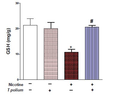
The GSH content of the nicotine-administered mice was significantly reduced (P<0.001) than that of the control mice. The GSH content of the nicotinic mice treated with TPLE increased significantly (P < 0.01) in comparison with the nicotine untreated group as shown in (Fig. 1).
The data shown in (Fig. 2) indicate that mice who received a subcutaneous injection of nicotine had significantly (P < 0.01) higher MDA levels than mice in the control group. The induction of TPLE treatment showed a significant (P < 0.05) reduction in MDA levels when compared with the group that received nicotine injections only.
The results in (Fig. 3) demonstrated a significant increase (P < 0.001) in NO level in nicotine-administered mice when compared with the control group. On the other hand, TPLE supplementation significantly (P < 0.001) decreased NO levels in comparison with nicotinic untreated mice group.

T. polium leaves extract increased spleen glutathione (GSH) content in nicotine-administered mice. Data are represented as mean ± SEM. *** P < 0.001 versus control and ## p < 0.01 versus nicotine.

TPLE ameliorated spleen malondialdehyde (MDA) level in nicotine-administered mice. Data are represented as mean ± SEM. ** P < 0.01 versus control and # P < 0.05 versus nicotine.

T. polium leaves extract improved spleen nitric oxide (NO) level in nicotine-administered mice. Data are represented as mean ± SEM. *** p < 0.001 versus control and
P < 0.001 versus nicotine.
The average spleen index in nicotine-TPLE- treated mice was lower, from 0.94 to 0.64, compared with nicotine administered mice (Fig. 4).
In the same line, microscopic observations of the control spleen (Fig. 5A, B) and TPLE (100 mg/kg)-treated spleen (Fig. 5C, D) showed regular structure of red pulp and white pulp, while in the nicotine-treated group (2.5mg/kg b.w), the spleen showed degeneration of red pulp and white pulp (Fig. 5E, F). Concomitantly, a normal spleen capsule was shown in the control (Fig. 6A, B) as well as the TPLE (100 mg/kg)-treated spleen (Fig. 6C, D). Although an observable thinning of the capsule was noticed due to (Fig. E, F), Supplementation of TPLE to nicotine-treated mice caused regeneration of red pulp and improved tissue damage in the spleen of experimental mice (Fig. 5G, H), besides recovering capsule thickness (Fig. 6G, H).

Changes in spleen index in a group of control, T. polium TPLE 100mg/kg, nicotine, and induced nicotine treated with TPLE 100mg/kg. Values are means ± SEM. ** Significant against the control group at P < 0.01. # Significant against nicotine group at P < 0.05.

Photomicrograph of mice spleen sections stained with hematoxylin and eosin (H&E). (A, B) Normal structure of control spleen. (C, D) TPLE 100mg/kg treated group (E, F), Nicotine-administered show spleen of mice with dilated and fused white and red pulps. (G, H) nicotine treated with TPLE shows a spleen with improved structure. Scale bar = 50 μm.

T. polium -improved changes in mouse spleen capsules induced- nicotine (A, B) control spleen with normal capsule. (C, D) TPLE 100mg/kg treated spleen with the normal capsule. (E, F) nicotinic spleen with thin capsule (G, H) nicotinic- TPLE treated (100 mg/kg) spleen with moderate thickness. Scale bar = 50 μm.
DISCUSSION
Tobacco use continues to be one of the world's major causes of disease and early death, despite a recent decline in its prevalence (West, 2017). According to Herxheimer et al., (1967) nicotine causes hemodynamic alterations identical to those caused by cigarette smoking. Studies by Schwartz et al. (2005) claim that nicotine simply diffuses into blood cells and changes them in several ways. Both innate and adaptive immunity are compromised by nicotine administration, although the underlying mechanisms are not well understood. Previous studies using rats suggested that exposure to nicotine or the equivalent of two to three packs of high-tar, high-nicotine cigarettes daily renders lymphocytes anergic by depleting intracellular Ca2 + reserves required for signal transduction (Kalra et al., 2000). Indeed, several research involve the administration of nicotine to mice that results in emphysema; in these studies, researchers found indicators of altered immunity, such as decreased pulmonary DC counts and decreased antigen presentation. reduced NK-cell mediated tumor surveillance, changed humoral responses, delayed clearance of lung bacteria, and compromised antiviral host defense (Zavitz et al., 2008).
Nicotine is a potential oxidant that can produce reactive oxygen species and free radicals, which damages organs like kidney, lung, liver, spleen, and heart through oxidative stress (Jung et al., 2001). Free radicals produced by nicotine interact with biomembranes to make cytotoxic aldehydes through lipid peroxidation and oxidative degradation of polyunsaturated fatty acids (Yildiz et al., 1998). Indeed, it has been observed that nicotine disrupts the mitochondria's respiratory chain and reaction to DNA, resulting in an increase in the generation of superoxide and hydrogen peroxide anions (Yildiz et al., 1998). According to earlier studies, the main causes of the free radical formation triggered by hydrogen peroxide and superoxide anion, as well as the depletion of antioxidants in cells brought on by nicotine (Mahapatra et al., 2009).
The results of the present study demonstrated that nicotine administration to mice causes alteration of oxidant-antioxidant status in the spleen, as evidenced by enhanced NO and MDA activity as well as decreased GSH level.
The disruption of cellular membrane integrity caused by lipid peroxidation is recognized to result in the release of cytoplasmic enzymes. In several tissues, increased lipid peroxidation is linked to antioxidant depletion and can produce a variety of damaging aldehydes that may disrupt membrane proteins (Husain et al., 2001). Furthermore, NO interacts with superoxide to generate the potent cytotoxic agent, peroxynitrite (Otunctemur et al., 2013). The major effect of peroxynitrite is the nitration of cellular proteins leading to nitrosative stress and tissue injury (Negrette-Guzmán et al., 2013). Glutathione is the essential cellular reductant that is involved as a line of defense within biological systems against dangerous chemicals, peroxides, and free radicals (Chakraborty et al., 2011). In our investigation, the amount of GSH in the mice's spleen was decreased due to the induction of nicotine. This observation agrees with a previous report by Xiao et al., (2011) that nicotine treatment significantly decreased the level of the antioxidant enzyme activities such as SOD and GSH.
Some studies have suggested that two-thirds of the plant species on Earth have medicinal value, with several medicinal plants having significant antioxidant potential (Krishnaiah et al., 2011).
Thus, using plants such as T. polium extract (TPLE), which is rich in antioxidant compounds, is an important strategy against these disorders. This finding may be due to the bioactive phytochemical ingredients present in the extract, such as essential oils, tannins, flavonoids, sterols, saponins, diterpenoids, and iridoids (Fatima, 2016). In this investigation, the utilization of TPLE improved the oxidative damage induced by nicotine administration in the spleen of mice, preventing the loss of GSH that results from nicotine and increasing MDA and NO generation. It was reported that significant lipid peroxidation inhibition, free radical scavenging activity, and protective effects of T. polium aqueous extracts on both erythrocyte and splenocyte were shown in rats treated with carbon tetrachloride (Rahmouni et al., 2018). Similar results were seen in an isolated rat heart against oxidative stress via increased levels of endogenous cardiac antioxidants (SOD, CAT, and thiol) and decreased levels of baseline lipid peroxidation (TBARS) during ischemia reperfusion damage, indicating T. polium's cardioprotective and antioxidant properties (Mahmoudabady et al., 2018).
Interestingly, TPLE treatment reduces the weight of the spleen in mice given nicotine, as evidenced by the finding that the spleen index of mice administered nicotine dropped from 0.94 to 0.64 when compared to mice administered nicotine treated with TPLE. These findings seem likely to be the consequence of both the direct effects of exposure to nicotine on splenic architecture and immune response, as well as the effects of a higher incidence of chronic infections. This would help to explain some of the mice's increased volume of white pulp following long-term nicotine treatment (Ayre et al., 1981). Together degeneration of red pulp and white pulp of the spleen was also observed due to nicotine-induced excess free radicals generation (Chakraborty et al., 2011).
The observable outcomes showed migration of progenitor cells from extramedullary hematopoiesis in the spleen after nicotine exposure may therefore modify the local immune responses in nicotine consumers. These changes may thereby enable progenitor cell proliferation at nonhematopoietic locations, which may result in a variety of serious diseases (Pandit et al., 2006).
As noted by Diniz et al., (2013), the splenic morphological structures in the current study seemed less organized, making it difficult to distinguish the white pulps region. Additionally, there was a recognizable variation in the thickness of the splenic capsules in the nicotine-induced mice in comparison with either experimental control or nicotine TPLE-treated mice.
Administration of T. polium extract at a dose of 100 mg/kg effectively reduced these pathological changes induced by nicotine damage showing no degeneration of red pulp and white pulp, in addition to a normal spleen capsule.
CONCLUSION
The current study shows that T. polium extract (TPLE) could considerably manage some of the splenic damage caused by nicotine's toxic effects in mice. The primary cause of T. polium extract's beneficial effects on splenic parameters may be attributed to its antioxidant properties. To precisely explain its mode of action, more research is recommended.
ACKNOWLEDGMENTS
This work was supported by the Researchers Supporting Project (RSP2024R3) at King Saud University (Riyadh, Saudi Arabia).
REFERENCES
- ADAM, H.; CAIHAK, G. Grosses zoologisches parktikum tell. In: ADAM, H.; Czihak, G. (Eds.). Arbeitsmethoden der makroskopischen und mikroskopischen anatomic Mit 283. [Alemanha]: [s.n.], 1964.
- ALATAWI, A.; AJAREM, J.; ALARIFI, S. et al. Teucrium polium extract ameliorates neurobehavioral neurochemical induced by nicotine in brain of mice. Indian J. Anim. Res., v.58, p.982-990, 2024.
- AL-SHAEBI, E.M.; AL-QURAISHY, S.; ABDEL-GABER,R. et al. Efficacy of Teucrium polium leaves extract as anticoccidial and anthelmintic: in vitro study. Arq. Bras. Med. Vet. Zootec., v.75, p.872-882, 2023.
- ALSHARARI, S.D.; KING, J.R.; NORDMAN, J.C. et al. Effects of menthol on nicotine pharmacokinetic, pharmacology and dependence in mice. PloS One, v.10, p.e0137070, 2015.
- ARCHER, S. Measurement of nitric oxide in biological models. FASEB J., v.7, p.349-360, 1993.
- AYRE, D.; KEAST, D.; PAPADIMITRIOU, J. Effects of tobacco smoke exposure on splenic architecture and weight, during the primary immune response of BALB/c mice. J. Pathol., v.133, p.53-59, 1981.
- BEUTLER E.; DURON, O.; KELLY, B.M. Improved method for the determination of blood glutathione. J. Lab. Clin., v.61, p.882-888, 1963.
- CHAKRABORTY, S.P.; MAHAPATRA, S.K.; SAHU, S.K.; PRAMANIK, P.; ROY, S. Antioxidative effect of folate-modified chitosan nanoparticles. Asian Pac. J. Trop. Biomed., v.1, p.29-38, 2011.
- COULADIS, M.; TZAKOU, O.; VERYKOKIDOU, E.; HARVALA, C. Screening of some Greek aromatic plants for antioxidant activity. Phytother. Res., v.17, p.194-195, 2003.
- DINIZ, M.F.; DOURADO, V.A.; SILVA, M.E. et al. Cigarette smoke causes changes in liver and spleen of mice newborn exposed during pregnancy. J. Cytol. Histol., v.4, p.1-5, 2013.
- FATIMA, N. A review on Teucrium oliveranum, a plant found abundantly in Saudi Arabia. Sci. Int., v.28, p.1229-1231, 2016.
- FOROUZANDEH, H.; AZEMI, M.E.; RASHIDI, I.; GOUDARZI, M.; KALANTARI, H. Study of the protective effect of Teucrium polium L. extract on acetaminophen-induced hepatotoxicity in mice. Iran. J. Pharm. Res., v.12, p.123, 2013.
- HERXHEIMER, A.; GRIFFITHS, R.; HAMILTON, B.; WAKEFIELD, M. Circulatory effects of nicotine aerosol inhalations and cigarette smoking in man. Lancet, v.290, p.754-755, 1967.
- HUSAIN, K.; SCOTT, B.R.; REDDY, S.K.; SOMANI, S.M. Chronic ethanol and nicotine interaction on rat tissue antioxidant defense system. Alcohol, v.25, p.89-97, 2001.
- JUNG, B.H.; CHUNG, B.C.; CHUNG, S.J.; SHIM, C.K. Different pharmacokinetics of nicotine following intravenous administration of nicotine base and nicotine hydrogen tartrate in rats. J. Control. Release, v.77, p.183-190, 2001.
- KALRA, R.; SINGH, S.P.; SAVAGE, S.M.; FINCH, G.L.; SOPORI, M.L. Effects of cigarette smoke on immune response: chronic exposure to cigarette smoke impairs antigen-mediated signaling in T cells and depletes IP3-sensitive Ca2+ stores. J. Pharmacol. Exp. Ther., v.293, p.166-171, 2000.
- KANDEIL, M.A.; MOHAMMED, E.T.; RADI, R.A. et al. Nanonaringenin and vitamin E ameliorate some behavioral, biochemical, and brain tissue alterations induced by nicotine in rats. J. Toxicol., v.2021, p.441131, 2021.
- KRISHNAIAH, D.; SARBATLY, R.; NITHYANANDAM, R. A review of the antioxidant potential of medicinal plant species. Food Bioprod. Proc., v.89, p.217-233, 2011.
- LI, Y.; YU, C.; SHEN, G. et al. Sirt3-MnSOD axis represses nicotine-induced mitochondrial oxidative stress and mtDNA damage in osteoblasts. Acta Biochim. Biophys. Sin., v.47, p.306-312, 2015.
- LIANG, G.; HE, Z.; CHEN, Y. et al. Existence of multiple organ aging in animal model of emphysema induced by cigarette Smoke extract. Tob. Ind. Dis., v.20, p.2, 2022.
- LIN, C.; LIN, Y.; WANG, S. et al. Bifidobacterium animalis subsp. lactis boosts neonatal immunity: unravelling systemic defences against Salmonella. Food Funct., v.15, p.236-254, 2024.
- MAHAPATRA, S.K.; CHAKRABORTY, S.P.; MAJUMDAR, S.; BAG, B.G.; ROY, S. Eugenol protects nicotine-induced superoxide mediated oxidative damage in murine peritoneal macrophages in vitro. Eur. J. Pharmacol., v.623, p.132-140, 2009.
- MAHMOUDABADY, M.; TALEBIAN, F.S.; ZABIHI, N.A.; REZAEE, S.A.; NIAZMAND, S. Teucrium polium L. Improved heart function and inhibited myocardial apoptosis in isolated rat heart following ischemia-reperfusion injury. J. Pharmacopuncture, v.21, p.159, 2018.
- MÜNZEL, T.; HAHAD, O.; KUNTIC, M. et al. Effects of tobacco cigarettes, e-cigarettes, and waterpipe smoking on endothelial function and clinical outcomes. Eur. Heart J., v.41, p.4057-4070, 2020.
- MUTHUKUMARAN, S.; SUDHEER, A.R.; NALINI, N.; MENON, V.P. Effect of quercetin on nicotine-induced biochemical changes and DNA damage in rat peripheral blood lymphocytes. Redox Rep., v.13, p.217-224, 2008.
- NEGRETTE-GUZMÁN, M.; HUERTA-YEPEZ, S.; TAPIA, E.; PEDRAZA-CHAVERRI, J. Modulation of mitochondrial functions by the indirect antioxidant sulforaphane: a seemingly contradictory dual role and an integrative hypothesis. Free Rad. Biol. Med., v.65, p.1078-1089, 2013.
- OHKAWA, H.; OHISHI, N.; YAGI, K. Assay for lipid peroxides in animal tissues by thiobarbituric acid reaction. Anal. Biochem., v.95, p.351-358, 1979.
- OMAR, N.A.A.; ALLITHY, A.; FALEH, F.M. et al. Apple cider vinegar (a prophetic medicine remedy) protects against nicotine hepatotoxicity: a histopathological and biochemical report. Am. J. Cancer Prev., v.3, p.122-127, 2015.
- OTUNCTEMUR, A.; OZBEK, E.; CEKMEN, M. et al. Protective effect of montelukast which is cysteinyl-leukotriene receptor antagonist on gentamicin-induced nephrotoxicity and oxidative damage in rat kidney. Renal Fail., v.35, p.403-410, 2013.
- PANDIT, T.S.; SIKORA, L.; MURALIDHAR, G.; RAO, S.P.; SRIRAMARAO, P. Sustained exposure to nicotine leads to extramedullary hematopoiesis in the spleen. Stem Cells, v.24, p.2373-2381, 2006.
- PIOZZI, F.; BRUNO, M.; ROSSELLI, S.; MAGGIO, A. Advances on the chemistry of furanoditerpenoids from Teucrium genus. Heterocycles Sendai Inst. Heterocyclic Chem., v.65, p.1221-1234, 2005.
- QABAHA, K.; HIJAWI, T.; MAHAMID, A. et al. Anti-inflammatory and antioxidant activities of teucrium poliumleaf extract and its phenolic and flavonoids Content. Asian J. Chem., v.33, p.881-884, 2021.
- RAHMOUNI, F.; SAOUDI, M.; AMRI, N. et al. Protective effect of Teucrium polium on carbon tetrachloride induced genotoxicity and oxidative stress in rats. Arch. Physiol. Biochem., v.124, p.1-9, 2018.
- SCHWARTZ, K.; WEIZMAN, A.; REHAVI, M. Decreased platelet vesicular monoamine transporter density in habitual smokers. Eur. Neuropsychopharmacol., v.15, p.235-238, 2005.
- WEST, R. Tobacco smoking: health impact, prevalence, correlates and interventions. Psychol. Health, v.32, p.1018-1036, 2017.
- WHO report on the global tobacco epidemic, 2013: enforcing bans on tobacco advertising, promotion and sponsorship. Rome: World Health Organization, 2013.
- XIAO, D.; HUANG, X.; YANG, S.; ZHANG, L. Antenatal nicotine induces heightened oxidative stress and vascular dysfunction in rat offspring. Br. J. Pharmacol., v.164, p.1400-1409, 2011.
- YILDIZ, D.; ERCAL, N.; ARMSTRONG, D.W. Nicotine enantiomers and oxidative stress. Toxicology, v.130, p.155-165, 1998.
- ZAVITZ, C.C.; GASCHLER, G.J.; ROBBINS, C.S. et al. Impact of cigarette smoke on T and B cell responsiveness. Cell. Immunol., v.253, p.38-44, 2008.