Ozone therapy as a potential enhancer of anti-measles and anti-mumps virus antibodies in goats for hyperimmune serum production: a pilot study
ABSTRACT
Goats have been proposed for hyperimmune serum production (HSP) in vaccine quality control, and ozone therapy may enhance immunoglobulin levels. This study aimed to establish an HSP protocol and evaluate whether major ozone autohemotherapy (O3-MAHT) could increase antibody titers in goats. A 2×2 crossover design was used, involving 10 goats inoculated with attenuated mumps (n=5) or measles (n=5) antigens. Twelve hours after booster inoculation, 240mL of whole blood were collected into bags, which either received 240mL of an O2/O3 gas mixture (20μg/mL; Gozone; n=5) or remained untreated (Gcontrol; n=5) before reinfusion. TBARS, MDA, complete blood count, and anti-measles and anti-mumps antibody titers were analyzed. A time effect was observed for TBARS, MDA, HCT, TPP, WBC, lymphocyte, segmented neutrophil, and monocyte indices. Antibody titers were affected by the study phase. In phase 1, there was a trend for time, treatment, and time × treatment interaction effects on viral particle inactivation. Additionally, Gozone tended to show greater viral particle inactivation at T21 compared to Gcontrol. All goats responded to the HSP protocol, and O3-MAHT showed a potential trend toward improving viral particle inactivation, supporting further investigation.
Keywords:
caprine species; diagnostic test; humoral response; ozone; whole blood count
RESUMO
Caprinos têm sido usados para produção de soro hiperimune (PSH) visando à qualidade vacinal, e a ozonioterapia pode incrementar os níveis de imunoglobulinas. Portanto, caracterizou-se um protocolo para PSH, além de se avaliar se a auto-hemoterapia maior ozonizada (AHTM-O3) poderia ser aplicada para aumentar os títulos de anticorpos. Adotou-se um delineamento cruzado 2x2, utilizando-se 10 cabras inoculadas com antígeno atenuado para caxumba (n=5) e sarampo (n=5). Doze horas após a inoculação do reforço, 240mL de sangue total foram coletados em bolsas, que receberam 240mL de gás medicinal O2/O3 (20μg/mL; Gozônio; n=5) ou não (Gcontrole; n=5) para serem posteriormente reinjetadas. Foram analisados TBARS, MDA, hemograma completo e anticorpos antissarampo e caxumba. Houve efeito de tempo nos índices TBARS, MDA, volume globular, proteína plasmática total, contagem global de leucócitos, linfócitos, segmentados e monócitos. Também houve efeito de fase nos títulos de anticorpos. Em relação à inativação de partículas virais, houve tendência de efeito de tempo, tratamento e interação tempo x tratamento na fase 1. Também houve tendência de o Gozônio apresentar maior inativação de partículas virais em T21 quando comparado ao Gcontrole. Todas as cabras responderam à PSH, e a AHTM-O3 tende a melhorar a inativação de partículas virais, o que encoraja estudos posteriores.
Palavras-chave:
espécie caprina; teste diagnóstico; resposta humoral; ozônio; hemograma completo
INTRODUCTION
Hyperimmune serum is produced by inoculating an antigen, typically an attenuated virus, to stimulate the immune system of the host animal (Keshavarz et al., 2007; Rabelo Netto et al., 2010). Goats are the preferred species for this purpose due to their high production of polyclonal antibodies and low allergenic potential for human use (Mitchell et al., 1967; Barenberg et al., 1930; Cadogan et al., 2001). Goat-derived polyclonal antibodies are widely used in vaccine quality control, diagnostic tests, and passive immunization in endemic scenarios, making them essential for public health (Hoyne and Gasul, 1926; Tunnicliff and Hoyne, 1926; Sattar et al., 1967; Zubavichene et al., 1999; Bellini and Helfand, 2003; Sutton and Kermode, 2006).
Ozone therapy, which involves administering a mixture of O2/O3 gases, has gained increasing attention in veterinary medicine, particularly in the treatment of small animals and horses (Aamer et al., 2023). However, its use in ruminants remains relatively unexplored. Some studies have suggested its potential in treating and preventing reproductive diseases while also reducing drug costs, withdrawal periods (as it leaves no residues), and the risk of bacterial resistance (Đuričić et al., 2015, 2016; Imhof et al., 2019).
Major ozone autohemotherapy (O3-MAHT) involves the reinfusion of a patient’s own blood after exposure to medicinal O2/O3 gas. This process enhances oxygenation by upregulating antioxidant enzymes during erythropoiesis in the bone marrow and improving blood circulation through the localized release of NO, CO, and platelet-derived growth factors (Bocci and Di Paolo, 2009). Additionally, low ozone concentrations (10-30µg/mL) have been employed due to their indirect role in modulating nuclear transduction mechanisms, particularly the Nuclear factor-erythroid 2-related factor 2 (Nrf2) and Nuclear factor kappa B (NF-κB) pathways (Pecorelli et al., 2013; Re et al., 2014; Yu et al., 2016). Ozone therapy also exhibits immunomodulatory effects on interleukins such as IFN-γ, IFN-β, IL-2, IL-6, IL-8, and IL-10 (Bocci et al., 1998; Ahmed et al., 2017; Wei et al., 2018), while enhancing both antioxidant capacity (Sagai and Bocci, 2011; Tsuzuki et al., 2015; Delgado-Roche et al., 2017; Galiè et al., 2019) and immunoglobulin levels (Mustafaev et al., 2007; Wei et al., 2018).
This study aimed to establish a protocol for hyperimmune serum production in goats and to evaluate whether the inclusion of O3-MAHT could enhance antibody titers within this protocol.
MATERIALS AND METHODS
All procedures in this study were approved by the Ethical Committee for Animal Use of Universidade Federal Fluminense (No. 7814150321) and conducted in accordance with the ethical principles of the Sociedade Brasileira de Experimentação Animal and the EU Directive 2010/63/EU for animal experiments.
The study was carried out at the Experimental Research Unit in Goats and Sheep (22°S, 42°W) at Universidade Federal Fluminense, located in Cachoeiras de Macacu, Rio de Janeiro, Brazil. The facility is classified as biosafety level 2 (BSL-2) and is designed for experimental infections. A total of ten crossbred Boer female goats (Capra hircus; 4.5±0.5 years old; 63.3±9.3kg; body condition score: 3.2±0.4 on a 1-5 scale) were used. All animals had undergone primary sensitization with attenuated mumps and measles antigens and were free from lentiviruses, brucellosis, and tuberculosis. The diet consisted of chopped elephant grass (Pennisetum purpureum; 5.0kg/goat/day) and a homemade concentrate (0.4kg/goat/day; 12% crude protein), provided twice daily. Water and mineral salt formulated for goats were available ad libitum. Throughout all handling procedures, animals were restrained in a quadrupedal position by a trained operator.
This trial aimed to enhance polyclonal antibody production in hyperimmune serum through O3-MAHT stimulation. The experimental design is illustrated in Figure 1. Briefly, before antigen booster administration, whole blood samples were collected from all animals for laboratory analyses, including complete blood count, anti-mumps, and anti-measles antibody assessment. Subsequently, a 4mL emulsion containing attenuated mumps (3.7 log10 CCID50/DH) or measles (3.0 log10 CCID50/DH) virus combined with an immunological adjuvant was injected subcutaneously at four different sites (behind the shoulder and paralumbar fossa; 1 mL per site, bilaterally), and an additional 1mL was administered intravenously, following the inoculation protocol of the Oswaldo Cruz Foundation. Twelve hours post-inoculation, 240mL of whole blood were collected from each animal into a transfusion bag and either treated with an O2/O3 gas mixture (Gozone; n=5) or left untreated (Gcontrol; n=5). Blood bags, whether ozonized or not, were refrigerated at 4°C for 12 hours. During this period, samples were collected from each bag at 0, 4, 8, and 12 hours to assess oxidative stress via thiobarbituric acid reactive substances (TBARS) and malondialdehyde (MDA) levels. After 12 hours, the treated or untreated blood was reinfused into the respective animals via the same venous route. Additional whole blood samples (10mL) were collected immediately after reinfusion (24 hours post-booster) and at 32, 36, 48, 72, and 84 hours, as well as on days 4, 5, 6, 7, 8, 14, and 21 for further laboratory analyses.

Experimental design. CBC: complete blood count; AA: antibody assessment; MDA: malondialdehyde; TBARS: thiobarbituric acid reactive substances.
After 21 days following the antigen booster, blood collection was performed on all goats by withdrawing 15% of their total blood volume (calculated as 15mL/kg BW). Blood withdrawal was conducted early in the morning for all animals. During the procedure, goats were manually restrained in sternal recumbency on a procedure table and underwent cervical antisepsis followed by jugular vein puncture using vacutainer tubes without anticoagulant (10mL; BD Vacutainer®; São Paulo, Brazil). The collected blood was allowed to clot for 20 minutes at room temperature, then centrifuged for 20 minutes at 1500×g, and stored in refrigeration for subsequent laboratory analysis. The blood collection process took approximately 25±5 minutes per animal.
The ozone used for medical purposes was generated by an ozone generator (O&L 1.5 - RM, Ozone & Life, São Paulo, Brazil), which consists of a high-voltage tube through which medical oxygen (O2) passes, splitting into molecules that generate ozone. A therapeutic ozone dose of 0.07mg/kg, as recommended by Schwartz et al. (2020), was adopted. Four 60mL syringes were connected to the generator output, and 240mL of the O2/O3 gas mixture was collected with the following parameters: O2 flow rate=0.125 L/min, and the feeder was set to position 3. This resulted in an ozone concentration of 20μg/mL. For ozonating the whole blood, four 60mL syringes (totaling 240mL of gas) were injected through a sterile needle (40x12mm; 18G) and mixed into a transfusion bag (500mL; CPDA-1; JP Indústria Farmacêutica S.A., São Paulo, Brazil), containing 240mL of goat blood. During the ozone generation process, the area was well-ventilated to facilitate the dispersal of any gas that might escape the system.
Blood was collected from each animal by jugular venipuncture into two vacuum tubes (Vacutainer®, BD, Juiz de Fora, Minas Gerais, Brazil): one containing ethylenediaminetetraacetic acid (EDTA) and the other without anticoagulant for serum collection. The blood with EDTA was sent for complete blood count analysis. The blood without anticoagulant was centrifuged (2600Xg), identified, placed in an Eppendorf microtube (São Paulo, São Paulo, Brazil), and stored at -20°C for antibody analysis.
The complete blood count included measurements of red blood cells (RBC), hemoglobin (HGB), hematocrit (HCT), mean corpuscular volume (MCV), mean corpuscular hemoglobin (MCH), mean corpuscular hemoglobin concentration (MCHC), red cell distribution width (RDW), total plasma protein (TPP), white blood cells (WBC), and differential count. Blood parameters were evaluated using an automatic hematological counter set for goat species (BC-2800Vet; Mindray, São Paulo, SP, Brazil). Hematological indices and WBC differential were conducted and interpreted according to Thrall et al. (2012)
To evaluate TBARS, 1.5mL aliquots of whole blood from the blood bag were placed into microtubes and centrifuged at 200g for 10 minutes at 4ºC. After centrifugation, the supernatant containing plasma and leukocytes was removed. Phosphate-buffered saline (PBS) was then added to each vial at a volume twice that of the RBC volume. The tube was closed, and the contents were resuspended by gently inverting the vials three times to avoid hemolysis. The tube was then centrifuged again at 1000g for 3 minutes, and this procedure was repeated twice. Next, 100µL of the RBC wash was added to 1700µL of Milli-Q water and stored in 2mL cryotubes at -80ºC for subsequent analysis of hemoglobin (HGB), TBARS, and MDA.
HGB values (in g/dL) were used as a reference factor to estimate the enzymatic activities of erythrocyte antioxidants. HGB was determined by spectrophotometry using commercial kits, as recommended by the manufacturer. The samples were analyzed in triplicate, and the average values were used to calculate the HGB content for each sample. TBARS, in each sample, were determined using a previously established technique (Ferreira et al., 1999) to evaluate the MDA reaction produced during lipid peroxidation. To measure TBARS concentration in mM/g of HGB, the following equation was used: TBARS = ((D/0.149)x4)/ HGB)x1000 (mM/g Hb), where D corresponds to the absorbance value of the sample, 0.149 is the extinction coefficient (µM) of TBA at 535nm, 4 is the hemolysate dilution factor (1:20), HGB concentration in the hemolysate in g/dL, and 1000 is the conversion factor from µM (from the extinction coefficient) to mM.
The dosing assay was based on specific cell lines and antisera to ensure the accurate determination of each virus's potency without interference from other vaccine components. Neutralizing antibodies were used for this purpose. The titration of polyclonal antibodies was performed using the measles and mumps vaccine neutralization assay, which involves inhibiting the development of the cytopathic effect (CPE) in the presence of increasing dilutions of the tested serum (adapted from ANVISA, 2019). Briefly, the vaccine titer was fixed, and serial dilutions of antibodies were prepared to neutralize the viral antigens. Each dilution was inoculated into 10 wells of a microplate containing a Vero cell suspension. The highest serum dilution capable of inhibiting more than 50% of the wells with CPE from measles or mumps in Vero cell cultures was selected, aiming for a reading profile similar to that of the standard immune serum. Antibody titers were calculated using a validated statistical method (Requirements…, 1994).
Data were analyzed using a statistical program (SAEG®, Arthur Bernardes Foundation, Viçosa, Brazil). The normality of the variables was assessed using the Lilliefors test, and the Bartlett test was applied to check for homogeneity of variances. The analysis considered the effects of treatment (Gozone and Gcontrol), time, treatment × time interaction, and phase (cross-over). Hematological and oxidative stress marker data were analyzed using one-way analysis of variance (ANOVA) for repeated (paired) measures. For comparison of individual means, Fisher least significant difference (LSD) test was used when the coefficient of variation (CV) was greater than 30%, and Tukey test was used when CV was less than 30%. Nonparametric analysis (Mann-Whitney test) was used to evaluate antibody titers and viral particle inactivation. A significance level of p<0.05 was considered statistically significant, and values between p=0.051 and p=0.11 were treated as trends and discussed accordingly.
RESULTS
The effects of treatment, time, and their interactions on hematological, oxidative stress, and antibody parameters in goats subjected to hyperimmune serum production are shown in Table 1. No differences were found between treatments (Gozone=Gcontrol) in the complete blood count; therefore, the data were aggregated and presented over time in Figures 2 and 3. A significant time effect was observed for the HCT and TPP indices (p<0.001; Fig. 2). Compared to baseline, HCT increased starting from the 14th and 21st days after the booster inoculation. In contrast, TPP showed lower values than baseline 24 hours after the booster inoculation. A significant time effect was also observed for WBC, lymphocytes, segs, and monocytes (p<0.001; Fig. 3). Higher values of WBC and segs were recorded between 24 and 60 hours, with a peak occurring between 32 and 36 hours after the booster inoculation. Lastly, monocytes increased after four days, peaking on the 5th day and returning to baseline values after the 7th day.

Red blood cell (RBC), hemoglobin (HGB), hematocrit (HCT), mean corpuscular volume (MCV), mean corpuscular hemoglobin (MCH), mean corpuscular hemoglobin concentration (MCHC), red cell distribution width (RDW) and total plasma protein (TPP) values over time in goats (n=20) subjected to hyperimmune serum production. Different lowercase letters indicate differences over time in each variable (a, b, c; p<0.05).

White blood cell (WBC), lymphocytes (LYMPH), segs (SEGS), monocytes (MON), eosinophils (EOS) and basophils (BAS) values over time in goats (n=20) subjected to hyperimmune serum production. Different lowercase letters indicate differences over time in each variable (a, b, c, d, e; p < 0.05).
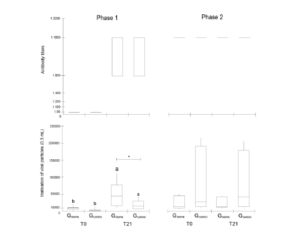
A significant time effect was observed for TBARS and MDA (p<0.01; Figure 4), along with a trend for a treatment effect on TBARS (Gozone: 0.0667±0.0043≠Gcontrol: 0.0683±0.0050; p=0.058; Table 1). A phase effect was noted on antibody titers (p<0.05; Fig. 5 and Supplementary Table 1) and the inactivation of viral particles (p<0.002; Fig. 5), thus the data were presented separately. Concerning the inactivation of viral particles, a trend for a time effect (p=0.065), treatment effect (p=0.054), and time x treatment interaction (p=0.114) was observed in phase 1 (Table 1). In both groups, the inactivation of viral particles at T0 was lower than at T21 (p<0.05). Additionally, there was a trend (p=0.114) indicating that Gozone had greater inactivation of viral particles at T21 compared to Gcontrol. In terms of frequency data, there was an average increase of 452% and 386% in the inactivation of viral particles in Gozone and Gcontrol, respectively, from T0 to T21. Notably, prior to data collection in phase 2 (T0) and the crossover of the experimental groups, Gozone showed a 75% increase in the inactivation of viral particles, compared to a 25% increase in Gcontrol. In phase 2, no effects related to time, treatment, or treatment x time interaction on antibody titers and inactivation of viral particles were observed (Fig. 5; Table 1).
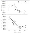
Thiobarbituric acid reactive species (TBARS) and malondialdehyde (MDA) values by treatment (Gozone: n=10 and Gcontrol: n=10) and over time in goats subjected to hyperimmune serum production. Different lowercase letters indicate differences over time in each experimental group (a, b; p<0.01).

Antibody titers and inactivation of viral particles in goats (n = 10/phase) subjected to hyperimmune serum production. Different lowercase letters indicate differences between time in each group (a, b; p<0.01). *Indicate differences between groups at the same time (tendency; p=0.114).
DISCUSSION
The first studies on the use of immune goat serum for measles prophylaxis date back to the late 1920s (Hoyne and Gasul, 1926; Tunnicliff and Hoyne, 1926). Using the adopted methodology, a strong immune response was achieved, with measles and mumps antibody titers reaching 1:1600. Other studies employing similar methods also reported comparable measles antibody titers (1:1024) from goats (Keshavarz et al., 2007). Interestingly, while the literature suggests that blood collection should occur 14 to 21 days after the antigen booster (Tunnicliff and Hoyne, 1926), higher antibody levels were observed 120 days after the first booster. Furthermore, a second booster administered at this point did not result in higher antibody titers or increased viral inactivation, which remained stable until the second blood collection 21 days later. As a result, following this protocol for hyperimmune serum production, a second booster application is unnecessary. Future studies should focus on monitoring the gradual increase in antibody production following booster stimulation until it stabilizes, in order to determine the optimal timing for blood collection.
It is believed that the humoral response observed in the present study may be due to the effects of O3, which interacts with polyunsaturated fatty acids to generate H2O2 and alkenals, including 4-hydroxynonenal (4-HNE). These compounds, in turn, alter the cysteine residues of Keap1, inhibiting ubiquitin conjugation to Nrf2 by the Keap1 complex and stimulating the release and nuclear translocation of Nrf2 (Pecorelli et al., 2013; Re et al., 2014). Similarly, Yu et al. (2016), using an O3 dose of 1.1mg/kg administered via rectal insufflation, showed that its beneficial biological effects may be partly attributed to the restoration of impaired Nrf2 activation and the downregulation of NF-κB activation in rats with adenine-induced chronic kidney disease. Therefore, such epigenetic modulation can initially increase the phagocytic count and activity of blood polymorphonuclear leukocytes (Mustafaev et al., 2007; Volkhovskaya et al., 2008). It can also trigger immunomodulatory effects by increasing serum cytokine levels, such as INF-γ, INF-β, IL-2, IL-6, IL-8, and IL-10 (Bocci et al., 1998; Sagai and Bocci, 2011; Tsuzuki et al., 2015; Delgado-Roche et al., 2017; Wei et al., 2018), as well as promoting the maturation of T-helper cells (CD4) and B-lymphocytes (CD20), which are related to the humoral immune response. Mustafaev et al. (2007) and Wei et al. (2018) demonstrated that ozone therapy can effectively improve blood immunoglobulin levels (IgG, IgA, and IgM) and immune function in human patients with prostatic adenoma and in rats with pelvic inflammatory disease, respectively
Regarding lipid peroxidation (oxidative stress) markers throughout the 12-hour incubation of ozonized and non-ozonized blood bags, our findings were intriguing. Greater TBARS and MDA values were expected in ozone-treated blood bags, as previously described in other studies using similar O3 concentrations ranging from 20 to 40μg/mL (Bocci et al., 1998; Travagli et al., 2007). However, MDA levels did not differ between groups, and TBARS levels showed a time effect in the control group (Gcontrol), with higher values at 4 hours compared to other time points. Recently, it has been demonstrated that ozone therapy, when combined with chemotherapy, can reduce tissue toxicity. It has been suggested that oxidative preconditioning may induce an effect similar to that observed in other phenomena such as exercise, ischemic preconditioning, and thermal or chemical preconditioning. A common feature of all these processes is that continuous and "moderate-controlled" stress can provide protection against prolonged and severe stress (Clavo et al., 2019). Therefore, a similar hormetic effect may have been achieved in the current study.
A slight increase in HCT was observed. Despite the low blood volume (250mL) collected per goat, which was returned to them 12 hours later, this may have been enough to trigger a spinal cord response, with HCT improvements occurring from 14 to 21 days later. In response to the booster application, neutrophilic leukocytosis was observed from 24 to 60 hours, peaking between 32 and 36 hours after inoculation. This finding was similar to what was previously reported by Tunnicliff and Hoyne (1926), who described an increase in WBC count between 24 and 48 hours (from 11,600 to 15,600 cells/mm³) following measles inoculation in goats.
Studies in both human and veterinary medicine have described inflammatory responses associated with blood transfusions (McMichael et al., 2010; Sousa et al., 2012, 2020). In Study 1, the autologous blood transfusion did not cause any disturbances detectable by complete blood count, even after the blood had been stored for 12 hours. However, an in vitro study showed a significant increase in seven markers of inflammation during prolonged blood storage (McFaul et al., 2009). Moreover, longer blood storage can promote leukocyte lysis, along with the release of cytokines and inflammatory immunomodulators, such as histamine, myeloperoxidase, and eosinophil cationic protein (Nielsen et al., 1997), which triggers an inflammatory response and leads to an increase in WBC count.
Finally, to our knowledge, this is the first study to feature hematological and antibody changes occurring throughout the production of hyperimmune serum in goats, highlighting the stages of booster stimulation and antibody production. Additionally, it is the first to evaluate the effects of an ozone therapy protocol, which yielded provocative results regarding the inactivation of viral particles in goats. Although the results may be limited by the small number of animals used per group, they serve as a pilot study and can inform future trials.
CONCLUSION
All goats responded effectively to the hyperimmune serum production following the Oswaldo Cruz Foundation protocol against measles and mumps. Additionally, a second booster, under the current conditions, did not result in improved humoral values. Finally, O3-MAHT showed potential in enhancing the inactivation of viral particles, warranting further investigation. Future studies could explore the long-term effects of ozone therapy on immune modulation and its potential applications in other viral infections or therapeutic contexts.
ACKNOWLEDGMENTS
The study was supported by FAPERJ. FZB and MFAB are CNPq and Faperj fellows. HGFP and ACSR were supported by CAPES (code 001). The authors also thank vet students from UFF and laboratory technicians from Oswaldo Cruz Foundation who helped in the data collection and antibody analysis, respectively, throughout studies.
REFERENCES
- AAMER, H.; NOUR, E.; REFAIE, A. et al. Current perspectives for the use of medical ozone in veterinary medicine. CABI Rev., v.18, n.1, 2023.
- AHMED, S.M.U.; LUO, L.; NAMANI, A. et al. Nrf2 signaling pathway: Pivotal roles in inflammation. Biochim. Biophys. Acta, v.1863, p.585-597, 2017.
- ANVISA. Brazilian Health Regulatory Agency Measles, mumps, rubella vaccine (PB061-00). Farmacopeia Bras., v.1, p.873, 2019. 2024.
- BARENBERG, L.H.; LEWIS, J.M.; MESSER, W.H. et al. Measles Prophylaxis. JAMA, v.95, p.4-8, 1930.
- BELLINI, W.J.; HELFAND, R.F. The Challenges and Strategies for Laboratory Diagnosis of Measles in an International Setting. J. Infect. Dis., v.187, p.S283-S290, 2003.
- BOCCI, V.; DI PAOLO, N. Oxygen-ozone therapy in medicine: an update. Blood Purif., v.28, p.373-376, 2009.
- BOCCI, V.; VALACCHI, G.; CORRADESCHI, F.; FANETTI, G. Studies on the biological effects of ozone: 8. Effects on the total antioxidant status and on interleukin-8 production. Mediators Inflamm., v.7, p.313-317, 1998.
- CADOGAN, M.; DALGLEISH, A.G.; HEENEY, J.; WHITE, S.D.T. Use of polyclonal anti-hiv goat serum as a therapeutic agent. European Patent Office EP1404367B1. 2001 Apr 07.
- CLAVO, B.; ESPARRAGÓN, F.R.; ABREU, D.R. et al. Modulation of oxidative stress by ozone therapy in the prevention and treatment of chemotherapy-induced toxicity: review and prospects. Antioxidants, v.8, p.588, 2019.
- DELGADO-ROCHE, L.; ROMO, M.R.; MESTA, F. et al. Medical ozone promotes Nrf2 phosphorylation reducing oxidative stress and pro-inflammatory cytokines in multiple sclerosis patients. Eur. J. Pharmacol., v. 811, p.148-154, 2017.
- ĐURIČIĆ, D.; VALPOTIĆ, H.; ŽAJA, I.Ž.; SAMARDŽIJA, M. Prophylaxis and therapeutic potential of ozone in buiatrics: Current knowledge. Anim. Reprod. Sci., v.159, p.1-7, 2015.
- ĐURIČIĆ, D.; VALPOTIĆ, H.; SAMARDŽIJA, M. Comparison of intrauterine antibiotics versus ozone medical use in sheep with retained placenta and following obstetric assistance. Reprod. Domest. Anim., v.51, p.538-540, 2016.
- FERREIRA, A.L.A.; MACHADO, P.E.A.. MATSUBARA, L.S. Lipid peroxidation, antioxidant enzymes and glutathione levels in human erythrocytes exposed to colloidal iron hydroxide in vitro. Braz. J. Med. Biol. Res., v.32, p.689-694, 1999.
- GALIÈ, M.; COVI, V.; TABARACCI, G.; MALATESTA, M. The role of nrf2 in the antioxidant cellular response to medical ozone exposure. Int. J. Mol. Sci., v.20, p.4009, 2019.
- HOYNE, A.L.; GASUL, B.M. Measles prophylaxis. JAMA, v.87, p.1185, 1926.
- IMHOF, S.; LUTERNAUER, M.; HÜSLER, J. et al. Therapy of retained fetal membranes in cattle: comparison of two treatment protocols. Anim. Reprod. Sci., v.206, p.11-16, 2019.
- KESHAVARZ, M.; SHAFYI, A.; TARIQI, G.H. et al. Production of high titre goat anti measles serum for quality control of MMR and MR vaccines and seroepidemiology researches. Vet. Res. Biol. Prod., v.20, p.70-77, 2007.
- MCFAUL, S.J.; CORLEY, J.B.; MESTER, C.W.; NATH, J. Packed blood cells stored in AS-5 become proinflammatory during storage. Transfusion, v.49, p.1451-1460, 2009.
- MCMICHAEL, M.A.; SMITH, S.A.; GALLIGAN, A.; SWANSON, K.S.; FAN, T.M. Effect of leukoreduction on transfusion-induced inflammation in dogs. J. Vet. Intern. Med.,v.24, p.1131-1137, 2010.
- MITCHELL, C.A.; GUERIN, L.F.; PASIEKA, A.E. Antibody production in milk serum after virus instillation of goat mammary gland: i. responses to influenza virus, mumps virus, and adenovirus 3. Can. J. Microbiol., v.13, p.1069-1078, 1967.
- MUSTAFAEV, E.M.; MARTOV, A.G.; NAUMOV, A.G. et al. The role of ozone therapy in prevention of pyoinflammatory complications after transurethral resection of prostatic adenoma. Urologia, v.27, p.18-23, 2007.
- RABELO NETTO, E.J.; LEAL, E.C.; DELGADO, I.F.; LEANDRO, K.C et al. Avaliação do controle da qualidade realizado nos produtos vacinais para sarampo, caxumba e rubéola utilizados no Programa Nacional de Imunizações do Brasil no período de 1999 a 2007. Rev. Inst. Adolfo Lutz, v.69, p.408-415, 2010.
- NIELSEN, H.J.; REIMERT, C.; PEDERSEN, A.N. et al. Leucocyte derived bioactive substances in fresh frozen plasma. Br. J. Anaesth., v.78, p.548-552, 1997.
- PECORELLI, A.; BOCCI, V.; ACQUAVIVA, A. et al. NRF2 activation is involved in ozonated human serum upregulation of HO-1 in endothelial cells. Toxicol. Appl. Pharmacol., v.267, p.30-40, 2013.
- RE, L., MARTÍNEZ-SÁNCHEZ, G., BORDICCHIA, M. et al. Is ozone pre-conditioning effect linked to Nrf2/EpRE activation pathway in vivo? A preliminary result. Eur. J. Pharmacol., v.742, p.158-162, 2014.
- REQUIREMENTS for measles, mumps and rubella vaccines and combined vaccines (live) (requirements for biological substance no. 47). Geneva: WHO, 1994.
- SAGAI, M., BOCCI, V. Mechanisms of action involved in ozone therapy: is healing induced via a mild oxidative stress? Med. Gas. Res., v.1, p.1-18, 2011.
- SATTAR, S.A.; MITCHELL, C.A.; GUERIN, F.L. Fluorescein-labeled goat milk antibodies: use in detection of mumps virus in tissue culture. Can. J. Comp. Med. Vet. Sci., v.10, p.265-267, 1967.
- SCHWARTZ, A.; SÁNCHEZ, G.M.; SABAH, F. Madrid declaration on ozone therapy. 3.ed. Madrid: ISCO, 2020. 104p.
- SOUSA, R.S.; CHAVES, D.F.; BARRÊTO-JÚNIOR, R.A. et al. Clinical, haematological and biochemical responses of sheep undergoing autologous blood transfusion. BMC Vet. Res., v.8, p.1-7, 2012.
- SOUSA, R.S.; MINERVINO, A.H.H.; OLIVEIRA, F.L.C. et al. Impact of blood storage duration on hematologic, blood gas, biochemical, and oxidative stress variables in sheep undergoing allogeneic blood transfusions. Vet. Clin. Pathol., v.49, p.545-556, 2020.
- SUTTON, I.; KERMODE, A.G. Hyperimmune goat serum (Aimspro): Hope or hype. J. Clin. Neurosci., v.13, p.1037-1038, 2006.
- THRALL, M.A.; WEISER, G.; ALLISON, R.; CAMPBELL, T. 2012. Veterinary hematology and clinical chemistry.ed. Hoboken, NJ: Wiley-Blackwel, 2012. 762p.
- TRAVAGLI, V.; ZANARDI, I.; SILVIETTI, A.; BOCCI, V. A physicochemical investigation on the effects of ozone on blood. Int. J. Biol. Macromol.,v.41, p.504-511, 2007.
- TSUZUKI, N.; ENDO, Y.; KIKKAWA, L. et al. Effects of ozonated autohemotherapy on the antioxidant capacity of Thoroughbred horses. J. Vet. Med. Sci., v.77, p.1647-1650, 2015.
- TUNNICLIFF, R.; HOYNE, A.L. Prevention of measles by immune goat serum. JAMA, v.87, p.2139-2140, 1926.
- VOLKHOVSKAYA, N.B.; TKACHENKO, S.B.; BELOPOLSKY, A.A. et al. Modulation of phagocytic activity of blood polynuclear leukocytes with ozonized physiological saline. Bull. Exp. Biol. Med., v.146, p.559-561, 2008.
- WEI, A.; FENG, H.; JIA, X.M. et al. Ozone therapy ameliorates inflammation and endometrial injury in rats with pelvic inflammatory disease. Biomed. Pharmacother., v.107, p.1418-1425, 2018.
- YU, G.; LIU, X.; CHEN, Z. et al. Ozone therapy could attenuate tubulointerstitial injury in adenine-induced CKD rats by mediating and NF-κB. Iran. J. Basic Med. Sci., v.19, p.1136-1143, 2016.
- ZUBAVICHENE, N.M.K.; SERGEYEV, N.N.; CHEPURNOV, A.A.; NETESOV, S.V. Preparation and use of hyperimmune serum for prophylaxis and therapy of ebola virus infections. J. Infect. Dis., v.179, p.S218-S223, 1999.