Treatment of anasarca in neonatal American Bully dogs - case report
ABSTRACT
Fetal anasarca is a congenital disorder commonly described in brachycephalic breeds, characterized by generalized edema, which may be associated with cardiac, lymphatic, hepatic or renal malformations. As it is considered a defect with often lethal prognosis, it is a condition that is often neglected in clinical routine, since many animals do not receive adequate treatment. Thus, the objective of this report was to describe the treatment of anasarca in newborn American Bully dogs and discuss the possible cause of this defect in the litter. Four neonates presented generalized edema at birth, and treatment was initiated with furosemide, subcutaneously, every three hours. After six administrations of the drug, the neonates reached the ideal weight, with no generalized edema. In the complementary exams, renal alterations were followed by ultrasound, demonstrating altered cortico-medullary relationships, with a report describing a possible congenital nephropathy, suggestive of renal dysplasia. Urea and creatinine were within normal limits for age. Quarterly monitoring of renal changes was recommended. This report demonstrates that treatment of anasarca is possible, and the clinical response of the animal will depend on the underlying cause. Furosemide was effective in treating the generalized edema in this litter. Immediate identification and intervention are essential for the survival of these patients.
Keyword:
canine neonatology; congenital malformation; fetal hydrops; renal dysplasia; puppy
RESUMO
A anasarca fetal é um distúrbio congênito comumente descrito em raças braquicefálicas, caracterizado por edema generalizado, podendo estar associado a malformações cardíacas, linfáticas, hepáticas ou renais. Por ser considerado um defeito de prognóstico reservado e muitas vezes letal, é uma condição frequentemente negligenciada na rotina clínica, e, por conseguinte, muitos animais não recebem o tratamento adequado. Assim, o objetivo deste relato foi descrever a terapêutica da anasarca em cães recém-nascidos e discutir a possível causa desse defeito na ninhada. Quatro neonatos da raça American Bully apresentavam edema generalizado no momento do nascimento, iniciando-se o tratamento com o diurético furosemida, por via subcutânea, a cada três horas. Após seis administrações do fármaco, os neonatos atingiram um peso ideal, com ausência de edema generalizado. Nos exames complementares, foi observada alteração renal em ultrassonografia, demonstrando relações corticomedulares alteradas, sendo a cortical adelgaçada, com laudo descrevendo uma possível nefropatia congênita, sugestiva de displasia renal. Ureia e creatinina estavam dentro dos padrões de normalidade para a idade. Foi recomendada a monitorização da alteração renal a cada três meses. Este relato demonstra que o tratamento da anasarca é possível, e a resposta clínica do animal dependerá da causa base. A furosemida foi eficaz na terapêutica do edema generalizado dessa ninhada. A identificação e a intervenção imediatas são fundamentais para a sobrevivência desses pacientes.
Palavras-chave:
neonatologia canina; malformação congênita; hidropisia fetal; displasia renal; filhote
INTRODUCTION
Anasarca, also known as congenital edema or fetal hydrops, is a defect characterized by the generalized accumulation of fluids in the subcutaneous space, which may be accompanied by thoracic and abdominal effusions. This disease is usually associated with cardiac, lymphatic, hepatic, and renal anomalies, nutritional deficiencies, or infections, which result in an imbalance in fetal fluid homeostasis (Smith, 2011; Siena et al., 2022).
This deformity has been described in humans, dogs, cats, cows, sheep, and other species (Monteagudo et al., 2002; Hopper et al., 2004; Çetinkaya et al., 2013; Brough et al., 2016; Alagar et al., 2017). In dogs, the incidence of anasarca is 0.7% (Pereira et al., 2019), with a high prevalence in brachycephalic breeds, mainly in English Bulldogs, French Bulldogs, Pugs and American Bullies, associated with a probable autosomal recessive genetic component of hereditary character, resulting in malformations in organic systems that lead to increased hydrostatic pressure or obstruction of fluid clearance (Hopper et al., 2004; Smith, 2011; Szaluś-Jordanow et al., 2024). However, teratogenic causes have also been described in dogs, such as infection during gestation by canine parvovirus type I (canine minute virus) and Ehrlichia canis, culminating in vasculitis, increased vascular permeability and generalized edema. Anasarca is also described in cases of hypoalbuminemia, resulting in decreased oncotic pressure (Carmichael et al., 1991; Bento et al., 2012).
Diagnosis can be made by gestational ultrasound, observing an anechoic image, with thoracic and abdominal cavities filled with fluids, and variable thickness of the subcutaneous tissue. Since this condition can develop in one or more fetuses in the litter and can cause obstructive dystocia, a cesarean section should be scheduled (Hopper et al., 2004; Silva et al., 2012; Siena et al., 2022).
Soon after birth, initial treatment consists of clinical management of respiratory distress, since these animals usually present pulmonary edema and dyspnea. Then, therapy with diuretics, such as furosemide, is initiated. The prognosis is described as reserved to poor (Cunto et al., 2015; Marçal et al., 2018; Vannucchi and Lourenço, 2015; Lourenço, 2023); however, the response to treatment and clinical improvement of newborns will depend on the cause of anasarca and the provision of immediate neonatal care (Cunto et al., 2015). Therefore, the objective of this report was to describe the treatment and clinical evolution of anasarca in neonatal puppies.
CASUISTRY
A pregnant American Bully dog, 1 year old, from a kennel, was attended at a veterinary hospital in Belo Horizonte - Minas Gerais, Brazil, for prenatal monitoring with gestational ultrasound after 60 days of intrauterine artificial insemination. During the ultrasound examination, an exacerbated accumulation of anechoic fluid was observed in the subcutaneous tissue and in the thoracic and abdominal cavities of some fetuses, diagnosing hydrops.
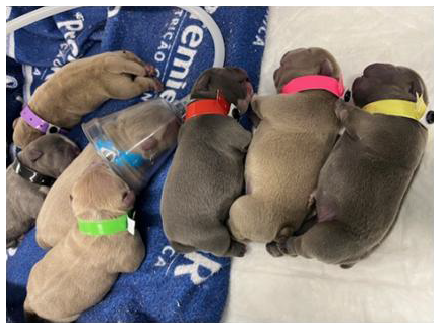
In the maternal history, the dog was primiparous, fed a super-premium commercial diet, vaccinated, dewormed, and had no history of diseases or use of medications during the gestational phase. The owner was unable to provide information on the history of malformations in the family line. The puppies' father had no birth defects. However, the bitch has a history of being diagnosed with the malformation aplasia cutis congenita, in the region of the frontal bone of the skull, when she was a puppy. After the diagnosis of fetal anasarca, the bitch underwent a cesarean section, resulting in the birth of seven puppies, four of which had mild anasarca (3 females and 1 male). The four neonates presented respiratory distress syndrome, manifesting cyanotic mucous membranes, dyspnea, bradycardia between 170 and 180 bpm, respiratory rate between 5 and 6 mpm, with an Apgar score of 3, absent muscle tone and reflex irritability, absent sucking reflexes, rooting and vestibular straightening, and hypothermia between 34.3 and 34.8 ºC. The weight of the puppies at birth ranged from 320 to 390 grams. Immediate neonatal care was provided with oxygen therapy by mask, warming in an incubator (32 ºC), and tactile respiratory stimulation with dorsal and thoracic massage, with the aid of dry and heated compresses. The neonates were then identified with colored ribbons around their necks (Fig. 1). After assistance, there was a significant improvement in neonatal parameters, with pink mucous membranes, heart rate >200 bpm, normothermia between 35.5 and 35.6 ºC, reflexes present, and improvement in the vitality score, with a score of 7.
Anasarca was treated with furosemide 2mg/kg, subcutaneously, every three hours; stimulation of urination (massage of the genital region with the aid of damp cotton) every 30 minutes; and monitoring of weight loss every 30 minutes. For every 30 grams of weight loss, five drops of 19.1% potassium chloride were empirically administered orally, due to potassium depletion caused by the diuretic. After 18 hours of treatment, the neonates reached an ideal weight, with no generalized edema, ranging from 270 to 330 grams. In total, six applications of furosemide were administered.

Pups in the incubator shortly after birth. Newborns with blue, black, pink, and yellow ribbons had anasarca. In the image, the pup with the blue ribbon is seen receiving oxygen therapy via mask.
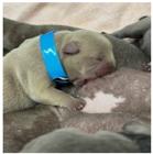
Soon after the start of treatment and clinical improvement, the neonates were placed to nurse on the bitch (Fig. 2). In addition to anasarca, the neonate with the pink ribbon presented a cleft palate malformation (Fig. 3). The nutritional management of this puppy was carried out by milking the mother's colostrum and feeding through an orogastric tube (using a number 6 urethral tube). This puppy was separated from the mother and subsequently fed with breast milk replacer (Support Milk Dog®) in a volume of 3 ml per 100 grams of weight, every three hours, through the orogastric tube.

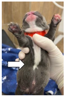
The puppy with the red ribbon had pectus excavatum malformation (Fig. 4), presenting clinical signs at seven days of age: dyspnea, cyanosis and substernal retraction during inspiration. Due to intense dyspnea, the neonate died before treatment began.
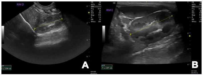
To investigate the possible cause of anasarca in the litter, an abdominal ultrasound was performed on a 20-day-old pup to diagnose possible alterations/malformations in organs. It was observed that the kidneys had regular surfaces, but with altered cortico-medullary relationships, with the cortex being thinned, with a report suggesting a possible nephropathy to be clarified. Despite this, the topographies and dimensions were normal, with the left kidney measuring approximately 2.97cm, and the right kidney 3.54cm. The distinction of the cortico-medullary junction was maintained and presented normal echogenicity of the layers (Fig. 5). No alterations were observed in other abdominal organs. The owner did not authorize further tests such as biochemical tests of renal function, urinalysis or other imaging tests in the other pups. However, the owner was advised about the need to investigate and monitor this alteration.

Red ribbon neonate with pectus excavatum, showing ventrodorsal narrowing in the sternal region (arrow).

At three months of age, tests were performed to reevaluate the kidneys of the same puppy: abdominal ultrasound, complete blood count, urea and creatinine. The ultrasound examination showed the kidneys with the same findings described previously, with an altered cortico-medullary relationship, with the cortex thinned (Fig. 6), suggesting nephropathy to be clarified. The complete blood count showed normocytic normochromic anemia (non-regenerative). Urea and creatinine were within the reference standards for the age.
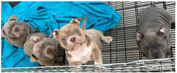
The four puppies are currently healthy and five months old (Fig. 7). Since anasarca is usually a hereditary defect in brachycephalic breeds, and congenital renal abnormalities were observed as a possible cause, the owner was advised to spay the puppies and the bitch in the future, avoiding breeding of these animals. A renal evaluation was recommended every three months to monitor the evolution of the observed abnormalities and to perform tests on the remaining puppies.

Left (A) and right (B) kidney with changes in relation to the cortico-medullary, demonstrating thinned cortex.

DISCUSSION
Malformations in dogs are related to prenatal events resulting from genetic factors, which may be hereditary, or to teratogenic factors, such as administration of drugs during pregnancy, exposure to toxic agents, chemicals, radiation, infectious diseases, unbalanced diet, among others (Smith, 2011 and Casal, 2016; Pereira et al., 2019). In this case, during the anamnesis of the bitch, the occurrence of teratogenic factors was not described. Since the bitch had congenital aplasia cutis and the litter had three types of malformations (anasarca, cleft palate and pectus excavatum), and it is known that these malformations occur mainly in purebred dogs, due to low genetic variability and increased homozygosity (Casal, 2016 and Pereira et al., 2019), it is possible that the cause of the deformities in this litter are related to genetic factors of the breed.
A diagnostic hypothesis for the cause of the anasarca in this report is renal dysplasia, which consists of the alteration between the cortex and the medulla, and disorganization of the renal parenchyma due to abnormalities during nephrogenesis. This deformity is congenital and hereditary, unilateral or bilateral, and can develop focally or disseminated throughout the kidneys, and can lead to chronic renal failure in young dogs. On ultrasound, the kidneys may be hyperechogenic, reduced in size, or with loss of the corticomedullary relationship (Hünning et al., 2009; Lima et al., 2017; Tusa and Levi, 2022). In this report, the corticomedullary relationship was altered, with a thinned cortex. Changes in laboratory tests will depend on the stage of renal failure, and normocytic normochromic anemia, azotemia, hyperphosphatemia, and metabolic acidosis may be observed (Tusa and Levi, 2022). In this report, normocytic normochromic anemia was observed. However, the definitive diagnosis is made by histopathological examination of the kidneys (Hünning et al., 2009; Tusa and Levi, 2022).
One report described a case of renal dysplasia in the American Bully breed (Homem et al., 2023). A seven-month-old dog presented with seizures, vomiting, hyperthermia, polyuria, and polydipsia. The complete blood count and biochemistry showed alterations such as normocytic normochromic regenerative anemia and increased creatinine. The ultrasound examination showed renal alterations (loss of the corticomedullary relationship and lack of definition of the corticomedullary junction of both kidneys). The animal was euthanized, and histopathological examination of the kidneys confirmed congenital renal dysplasia (Homem et al., 2023). This demonstrates that renal dysplasia may have a genetic nature in this breed.
In humans, renal dysplasia is described as a cause of anasarca in newborns (Çetinkaya et al., 2013; Mardy et al., 2019; Teles-Silva et al., 2020). The pathophysiology is associated with disorders of urinary flow obstruction or venous return, which may play a role in increased hydrostatic pressure or central venous pressure, increased vascular permeability and consequent increase in interstitial fluid (Bellini et al., 2009; Çetinkaya et al., 2013). This may have possibly occurred in the litter of this report. Despite the renal immaturity observed in neonatal dogs (Grundy, 2006), therapy with the diuretic furosemide was successful in the puppies of this report, with a reduction in generalized edema observed over the course of hours. No adverse effects were observed. In two reports of English Bulldog litters with anasarca, furosemide was also used as treatment, with clinical improvement observed (Cunto et al., 2015; Marçal et al., 2018). The duration of treatment and the number of doses administered vary according to the degree of anasarca in each neonate. Treatment should be terminated when clinically no generalized edema is observed (Lourenço, 2023). In this report, six administrations were necessary.
Anasarca is commonly described as a lethal disease or with a poor prognosis. Consequently, this disease is still largely neglected in clinical practice. It is essential that veterinarians are aware of the treatment, and that the patient's clinical improvement will depend on the cause of the anasarca. Puppies with heart disease and other congenital defects may have poor prognosis. However, it is necessary to start treatment of all animals and provide other care.
CONCLUSION
Anasarca should not be neglected and considered lethal in all cases. Treatment is possible and the prognosis will depend on the underlying cause. Furosemide was effective in reducing the generalized edema of the puppies in this report. Assessment of newborns and early intervention are essential for adequate treatment and survival of patients. In this case, a possible congenital renal malformation may be associated with the cause of anasarca, which should be monitored with additional tests to assess the progression of nephropathy.
REFERENCES
- ALAGAR, S.; VELLADURAI, C.; MANIVANNAN, S.; SELVARAJU, M. Succesfull management of dystocia due to foetal anasarca in Holstein friesian crossbred cows. Am. J. Rep. Anim., v.7, p.205-207, 2017.
- BELLINI, C.; HENNEKAM, R.C.; FULCHERI, E. et al. Etiology of nonimmune hydrops fetalis: a systematic review. Am. J. Med. Gen. A., v.149A, p.844-851, 2009.
- BENTO, D.D.; CHACAR, F.C.; ARRUDA, V.D. et al. Anasarca associated to vasculite caused by canine erlichiose: a case report. v.17, p.96-98, 2012.
- BROUGH, A.; LLEWELLYN, R.; NAKAHARA, N.; AVESTON, F. Fetal anasarca in an Abyssinian Kitten. Clin. Theriogenology. v.8, p.425-429, 2016.
- CARMICHAEL, L.E.; SCHLAFER, D.H.; HASHIMOTO, A. Pathogenicity of minute virus of canines (MVC) for the canine fetus. Cornell Vet. v., 81, p.151-171, 1991.
- CASAL, M.L. Congenital and genetic diseases of puppies before the weaning: can we prevent them?. In: INTERNATIONAL SYMPOSIUM ON CANINE AND FELINE REPRODUCTION, 8., 2016, Paris. Proceedings.... Paris: IVIS, 2016.
- ÇETINKAYA, M.; DURMAZ, O.; BÜYÜKKALE, G. et al. Neonatal Bartter syndrome and unilateral ectopic renal cyst as new renal causes of hydrops fetalis: two case reports and review of the literature. J. Mat. Neo. Med., v.26, p.1147-1150, 2013.
- CUNTO, M.; ZAMBELLI, D.; CASTAGNETTI, C. et al. Diagnosis and treatment of foetal anasarca in two English bulldog puppies. Pak. Vet. J., v.35, p.251-253, 2015.
- GRUNDY, S. A. Clinically relevant physiology of the neonate. Vet. Clin. North Am. Small Anim. Pract., v.36, p.443-459, 2006.
- HOMEM, R.T.; STACKE, F.C.; BUDIN, M.C. Displasia renal em cão - relato de caso. Scient. Eletr. Arch., v.16, p.73-77, 2023.
- HOPPER, B.J.; RICHARDSON, J.L.; LESTER, N.V. Spontaneous antenatal resolution of canine hydrops fetalis diagnosed by ultrasound. J. Small. Anim. Pract., v.45, p.2-8, 2004.
- HÜNNING, P.S; AGUIAR, J.; LARCERDA, L.A. et al. Displasia renal em um cão. Acta. Sci. Vet., v.37, p.73-77, 2009.
- LIMA, S.R.; SILVA, L.A.; DIAS, G.B.G. et al. Displasia renal em cães: estudo retrospectivo (2008-2013). Acta. Sci. Vet., v.45, p.5, 2017.
- LOURENÇO, M.L.G. Cuidados com neonatos e filhotes. In: JERICÓ, M.M.; KOGIKA, M.M.; ANDRADE, N.J.P. Tratado de medicina interna de caes e gatos. 2.ed. Rio de Janeiro: Guanabara Koogan, 2023.
- MARÇAL, A.P.; SILVA, T.L.; FERRAZ, A.F. et al. Anasarca congênita em cães da raça buldogue inglês: relato de caso. Nosso Clín., v.21, p.12-16, 2018.
- MARDY, A.H.; CHETTY, S.P.; NORTON, M.E.; SPARKS, T.N. A system‐based approach to the genetic etiologies of non‐immune hydrops fetalis. Prenat Diagn., v.39, p.732-750, 2019.
- MONTEAGUDO, L.L.L.; TEJEDOR, T.; CLIMENT, S. et al. Fetal anasarca (hydrops foetalis) associated with lymphoid tissue agenesis possibly due to an autosomal recessive gene defect in sheep. Theriogenology, v.58, p.1219-1228, 2002.
- PEREIRA, K.H.N.P.; CORREIA, L.E.C.S.; OLIVEIRA, E.L.R. et al. Incidence of congenital malformations and impact on the mortality of neonatal canines. Theriogenology, v.140, p.52-57, 2019.
- SIENA, G.; CORRÒ, M.; ZANARDELLO, C. et al. A case report of a rapid development of fetal anasarca in a canine pregnancy at term. Vet. Res. Commun., v.46, p.597-602, 2022.
- SILVA, T.M., ZALKIMI, R.S.; GARCIA, P.D. et al. Diagnóstico ultrassonográfico de hidropisia fetal intrauterino-relato de caso. Rev. Educ. Contin. Med. Vet. Zootec. CRMV-SP., v.10, p.26-31, 2012.
- SMITH, F.O. Prenatal care for the dog and the cat. In: PETERSON, M.E.; KUTZLER, M.A. (Eds.). Small animal pediatrics. Saint Louis, MO: Elsevier, 2011. p.1-10.
- SZALUŚ-JORDANOW, O., BARSZCZ, K., MĄDRY, W. et al. Hydrops fetalis caused by a complex congenital heart defect with concurrent hypoplasia of pulmonary blood vessels and lungs visualized by micro-CT in a French Bulldog. BMC Vet. Res. v.20, p.189, 2024.
- TELES-SILVA, C.; MARTINS, F.; COSTA, S.; SOARES, P. et al. renal dysplasia and progressive renal failure in a newborn with interstitial chromosome 4 deletion 4q25-28.3: a new phenotype?. J. Pediatr. Genet., v.10, p.39-44, 2020.
- TUSA, N.V.; LEVY, N.A. Congenital bilateral renal dysplasia in a Cavalier King Charles Spaniel. Vet. Rec. Case. Rep., v.10, p.1-3, 2022.
- VANNUCCHI, C.I.; LOURENÇO, M.L.G. Neonatologia. In: CRIVELLENTI, L.Z.; BORIN-CRIVELLENTI, S. Casos de rotina em medicina veterinária de pequenos animais. 3.ed. São Paulo: Medvet, 2015. 1248p.