Limb amputation in ruminants: report of six cases
ABSTRACT
Amputation is a rare practice in the treatment of the ruminants due to the complexity of the procedure, high costs, and unpredictable prognosis. The decision to proceed with amputation is made after evaluating factors such as the severity of the fracture, available treatment options, the economic and genetic value of the animal, prognosis, and costs. Performing amputation in cases with favorable prognosis can offer several financial benefits to the producer and welfare benefits to the animal. Thus, this study reports on six amputation surgeries in ruminants, including four cattle, one goat, and one sheep. The procedures were performed both in a hospital setting and in the field, using appropriate anesthetic protocols and surgical techniques. All animals recovered well, returning to locomotion and gaining weight during the postoperative period. The results show amputation as a viable surgical approach with a favorable prognosis, aligning with animal welfare standards. It serves as an alternative to euthanasia, allowing for good recovery and adaptation of the patient, which in turn consequently promotes financial return to the producer.
Keywords:
surgery; locomotor; fracture; anesthesia
RESUMO
A amputação é uma prática rara no tratamento de ruminantes, devido à complexidade do procedimento, aos altos custos e ao prognóstico imprevisível. A decisão é tomada após avaliação de fatores como gravidade da fratura, opções de tratamento, valor econômico e genético do animal, prognóstico e custos envolvidos. A realização da amputação em casos indicados com prognóstico favorável pode trazer diversos benefícios econômicos ao produtor e bem-estar ao animal. Desse modo, este estudo tem o intuito de relatar seis cirurgias de amputação em ruminantes, abrangendo quatro bovinos, um caprino e um ovino. Os procedimentos foram realizados em ambientes hospitalares e a campo, com protocolo anestésico e técnica cirúrgica adequados. Todos os animais apresentaram boa recuperação, retornando à locomoção e ganhando peso no pós-operatório. Os resultados evidenciam a amputação como uma abordagem cirúrgica viável e com prognóstico favorável dentro das normas de bem-estar animal, sendo uma alternativa à eutanásia, com boa recuperação e adaptação do paciente, permitindo-lhe mobilidade e conforto e, consequentemente, retorno financeiro ao produtor.
Palavras-chave:
cirurgia; locomotor; fratura; anestesia
INTRODUCTION
Cattle farming is one of the main economic activities in Brazil. The country is among the world's leading exporters of beef and milk (Beef…, 2023). However, producers face several challenges, especially regarding the health and management of the animals. Clinical and surgical issues, such as fractures and traumatic injuries, are common in young cattle due to inadequate management, poor transportation practices, and other factors (Spadeto Júnior et al., 2010), leading to significant economic losses. Fractures can be serious determinants of animal welfare, sometimes necessitating limb amputation to avoid euthanasia. Limb amputation may also be indicated in cases of osteomyelitis, poor circulation, severe muscles, nerves, or tendons ruptures, among other conditions (Peixoto Braga et al., 2017; Spadeto Jr et al., 2010; Gamsjaeger and Chigerwe, 2018). However, this procedure is rarely performed in ruminants as the process is complex, costs are usually high, and the prognosis is unpredictable (Mulon, 2013). Veterinarians also face challenges when conducting amputations in the field, such as exposure to weather conditions and lack of infrastructure, among others. Moreover, there are risks of complications, including rupture of the contralateral limb, prolonged decubitus, persistent claudication, low return to production, and a poor quality of life (Gamsjaeger and Chigerwe, 2018). Therefore, the decision to perform an amputation should be taken after evaluating several factors, such as the severity of the fracture, alternative treatments, the economic and genetic value of the animal, the prognosis, and treatment costs (Desrochers et al., 2014). Studies on amputation cases in cattle allow for an evaluation of the procedure's effectiveness and potential drawbacks. However, in Brazil, research on this topic is lacking (Peixoto Braga et al., 2017), and cases of amputation have only been reported at conferences within the country. Thus, this study is significant as it reports six cases of appendicular limb amputation in ruminants: four in cattle (one male and three females) (Bos taurus), one female goat (Capra hircus), and one female sheep (Ovis aries).
CASUISTRY
This study reports six cases of appendicular limb amputation in ruminants, including four bovines (one male and three female) (Bos taurus), one female goat (Capra hircus), and one female sheep (Ovis aries) of various ages. Two of the cases were treated at the Veterinary Hospital of the Federal Rural University of Rio de Janeiro (HV/UFRRJ), one at the Veterinary Hospital of the Federal University of Tocantins (HV/UFT), one in the municipality of Valença (RJ) and two in the municipality of Araguaína (TO) (see Table 1).
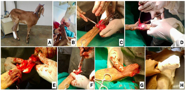
Two cases were treated at the Veterinary Hospital of the Federal Rural University of Rio de Janeiro (HV/UFRRJ). The first case involved an exposed fracture in the right metatarsophalangeal joint caused by the limb being trapped between the wooden slats of the barn 20 days prior (C1) (Fig. 1). The second case involved an exposed fracture of the left radius and ulna after the animal was run over (C2) (Fig. 2).

A female goat with an exposed fracture in the right metatarsophalangeal joint, treated at the Veterinary Hospital of the Federal Rural University of Rio de Janeiro (HV/UFRRJ), underwent amputation via tibiotarsal disarticulation. A) Animal in the pre-surgical period, showing claudication and lack of support on the right pelvic limb is observed. B) Local anesthesia was administered with a ring block using 2% lidocaine without vasoconstrictor (Lidovet®), at a dose of 10 ml (5 mg/kg), applied above the tibiotarsal joint. C) Two skin incisions (lateral and medial) were made approximately 5 cm below the hock. D) Blunt divulsion of the skin and subcutaneous tissue. E) Incision of the joint capsule and ligaments (long, short, medial, and lateral collateral) of the tibiotarsal joint with a scalpel blade no. 24. F) Correction of bone irregularities of the distal tibia with the aid of a gouge to prevent dehiscence of the skin suture. G) Dermorrhaphy was performed using no. 1 nylon thread in a simple interrupted pattern. H) Bandaging in three layers (primary layer with gauze; secondary layer with hydrophilic cotton; tertiary layer with bandage and adhesive tape) of the operated region.

Female sheep with exposed fractures of the left radius and ulna was treated at the Veterinary Hospital of the Federal Rural University of Rio de Janeiro (HV/UFRRJ) and underwent amputation via scapulohumeral disarticulation. A) Preoperatively, the animal exhibited grade V claudication with support on the left thoracic limb, alongside extensive soft tissue injuries, an exposed fracture of the radius and ulna, and exposure of ligaments and tendons. B) Dermoscopy following scapulohumeral disarticulation, using no. 1 nylon thread in a simple interrupted pattern. C) In the immediate postoperative period, the animal was observed standing in the stall with three-layer bandaging (primary layer with gauze; secondary layer with hydrophilic cotton; tertiary layer with bandage and adhesive tape) applied to the operated region.
A case was treated at the Veterinary Hospital of the Federal University of Tocantins (HV/UFT). It was an amputation via tibiotarsal disarticulation in a female bovine with an exposed fracture in the left metatarsal due to unknown etiology (C3) (Fig. 3).

A female bovine with an exposed fracture in the left metatarsal present for seven days due to unknown etiology, was treated at the Federal University of Tocantins (HV/UFT) and underwent amputation via tibiotarsal disarticulation. A) Pre-surgery: immobilization of the distal region below the hock of the left pelvic limb performed using a PVC pipe is observed. B) Radiographic projections (lateromedial: image on the left/anteroposterior: image on the right) of the affected limb, showing multiple fractures in the proximal and middle diaphysis of the left metatarsal with lateral displacement. C) Two skin incisions (lateral and medial) were made approximately 5 cm below the hock. D) Blunt dissection of the skin, subcutaneous tissue, incision of the joint capsule, and ligaments (long, short, medial and lateral collateral) of the tibiotarsal joint with a scalpel blade number 24. E) Hemostasis of the remaining blood vessels immediately after amputation of the limb. F) Dermoscopy using nylon thread number 1 in a simple interrupted pattern. G) Three-layer bandaging of the operated region protected externally with plastic film. H) Immediate postoperative period, showing the animal grazing in a stationary position.
A case was treated in the field on a farm located in the municipality of Valença-RJ, involving amputation due to right tibiotarsal disarticulation in a male calf, who had an exposed fracture in the right metatarsal after being trampled by its mother (C4) (Fig. 4).

A male calf with an exposed fracture in the right metatarsal after being trampled by its mother, treated in the field on a farm located in the city of Valença-RJ, underwent amputation via tibiotarsal disarticulation. A) The animal in the pre-surgery period, showing an exposed fracture of the right metatarsal. B) Two skin incisions (lateral and medial) were made approximately 5 cm below the hock. C) Blunt divulsion of the skin and subcutaneous tissue, with hemostasis of the blood vessels achieved using a hot iron. D) Incision of the joint capsule and ligaments (long, short, medial and lateral collateral) of the tibiotarsal joint with a scalpel blade no. 24. E) Dermoscopy using no. 1 nylon thread in a simple interrupted pattern. F) Immediate post-surgery, showing the animal standing in the stall with a three-layer bandage over the operated region, externally protected by a surgical glove.
Two cases were treated in the field on farms in the municipality of Araguaína-TO. Both involved amputation via tibiotarsal disarticulation due to laceration caused by fence wire. One case involved a female calf with an injury in the right metatarsophalangeal joint that occurred three days prior (C5) (Fig. 5), and the other involved a female bovine with a laceration to the left metatarsophalangeal joint that occurred four days prior (C6) (Fig. 6).

A female calf with a laceration caused by fence wire in the right metatarsophalangeal joint three days prior was treated in the field on a farm in the municipality of Araguaína-TO and underwent amputation via tibiotarsal disarticulation. A) Pre-surgery: The animal exhibited grade V claudication, with support of the right pelvic limb, along with an extensive laceration of soft tissues, bone exposure distal to the hock, and exposure of ligaments and tendons (lateral view). B) Medial view of the injury. C) Three-layer bandage protected externally by a surgical glove. D) Amputated right pelvic limb.

A female bovine with a laceration caused by fence wire in the left metatarsophalangeal joint, treated in the field on a farm in the municipality of Araguaína-TO four days after the injury, underwent amputation via tibiotarsal disarticulation. A) Pre-surgery: The animal exhibited grade V claudication with support on the left pelvic limb. B) Lateral view of the left pelvic limb, showing extensive laceration of soft tissues, with exposure of bones distal to the hock, ligaments, and tendons. C) Medial view of the lesion.
In the six cases, the animals underwent a physical evaluation, revealing clinical parameters within normal limits for each species. Notably, the animals exhibited either claudication without the support of the affected limb (C2) or grade V support claudication (other cases). Upon inspection, all cases presented extensive soft tissue injuries and/or fractures with exposure of bones, ligaments, and tendons (C1, 2, 3, and 4).
For patients treated at hospitals (C1, 2, and 3), blood was collected from the jugular vein for blood count and serum biochemistry, both of which showed parameters within normal limits. Additionally, radiographs in two projections (anteroposterior and lateral) were taken to evaluate the bone lesions and their respective prognosis in more detail. In C3, the distal region (below the hock) of the left pelvic limb was immobilized using a PVC pipe (Fig. 3A) to stabilize the fracture, allowing for radiographic projections (Fig. 3B). In contrast, for animals treated in the field (C4, 5 and 6), imaging and laboratory tests could not be performed due to transportation issues and difficulty accessing laboratories.
Due to the severity of the soft tissue and bone injuries presented by the six patients in this report, it was decided to perform amputation procedures on the affected appendicular limb regions. The bovine patients (C3, 4, 5, and 6) were subjected to a 12-hour fast from food and 8-hour fast from water, while the small ruminants (C1 and 2) were subjected to a 24-hour fast from food and 12-hour fast from water.
Different anesthetic protocols were used in the cases (Table 2). In C1 and C2, orotracheal intubation was performed, followed by connection to an anesthetic circuit for maintenance, enabling the animals to inhale anesthesia with isoflurane (Forane®) at an effective dose, along with oxygen therapy. The animals were positioned in lateral decubitus. The right tibiotarsal region of the goat (C1) and the left scapulohumeral region of the sheep (C2) were trichotomized, and antisepsis was performed using degerming and chlorhexidine.
In C1, two half-moon skin incisions were made approximately 5cm below the hock, followed by blunt divulsion of the subcutaneous tissue (Fig. 1C and 1D). The muscles (cranial tibial, peroneus tertius, long and short, long, lateral and short digital extensor, gastrocnemius, soleus, superficial and deep digital flexor, and proximal sesamoid) were incised at their insertion in the tibiotarsal joint using a scalpel blade number 24. The hemostasis of the regional vessels (saphenous, cranial, and caudal tibial arteries; cranial tibial and lateral saphenous, and medial saphenous veins) was achieved through ligation and, when necessary, transfixation using absorbable multifilament suture no. 2-0. The joint capsule and ligaments (long and short collateral medial and lateral ligaments) of the tibiotarsal joint were incised using a scalpel blade number 24. After disarticulation, the tarsometatarsal region of the limb was amputated (Fig. 1E). Bone irregularities of the distal tibia were corrected using a gouge to avoid dehiscence of the skin suture (Fig. 1F). The subcutaneous tissue was sutured using absorbable multifilament suture no. 2-0 in a mattress pattern, and dermoscopy was performed using nylon suture no. 1 with a simple interrupted pattern (Fig. 1G). The same surgical technique for tibiotarsal disarticulation was used in C3, 4, 5, and 6.
In C2, two half-moon skin incisions were made approximately 3 cm below the greater tubercle of the humerus, followed by blunt divulsion of the subcutaneous tissue. The muscles (brachiocephalic, superficial and deep pectoralis, deltoid, supraspinatus, infraspinatus, teres minor, latissimus dorsi, teres major, cutaneous trunk, biceps brachii, coracobrachialis, subscapularis, and triceps brachii) were incised at their insertion in the scapula and humerus using a scalpel blade no. 24. Hemostasis of the regional vessels (brachial artery; cephalic and brachial veins) was achieved through ligation and, when necessary, transfixation with absorbable multifilament suture number 2-0. The joint capsule of the scapulohumeral joint was incised with a scalpel blade number 24, and after disarticulation, the thoracic limb was amputated. The superficial and deep pectoralis muscles were sutured to the longissimus dorsi, teres major, infraspinatus, and brachiocephalic muscles using an absorbable multifilament suture number 2-0 in an “X” pattern. The subcutaneous cellular tissue was sutured with absorbable multifilament suture No. 2-0 in a mattress pattern, and dermoscopy was performed using nylon suture number 1 with a simple interrupted pattern (Fig. 2B).
In the postoperative period, C1, 2, and 4 received flunixin meglumine (Flunixin®; 1.1mg/kg; IM; SID; 3 days) and oxytetracycline hydrochloride 20% (Oxytetracycline LA®; 10mg/kg; IM; 5 applications on alternate days). For C3, 5, and 6, the postoperative regimen included meloxicam 2% (Maxican®; 0.5 mg/kg; IM; SID; 15 days) and enrofloxacin 10% (Chemitril®; 2.5mg/kg; IM; SID; 15 days). A three-layer bandage (primary layer with gauze; secondary layer with hydrophilic cotton; tertiary layer with bandage and adhesive tape) was applied to the operated regions of all animals (Figs. 1H, 2C, 3H, 4F, and 5C). The bandage was changed daily, and the wounds were cleaned with saline solution and topical povidone until complete healing. The animals were kept in restricted space with limited exercise for 15 days postoperatively, at which time the sutures were removed, and the patients were discharged. There were no postoperative complications reported in any of the cases. All animals displayed autonomy of locomotion within the first few hours postoperatively (Figs. 2C, 3H, and 4F), accompanied by an increasing appetite and weight gain during the recovery period.
DISCUSSION
This report is the first to document cases of limb amputation in ruminants in both surgical and field settings in Brazil. Severe traumatic injuries are common in ruminants (Mulon, 2013). However, limb amputation in these animals is not common, and has only been described in a few articles (Peixoto Braga et al., 2017) due to concerns regarding the animal's productivity and economic value, as well as the complexities associated with postoperative care and recovery, which can be further complicated by the animal's environment, size, and temperament (Fernandes and Terceiro, 2021; Rocha et al., 2021); this reinforces the importance of this work that reports successful cases of limb amputation in ruminants both in the surgical center and in the field.
All animals in this study exhibited a good recovery trajectory. Both young and adult ruminants adapted to the amputation procedure almost spontaneously, and the postoperative period lasted approximately 15 days. These findings suggest that amputation is a viable and safe alternative for managing severe limb injuries in ruminants, warranting consideration as an option to preserve the animal's life and welfare (Peixoto Braga et al., 2017).
In the reported cases, the animals had serious injuries to their limbs with no possibility of reconstruction or correction, leaving euthanasia or amputation as the only alternatives, as reported in the literature (Spadeto Júnior et al., 2010). Based on the standards of ethics and animal welfare (Ministério..., 2016), and in agreement with the animals' guardians, the best therapeutic option was executed to alleviate suffering while ensuring survival.
Radiographic examination is essential for assessing the extent of bone injuries and determining appropriate therapeutic options and prognosis (Grant and Olds, 2007). However, due to the differing contexts of the cases- some occurring in hospitals and others as emergency field care- radiographic examinations were not possible for all animals. In instances of exposed, severe, and/or infected fractures in the field, radiography can be unnecessary and difficult to access, making clinical evaluation sufficient for decision-making. It is expected that animals with severe fractures and infections would present specific changes in the blood count and serum biochemistry (Weiser et al., 2015). However, no changes were observed in the animals included in the study.
The anesthetic protocols used, both in the surgical center and the field, were efficient and allowed the safe performance of the procedures (Muir et al., 2008). The protocols used in the field were low-cost, easy to perform, and did not present complications. Despite the advantages, general anesthesia procedures require adequate respiratory support, which can pose risks to the animals, and expensive equipment that is difficult to transport to the field and not accessible to most owners. This highlights the importance of searching for safe, economically viable solutions and alternatives that can be performed in the field for the most different types of treatment of farm animals, ensuring their life, well-being, and, if possible, the productivity of these animals (Rocha et al., 2021).
The techniques employed for amputation are associated with better patient recovery (Desrochers and Anderson, 2014). The reported cases in this article followed all the recommendations described in the literature above and demonstrated a favorable prognosis. All animals regained locomotion autonomy and showed progressive increases in appetite and weight within hours post-surgery. No significant complications were recorded in the immediate postoperative period, indicating the effectiveness of the protocols adopted for perioperative management (Peixoto Braga et al., 2017; Desrochers and Anderson, 2014). Long-term prognosis depends on the animal's weight and subsequent management strategies. Research indicates that lighter animals generally experience more favorable outcomes (Gangl et al., 2006) and animal owners frequently fail to recognize the significance of long-term maintenance conditions. Consequently, the success rate of postoperative outcomes diminishes due to insufficient enhancements in the postoperative environment (Arican et al., 2024).
CONCLUSIONS
The results of the reported cases were favorable, as all animals successfully regained their autonomy of locomotion and demonstrated progressive weight gain and improved appetite during the first few hours of the postoperative period. No significant complications were noted during the immediate postoperative phase, indicating the effectiveness of the protocols adopted for perioperative management. Therefore, complete limb amputation emerges as a viable and satisfactory alternative to euthanasia, yielding positive outcomes in terms of recovery and adaptation for the patient. This approach not only facilitates mobility and comfort for the animals but also offers potential financial benefits to the producers.
REFERENCES
- ARICAN, M.; ALTAN, S.; PARLAK, K.; ALKAN,F. Interlocking Nail Stabilization Technique for Long Bone Fractures in Calves. Thai J. Vet. Med., v.53, p.213-220, 2024.
-
BEEF report: perfil da pecuária no Brasil. São Paulo: ABIEC, 2023. Disponível em: https://www.abiec.com.br/catpub/impressos/. Acessado em: 17 set. 2024.
» https://www.abiec.com.br/catpub/impressos - DESROCHERS, A.; ST-JEAN, G.; ANDERSON, D.E. Limb amputation and prosthesis. Vet. Clin. North Am. Food Anim. Pract., v.30, p.143-155, 2014.
- FERNANDES, A.S.S.; TERCEIRO, I.B. Fatores de morbimortalidade na amputação das extremidades inferiores de etiologia vascular na cidade de Marabá, Pará. Braz. J. Health Rev., v.4, p.29264-29276, 2021.
- GAMSJAEGER, L.; CHIGERWE, M. Indications for and outcomes of limb amputation in goats and sheep. Am. Vet. Med. Assoc., v.252, p.860-863, 2018.
- GANGL, M.; GRULKE, S.; SERTEYN, D.; TOUATI, K. Retrospective study of 99 cases of bone fractures in cattle treated by external coaptation or confinement. Vet. Rec., v.158, p.264-268, 2006.
- GRANT, G.R.; OLDS, R.B. Tratamento das fraturas expostas. In: SLATTER, D. (Ed.). Manual de cirurgia de pequenos animais. 3.ed. Barueri: Manole, v.2, cap.127, p.1793-1798, 2007.
- MINISTÉRIO DA CIÊNCIA, TECNOLOGIA E INOVAÇÃO. Diretriz brasileira para cuidado e uso de animais em atividades de ensino ou pesquisa científica. Brasília: MCTI, 2016.
- MUIR, W.W.; HUBBE, J.A. E.; SKARDA, R.T.; BEDNARSKI, R.M. Anestesia local em bovinos, ovejas, cabras y cerdos. In: AMBRÓSIO, A.M. Manual de anestesia veterinária. 4.ed. Madrid: Elsevier, 2008.
- MULON, P. Management of long bone fractures in cattle. In Practice, v.35, p.265-271, 2013.
- PEIXOTO BRAGA, G.; MOREIRA, I.L.; SOUZA, C.O. et al. Amputação de membro pélvico para tratamento de avulsão da articulação metatarso falangeana em um bovino. Rev. Acad. Ciênc. Anim., v.15, Suppl. 2, p.469-470, 2017.
- ROCHA, L.R.; OTT, L.C.; CHENARD, M.G. et al. Partial penectomy in a horse with paraphimosis. Acta Sci. Vet., v.49, Suppl., 2021.
- SPADETO JUNIOR, O.; FALEIROS, R.R.; ALVES, G.E.S. et al. Falhas na utilização de poliacetal e poliamida em forma de haste intramedular bloqueada para imobilização de fratura femural induzida em bovinos jovens. Ciênc. Rural, v.40, p.907-912, 2010.
- WEISER, G. Parte 1: Tecnologia laboratorial em medicina veterinária. In: THRALL, M.A.; WEISER, G.; ALLISON, R.W.; CAMPBELL, T.W. Hematologia e bioquímica clínica veterinária. 2.ed. Rio de Janeiro: Guanabara Koogan, 2015. p.868.